

 PDF
PDF Citation
 Print
Print


Introduction
Aluminum is the most abundant metal in the earth’s crust, and it is considered the second most important metallic material after steel. Aluminum has been associated with developmental neurotoxicity and its impact on the urinary system has been previously documented [1]. Aluminum is used in the processing and production of products such as cooking pots, frying pans, aluminum foil, beverage cans, car parts, and so on [1]. Thousands of aluminum products are produced annually thus making human and animal exposure to aluminum inevitable [2]. Exposure to aluminum is majorly through ingestion of contaminated food which could be from using aluminum cooking pots or any other household products made from aluminum [1]. Upon ingestion of aluminum contaminated food, the aluminum is absorbed and distributed to other tissues of the body, and it is excreted majorly by the kidney and partly through bile [3-5]. It has been established that aluminum can assess the fetal tissue due to its ability to cross the placental membrane [5]. Other studies have reported the ability of aluminium to cross the blood-brain barrier, thus its subsequent accumulation in the brain [6]. Aluminum exposure has been linked with behavioral disorders in laboratory animals, even in the absence of histopathological lessons [7]. Prenatal aluminium exposure is linked with delayed sensory-motor development, altered grip strength, and escape behaviour [1]. Prenatal aluminium exposure is also linked with learning and memory deficit [6, 8]. Mechanism of aluminum-induced neurotoxicity is through depletion of mitochondrial integrity and function, depletion of the glutathione pool, potentiation of iron induced oxidative pathway, and dysfunction of glial cells [9].
The cerebellar cortex controls motor function and is susceptible to impacts of neurotoxicants such as aluminum. The impact of aluminum exposure extends into adulthood [8]. Considering the documented impact of aluminum exposure on humans and animals, there is a need for a multidisciplinary approach to finding a remedy against its reported neurotoxic effects.
Alternative medical approach depends majorly on the use of some plant and animal products in the management of diverse health conditions [10]. Medicinal plants particularly play important role in the management of some complex animal and human disease conditions [11, 12]. Tamarindus indica is a plant mostly found in the dry savannas of tropical Africa [13]. Phytochemical examination by Panara et al. [14] revealed that Tamarindus indica contains phenols, glycoside, amino acid, alkaloids, and carbohydrates. Our recent independent studies on the ethyl acetate leaf fraction of Tamarindus indica (EATI) indicated the presence of flavonoids and carbohydrate based on the preliminary phytochemical screening, major components detected based on the gas chromatography–mass spectrometry screening of EATI included oleic acid, n-Hexadecanoic acid, Phenol, 3,5-bis (1,1-dimethyl ethyl), and cis-9-Hexadecenal (unpublished observation). Tamarindus indica is reported to possess antioxidant activities, antipyretic, antimicrobial, and hypolipidemic properties [15]. Administration of ethanol pulp extract of Tamarindus indica during prenatal ethanol exposure revealed some protective potentials on the cerebral cortex of wistar rats [15, 16]. A previous study on the effect of EATI during prenatal aluminum chloride (AlCl3) exposure revealed, improvement in brain metal concentration, oxidative stress parameters, GFAP expression in the cerebral cortex and the hippocampus (unpublished observation). To date, there is limited data available on the ameliorative effect of Tamarindus indica on the cerebellum following prenatal AlCl3 exposure. Hence the aim of the present study.
Go to : 
Materials and Methods
Tamarindus indica leaves were collected from the Botanical garden of Ahmadu Bello University, Zaria. The leaves were authenticated and assigned voucher number (2,417) in the Herbarium unit of the Department of Botany, Faculty of Life Sciences, Ahmadu Bello University, Zaria.
Fresh leaves of Tamarindus indica collected were shade dried, pulverized using an electric blender, the extracted by maceration method using ethanol as the solvent, followed by subsequent fractionation [16]. Pulverized powder of Tamarindus indica leaves (100 g) was suspended in in 500 ml ethanol for four days with continuous shaking. The mixture was then filtered using Whatman filter paper (no. 41). The collected filtrate was condensed at 60 °C in a rotary evaporator, then dried, with a percentage yield of 6.95 g. The different fractions (n-butanol, n-hexane, and ethyl acetate) of Tamarindus indica leaves was prepared by dissolving 6 g of the ethanol extract in 200 ml of warm water and successively partitioned with n-hexane (3×100 ml), ethyl acetate (3×100 ml), and n-butanol (3×100 ml). A Rotary evaporator was used to dry the different fractions at 40°C. The fractions were weighed separately, transferred into universal bottles for storage at a low temperature (4°C), and subsequent analysis. EATI was selected for the present study due to its rich phytochemical composition and oil-like composition.
Aluminum chloride (CAS Number: 7446-70-0) was obtained from Sigma-Aldrich (St. Louis, MO, USA). Aluminum chloride stock solution was prepared by dissolving 1g of aluminum chloride in 10 ml of distilled water to produce a working stock.
Capsules of vitamin E (Gujarat liquid pharmacaps Pvt; Gujarat, India) were obtained from a reputable drug store. The obtained capsules were cut open and emptied into a neat container. The stock solution was prepared using 0.01% w/w, Tween 80 (CAS Number: 9005-65-6) from Sigma-Aldrich (St. Louis, MO, USA), ensuring 0.2 ml of suspension contained 60 mg of vitamin E. The stock solution containing vitamin E was then shielded from direct light to avoid photodegradation.
Twenty (20) adult female rats and 10 adult male rats of Wistar strain were acclimatized for two weeks in the animal house of the Department of Anatomy, Ahmadu Bello University. The vaginal smear was taken from all the female rats and examined under a light microscope for the observation of their estrous cycle. The female rats in the proestrus phase were mated overnight with the male rats (ratio of 2 females to 1 male); the presence of vaginal plugs the following morning indicated mating and was taken as day zero of pregnancy [17-19]. Animals were allowed free access to feed and water before and during the experiment, and kept in a well ventilated housing condition. Ethical approval number ABUCAUC/2019/001 was obtained from the Ahmadu Bello University Committee on Animal Use and Care before the commencement of the experiment.
Gestational rats on day 7 of post-coitus (day 0 of the experiment) were administered the extracts for two weeks i.e. day 7 to day 21 of gestation. To reduce biases, animals were assigned random numbers and independently assigned to groups (n=4) in line with the ARRIVE guidelines on experimental animals. Group 1: distilled water (negative control), Group 2: 200 mg/kg bw of AlCl3 (positive control), Group 3: 200 mg/kg bw of AlCl3 and 400 mg/kg bw EATI, Group 4: 200 mg/kg bw of AlCl3 and 800 mg/kg bw EATI, Group 5: 200 mg/kg bw of AlCl3 and 300 mg/kg bw of vitamin E. A dosage of 200 mg/kg bw was adopted for AlCl3 based on previous studies in Wistar rats [20, 21]. The adopted dosage for EATI were 400 and 800 mg/kg bw (low and high dose respectively). A dosage of 300 mg/kg bw was adopted for vitamin E based on previous studies using the Wistar rat model [15, 16].
Beam walking test was performed as outlined by Stanley et al. [22], on postnatal days 17 to 20. The pups were trained to walk from a start platform along a ruler (100 cm long, 3 cm wide) elevated 50 cm to a goal box (enclosed hamster house) for 3 days, morning and evening. On the final day of the test, the pups were placed on the beam at one end and allowed to walk to the goal box. Pups that fell from the meter rule were returned to the position they fell from, each trial lasted for 1minutes (60 seconds). The time taken to traverse the beam and number of foot slips during the test were then recorded.
Neonatal rats on post gestation days 7 and 21 were euthanized by intraperitoneal injection of 5 mg/kg thiopental sodium since it was ethically acceptable in experimental animals [23, 24]. The skull was dissected and the brain tissues from the pups were harvested and fixed in 10% neutral buffered formalin for histological and immunohistochemical studies.
The fixed brain tissues were trimmed and processed with the aid of Automatic Benchtop Tissue Processor (TP1020; Leica, CA, USA). The processed tissues were cut using rotary microtome at a thickness of 6 μm, then stained for hematoxylin and eosin (H&E), Cluver berrera, and GFAP reactivity.
The tissue sections for H&E were stained as outlined by [25]. The sections were stained in hematoxylin for about 30 minutes after dewaxing and rehydration. The stained slide was rinsed in tap water for 5 minutes until the section turn blue. The sections were differentiated in 70% ethanol containing 1%HCl for about 5 seconds, to remove the excess stain. The sections were rinsed for 5 minutes in tap water, then stained in eosin solution for about 10 minutes. The sections were then rinsed for 5 minutes in tap water. Finally, the stained sections were dehydrated, cleared, and coverslipped for light microscopy [25, 26].
Tissue sections for Cluver berrera staining were cut at 10 microns thickness. Xylol was used for deparaffinization followed by subsequent running in absolute alcohol and several changes of 95% alcohol. The section was stained overnight in 0.1% solution of Luxol Fast Blue MBS (Essex, UK) at 57°C. The following morning, stained slides were dipped in 95% alcohol to wash off excess stain, followed by rinsing in distilled water. Differentiation was done by rapid immersion in 0.05% lithium carbonate. Differentiation was continued in several changes of 70% alcohol until gray and white matter can be distinguished, then subsequent rinsing in distilled water. The differentiated tissues slides were then stained in 0.25% solution of Coleman and Bell’s cresyl violet at 57°C for 6 minutes, followed by differentiation in several changes of 95% alcohol, and clearing in xylol-terpineol mixture, then mounted for microscopic examination. The cleared slides were mounted with synthetic resin for light microscopy.
Staining for GFAP was evaluated using a GFAP antibody (DAKO, Carpinteria, CA, USA). Sections were treated with 0.01 M citrate buffer (pH 6.0) for 10 minutes to unmask the antigen. The sections were then incubated in 0.3% H2O2 for 30 minutes to get rid of tissue endogenous peroxidase activity followed by blocking with 5% horse serum for at least one to two hours. The sections were then incubated with the primary antibody (1:500 mouse monoclonal anti-GFAP) for 18 to 20 hours at a temperature of at 4°C. The sections were washed, then incubated with biotinylated secondary antibodies (ABC kit, 1:200), and then with avidin-biotin complex. The sections were finally developed with 0.05% diaminobenzidine, followed by counterstaining with hematoxylin before mounting [27]. Klein’s semiqualitative immune-reactive scoring approach was adopted as outlined by Fedchenko and Reifenrath (2014) [28] and Archibong et al. (2020) [29] and slightly modified for the sake of this study. In brief, the scoring was established by scoring each sample twice by two independent pathologist. The independent pathologist were both blinded on the experimental grouping to minimize possible bias. Five photomicrographs were snapped at random per section from individual rats for examination by the independent pathologist. For the percentage labeling; 0 (absence of astrocyte labeling), 1 (<30% astrocyte labeling), 2 (30%–60% astrocyte labeling), and 3 (>60% astrocyte labeling). For the intensity of the immunostaining; 0 (no staining), 1 (weak), 2 (mild), and 3 (strong staining). The immunohistochemical staining intensity was multiplied by the percentage labeling, which gave a range of scores between 0 and 9. The mean of the immunohistochemical scores from the independent pathologists for the different groups was computed and presented as the final scores, then entered into for onward statistical analysis.
The obtained data were analized using statistical package for social science (version 20; IBM Corp., Armonk, NY, USA) and Graphpad Prism (version 8.3; San Diego, CA, USA). Quantitave data from multiple (more than two) groups were analyzed using a one-way analysis of variance (ANOVA), followed by Tukey post hoc test where necessary. P-value equal or less than 0.05 were considered significantly different.
Go to :
Results
All the experimental animals littered on prenatal day 21, with no record of maternal cannibalism; although stillbirths were recorded in the positive control (administered 200 mg/kg bw of AlCl3) and 400 mg/kg EATI, with one of the dams in the positive control group losing all the litters (Table 1). Prenatal AlCl3 exposure was associated with marked interference in mean litter size per dam (life and dead pups) when compared with the normal control group administered distilled water (Table 2). The administration of EATI (400 and 800 mg/kg of EATI) and 300 mg/kg of vitamin E were associated with marked improvement.
Table 1
Dead pups per group (DPG) on PoND 1 following the administration of EATI during prenatal AlCl3 exposure (n=4)
| Group | DPG |
|---|---|
| 2 ml/kg of H2O | 0 |
| 200 mg/kg bw of AlCl3 | 8 |
| 200 mg/kg bw of AlCl3+400 mg/kg bw EATI | 2 |
| 200 mg/kg bw of AlCl3+800 mg/kg bw EATI | 0 |
| 200 mg/kg bw of AlCl3+300 mg/kg bw of vitamin E | 0 |
![]()
Table 2
Mean litter size and number of pups per group following the administration of EATI during prenatal AlCl3 exposure (n=4)
![]()
Prenatal aluminum exposure significantly interfered with body weight gain when compared to the distilled water-treated group. The administration of 400 and 800 mg/kg EATI, and 300 mg/kg of vitamin E was marked with improvement in body weight when compared to the group administered 200 mg/kg of AlCl3 (Table 3).
Table 3
Mean body weight change in pups on PoND 1, 3, 6, 7, 12, 15, 21 following the administration of EATI during prenatal AlCl3 exposure (n=6)
| Group | BW D1 | BW D3 | BW D6 | BW D7 | BW D12 | BW D15 | BW D21 |
|---|---|---|---|---|---|---|---|
| 2 ml/kg of H2O | 7.01±0.07 | 7.84±0.65 | 12.41±0.54 | 14.23±0.10 | 18.81±0.08 | 22.90±0.32 | 38.34±0.75 |
| 200 mg/kg bw of AlCl3 | 5.90±0.15a) | 7.81±0.17 | 10.38±0.11 | 10.86±0.24a) | 16.86±0.54a) | 21.05±1.18 | 29.26±0.63a) |
| 200 mg/kg bw of AlCl3+400 mg/kg bw EATI | 5.79±0.13a) | 9.43±0.86 | 14.60±0.95 | 12.26±0.30 | 18.19±0.70 | 23.57±0.57 | 34.74±1.81 |
| 200 mg/kg bw of AlCl3+800 mg/kg bw EATI | 6.07±0.03a) | 9.06±0.16 | 10.90±0.19 | 14.98±0.80 | 19.37±0.13b) | 25.01±0.28b) | 30.84±1.00 |
| 200 mg/kg bw of AlCl3+300 mg/kg bw of vitamin E | 5.82±0.18a) | 8.46±0.40 | 10.89±0.13 | 12.78±0.63 | 15.33±0.43 | 23.51±0.57 | 33.54±0.97 |
Values are presented as mean±SEM. PoND: post-natal day; EATI, ethyl acetate leaf fraction of Tamarindus indica; BW D1, body weight of pups on PoND 1; BW D3, body weight of pups on PoND 3; BW D6, body weight of pups on PoND 6; BW D7, body weight of pups on PoND 7; BW D12, body weight of pups on PoND 12; BW D15, body weight of pups on PoND 15; BW D21, body weight of pups on PoND 21. a,b)Significance difference (P<0.05) compared to 2 mg/kg bw of distilled water and 200 mg/kg bw of AlCl3 treated groups respectively.
![]()
The results of the beam walking test revealed that prenatal AlCl3 exposure significantly interfered with motor coordination, marked by the observed higher mean latency time to cross the beam to the dark area and number of foot slips when compared to the normal control. The administration of 400 and 800 mg/kg EATI, and 300 mg/kg of vitamin E was marked with improvement in motor coordination when compared to the group administered 200 mg/kg of AlCl3 (Tables 4 and 5).
Table 4
Changes in time taken to cross the beam during the beam walking test in the different experimental groups (n=6)
| Group | Training (sec) | T1 (sec) | T2 (sec) |
|---|---|---|---|
| 2 ml/kg bw of H2O | 9.53±1.13 | 7.72±0.88 | 4.07±0.48 |
| 200 mg/kg bw of AlCl3 | 9.60±1.35 | 7.83±0.78 | 7.32±0.34a) |
| 200 mg/kg bw of AlCl3+400 mg/kg bw EATI | 10.71±0.82 | 6.90±1.14 | 4.77±0.53 |
| 200 mg/kg bw of AlCl3+800 mg/kg bw EATI | 12.70±2.69 | 5.99±0.77 | 4.55±0.49 |
| 200 mg/kg bw of AlCl3+300 mg/kg bw of vitamin E | 10.89±1.20 | 7.78±0.85 | 4.18±0.68 |
![]()
Table 5
Mean number of foot slip during the beam walking test in the different experimental groups (n=6)
| Group | Training (sec) | T1 (sec) | T2 (sec) |
|---|---|---|---|
| 2 ml/kg bw of H2O | 0.83±0.31 | 0.67±0.33 | 0.83±0.31 |
| 200 mg/kg bw of AlCl3 | 3.83±0.31a) | 3.50±0.22a) | 3.00±0.73a) |
| 200 mg/kg bw of AlCl3+400 mg/kg bw EATI | 3.17±0.48a) | 2.33±0.21b) | 1.67±0.42 |
| 200 mg/kg bw of AlCl3+800 mg/kg bw EATI | 1.00±0.37b) | 0.83±0.31b) | 1.00±0.37b) |
| 200 mg/kg bw of AlCl3+300 mg/kg bw of vitamin E | 1.83±0.40b) | 0.83±0.31b) | 1.00±0.37b) |
![]()
The administration of 200 mg/kg bw of AlCl3 interfered with the development of the cerebellar cortex. This was evident by wider fissure, thinner external granular layer, the lesser density of granular cells in the internal granular layer, with multiple layers of Purkinje cells on post-natal day (PoND) 7 (Fig. 1B) when compared to the normal control (Fig. 1A). A dose-dependent improvement was observed in the group administered 400 and 800 mg/kg bw EATI (Fig. 1C, D). The group administered 300 mg/kg of vitamin E and showed marked developmental improvement in the cerebellar cortical histo-architecture (Fig. 1E). On PoND 21, the external granular layer was absent in all the treatment groups. Purkinje cells in all treatment groups were arranged in single rows. Purkinje cells appeared peared-shape in the distilled water treated group (Fig. 2A) and some spindle-shaped in the different treatment groups (Fig. 2B–E). The Purkinje cells in the different treatment groups possessed one or more main apical dendrites. The dendrites arise from the upper ends of the cell body with branches extending through the molecular layer, forming the dendritic arborization. The distilled water treated group (Fig. 3A) showed well-developed dendritic arborization, normal distribution of Nissl substance within cell bodies of Purkinje cells, and normal myelin staining appearing as the blue background. The 200 mg/kg of AlCl3 treated group (Fig. 3B) showed sparse dendritic arborization, decreased myelin staining, and degenerating Purkinje cells, with regions of the reduced linear distribution of Purkinje cells. The groups administered 400 mg/kg EATI and 300 mg/kg vitamin E during prenatal AlCl3 exposure (Fig. 3C, E; respectively) showed sparse dendritic arborization, improved myelin staining, and degenerating Purkinje cells, with a reduced linear distribution of Purkinje cells. The groups administered 800 mg/kg EATI during prenatal AlCl3 exposure (Fig. 3D) showed sparse dendritic arborization, improved myelin, and Nissl staining.
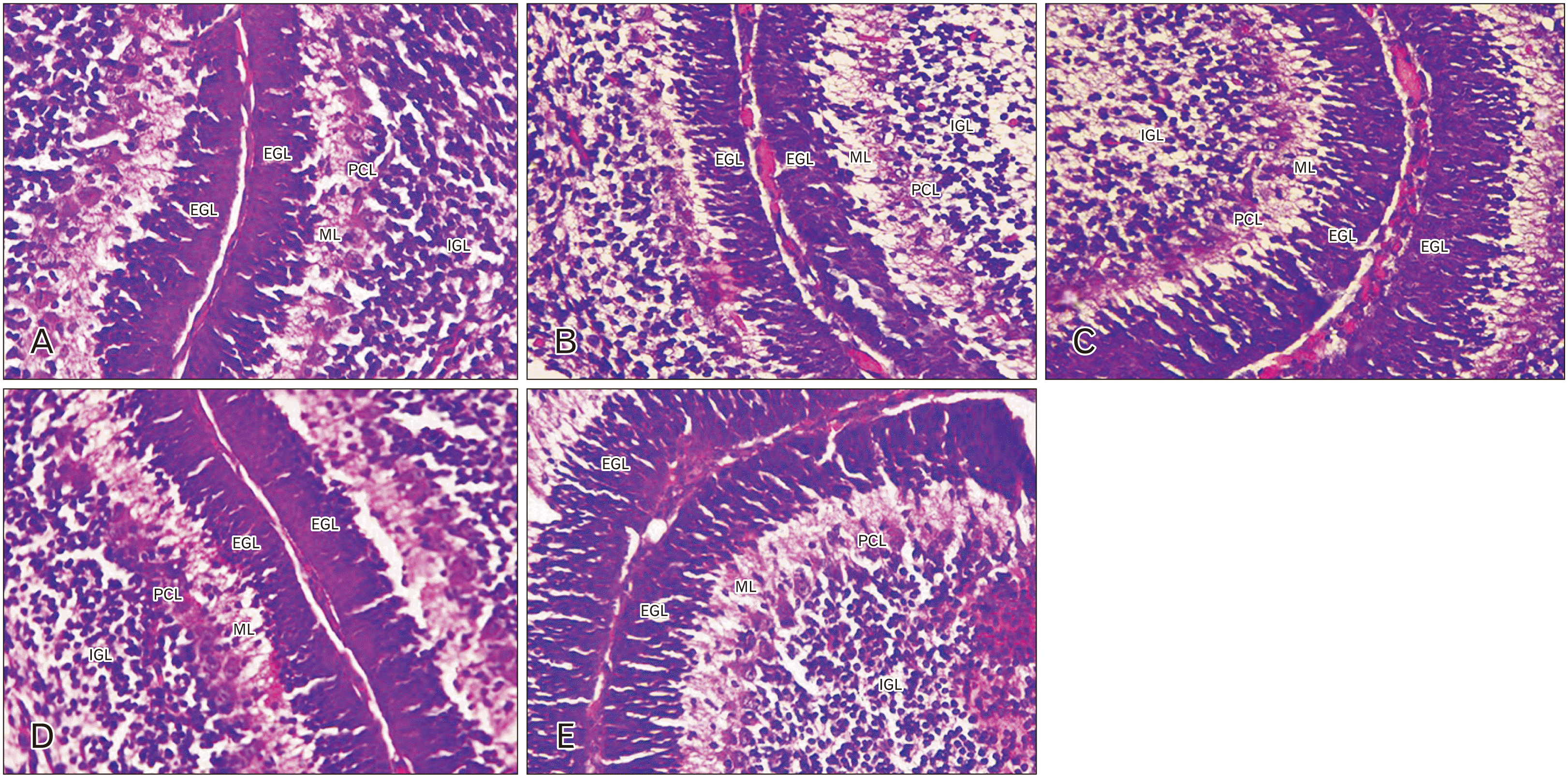 | Fig. 1Photmicrograph of the cerebellar cortex on post-natal day 7 from the treatment groups (H&E, ×400). Negative control (distilled water) (A), positive 2 control (200 mg/kg bw of AlCl3) (B), 200 mg/kg bw of AlCl3+400 mg/kg bw ethyl acetate leaf fraction of Tamarindus indica (EATI) (C), 200 mg/kg bw of AlCl3+800 mg/kg bw EATI (D), and 200 mg/kg bw of AlCl3+300 mg/kg bw of vitamin E (E). EGL, external granular layer; ML, molecular layer; PCL, Purkinje cell layer; IGL, internal granular layer; GC, granular cells.
|
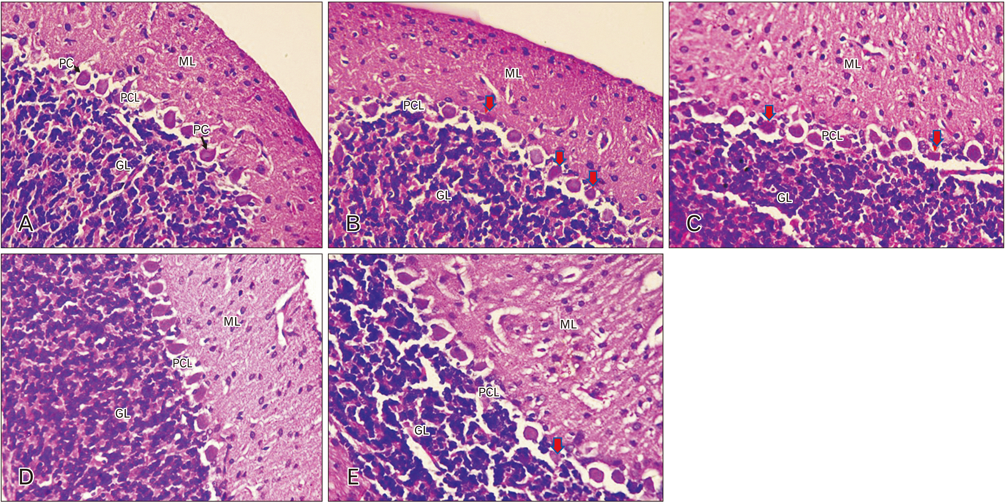 | Fig. 2Photmicrograph of the cerebellar cortex on post-natal day 21 from the treatment groups (H&E, ×400). Negative control (distilled water) (A), positive 8 control (200 mg/kg bw of AlCl3) (B), 200 mg/kg bw of AlCl3+400 mg/kg bw ethyl acetate leaf fraction of Tamarindus indica (EATI) (C), 200 mg/kg bw of AlCl3+800 mg/kg bw EATI (D), and 200 mg/kg bw of AlCl3+300 mg/kg bw of vitamin E (E). Red arrow indicates degenerating cells. GL, granular layer; ML: molecular layer; PCL, Purkinje cell 10 layer; PC, Purkinje cell.
|
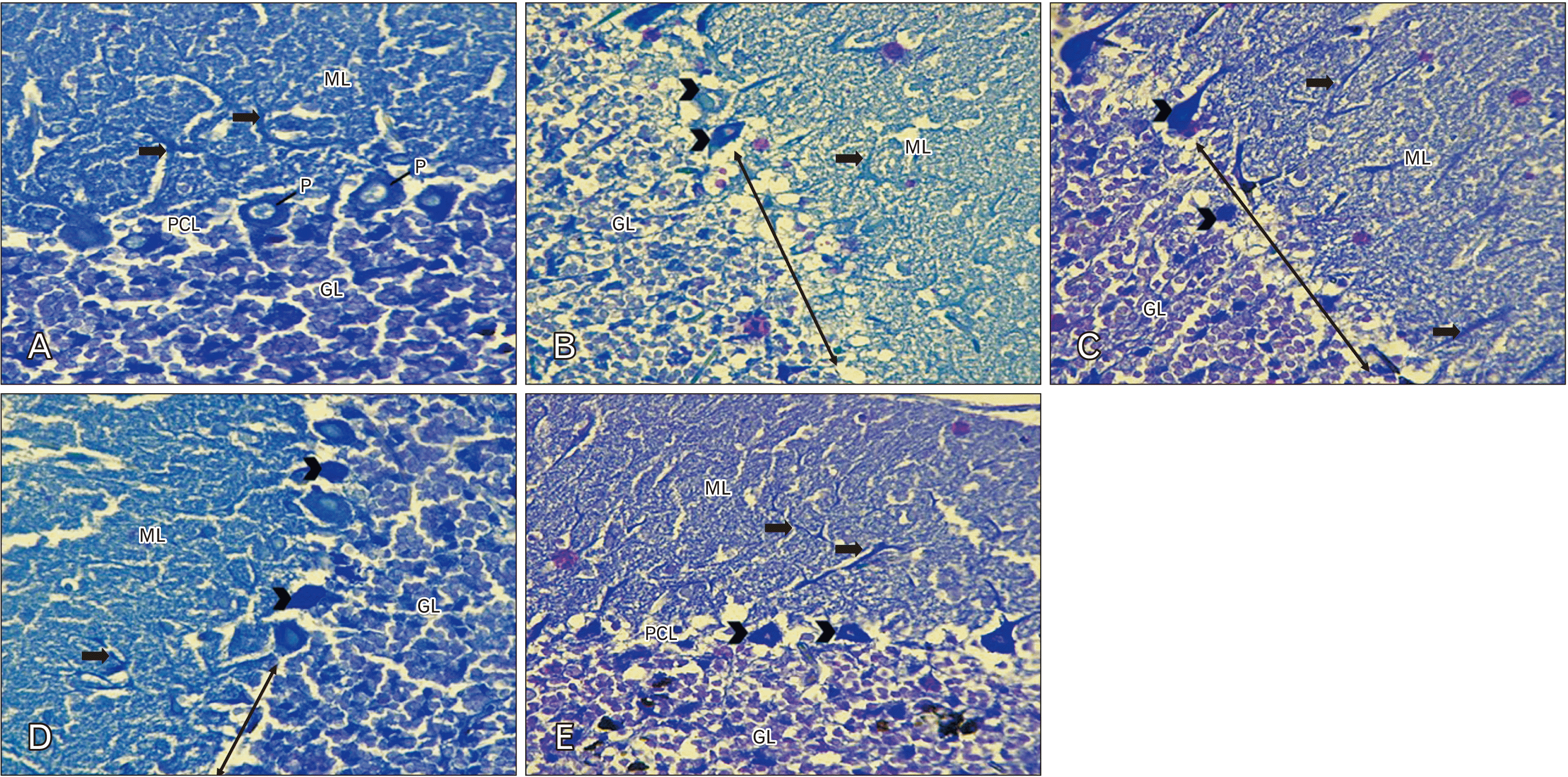 | Fig. 3Photmicrograph of the cerebellar cortex on post-natal day 21 from the treatment groups (Cluver 17 berrera; ×400). Negative control (distilled water) (A), positive 14 control (200 mg/kg bw of AlCl3) (B), 200 mg/kg bw of AlCl3+400 mg/kg bw ethyl acetate leaf fraction of Tamarindus indica (EATI) (C), 200 mg/kg bw of AlCl3+800 mg/kg bw EATI 15 (D), and 200 mg/kg bw of AlCl3+300 mg/kg bw of vitamin E (E). Black arrows indicate dendritic arborization. GL, granular layer; ML, molecular layer; PCL, Purkinje cell 16 layer; PC, Purkinje cell; RLD, region of reduced linear distribution of Purkinje cell.
|
The results of the GFAP stained sections of the cerebellar cortex showed significantly higher GFAP reactivity in the positive control (administered 200 mg/kg of AlCl3) and the 400 mg/kg EATI treated groups (Fig. 4B, C, E) when compared to the distilled water treated group (Fig. 4A, E). There was a significant improvement in GFAP reactivity in the groups administered 400 mg/kg EATI and 300 mg/kg of vitamin E (Fig. 4D, E) when compared to the positive control group.
 | Fig. 4Photomicrograph of Wistar rat cerebella cortex on post-natal day 21, showing immune-positive cells (black arrow head) (GFAP, ×400). (A) 2 ml/kg bw of distilled water, (B) 200 mg/kg bw of AlCl3, (C) 200 mg/kg bw of AlCl3 and 400 mg/kg bw ethyl acetate leaf fraction of Tamarindus indica (EATI), (D) 200 mg/kg bw of AlCl3 and 800 mg/kg bw EATI, (E) 200 mg/kg bw of AlCl3 and 300 mg/kg bw of vitamin E, and (F) the immuno-histochemical score (IHS) using Klein’s semiqualitative approach; with values presented as mean±SEM. a,b)Significance difference (P<0.05) compared to the negative control (group 1). GL, granular layer; ML, molecular layer. GFAP is the staining method in this case.
|
Go to :
Discussion
Aluminum is a heavy metal that has received lots of attention in recent times, majorly due to its reported neurotoxic potentials. The result from the present study revealed that prenatal aluminum chloride exposure impacted neurodevelopment and body weight in neonatal rats. Prenatal AlCl3 exposure was marked with impaired motor coordination, delayed cytoarchitectural development, histopathological changes, and increased cerebellar cortical GFAP expression. On the other hand, treatment with EATI (400 and 800 mg/kg of EATI) and 300 mg/kg of vitamin E was associated with significant improvements.
The observed effects following prenatal AlCl3 exposure in the present study may be due to the reported developmental neurotoxicity associated with aluminum exposure both during intrauterine and early childhood. This is in line with the study done by Gonda and Lehotzky (1996) [30] who reported that prenatal aluminum exposure was found to significantly reduce postnatal body weight in animals. Prenatal exposure of mice to aluminum sulfate (from the 10th day to the 13th day of gestation) revealed marked growth retardation in pups [31]. On the contrary, Muller et al. [32] did not notice changes in body weight among pups prenatally exposed to aluminum lactate when compared to the pups in the control group. The degree of aluminum-induced toxicity may vary according to its chemical form and route of exposure [1]. The observed changes in motor coordination was marked with higher mean latency/time to cross the beam to the dark box and an increase in the number of footslip during the beam walking test. Prenatal effect of aluminium exposure is usually irreversible, hence the observed alteration in motor function of the cerebellar cortex [33]. Previous studies have linked aluminum exposure with behavioral disorders in experimental animals, even in the absence of histopathological lesions [7]. Other studies have reported delayed sensory-motor development, learning and memory deficit, altered grip strength, and escape behavior being associated with prenatal aluminum exposure [1, 7, 9]. The observed alteration in motor coordination in the present study is in line with the finding of Fernandes et al. [34] who reported that prolong of aluminium exposure in wistar rats was associated with impairement in motor coordination, evident by the observed higher number of foot slip in the exposed groups.
The observed delayed cytoarchitectural development and increase in GFAP expression following prenatal AlCl3 exposure supports the reports from previous studies. Thus, the finding from the present study is in line with the studies by Reichert et al. [35] who reported that exposure to AlCl3 interferes with neuronal cell proliferation, migration, and differentiation in neural progenitor cells isolated from embryonic telencephalons, cultured as neurospheres. The observed variation in the thickness of the external granular and the density of cells in the internal granular layer in the groups exposed to AlCl3 supports reports of the toxic effects of prenatal AlCl3 exposure. The external granular layer serves as the external germinal layer where the granular cells germinate, then they migrate through the Purkinje cell layer at postnatal day 4 to form the internal granular layer [36]. It is therefore not out of place to observe the interference in the internal granular layer associated with interference of the external granular layer. The reduced Nissl staining observed in the present study is a biomarker of reduced cellular activity [37].
Treatment with Tamarindus indica showed a dose-dependent effect during prenatal aluminum exposure. The observed improvement may be linked with metal-chelating effects and the ability to counter processes involved in the generation of reactive oxygen species by some phytochemicals available in EATI. Studies have shown that Tamarindus indica possesses antioxidant activities, antipyretic, antimicrobial, and hypolipidemic properties [15]. Administration of ethanolic pup extract of Tamarindus indica during prenatal ethanol exposure revealed some protective potentials on the cerebral cortex of Wistar rats [15, 16]. A previous study on the effect of EATI during prenatal AlCl3 exposure revealed, improvement in brain metal concentration, oxidative stress parameters, GFAP expression in the cerebral cortex and the hippocampus (unpublished observation).
The observed improvement in the group treated with vitamin E following intrauterined AlCl3 exposure may be linked with its neuroprotective potential. Vitamin E is a known antioxidant [38], and reported to be neuroprotective in heavy metal exposure [39]. Although, the administration of EATI especially at 800 mg/kg was more effecacious than vitamin E at 300 mg/kg in the present study. Therefore, EATI could be promising for use in complementary and alternative medicine.
The present study though basic seems to offer an insight into the fact that treatment with Tamarindus indica during prenatal AlCl3 exposure has some protective potential. However, the authors recommend a more detailed investigation into the mechanism via which Tamarindus indica exerts its protective potentials, therefore TUNEL, cleaved caspase-3 or other markers may be used to demonstrate neuronal damage and possible improvements; neuronal nuclear protein may be used to demonstrate matured neuronal cells, as the authors were constrained by resources and time.
The present study concludes that prenatal AlCl3 exposure impacted neurodevelopment, body weight, neurobehavioural functions, and cytoarchitecture of the cerebellum in neonatal rats. However, treatment with EATI showed significant improvements.
Go to :
 XML Download