Introduction
The frontal sinus is a pneumatised cavity that is present inside the frontal bone of the skull [
1,
2]. The frontal sinus often appears as two irregular cavities extending upward and laterally into squama frontalis [
3]. It is seldom symmetrical, as the left and right lobes are separated by a central septum causing both lobes to develop independently and deviate from the midline [
4]. The sinus is divided into several lobes by the formation of partial bony septa. The frontal sinus is not visible at birth and begins its development during the second year of life [
5]. It is radiographically detected after further pneumatisation that takes up to five to eight years of life [
6]. The development is completed by the 20th year of life, and it occurs earlier in girls than boys. The frontal sinus development remains stable throughout adult life until further slight enlargement which may occur from bone resorption during advanced age [
6].
The morphological variation of the frontal sinus has both surgical and forensic significance [
7]. In the case of the surgical field, frontal sinus is frequently accessed during neurosurgical procedures on the frontal bone (
i.e., supraorbital and pterional craniotomies) however, due to the close relationship between the frontal sinus and the anterior skull base or orbit, the frontal sinus may be susceptible to surgical complications [
7]. Breaching of the frontal sinus should be avoided to reduce the risk of postoperative complications such as a cerebrospinal fluid leak, sinusitis and mucoceles development [
8]. For this reason, a thorough knowledge of the complex anatomy, anomalies and variations of the frontal sinus is vital for neurosurgeons to avoid complications and maximise the success of neurosurgical procedures [
8]. This can be particularly helpful in preoperative planning and deciding on the appropriate neurosurgical approach [
7].
In the forensic field, the use of frontal sinus in identifying human skeletal remains is increasingly applied and recognised in forensic investigation [
3,
9]. The significance of frontal sinus in forensic identification lies in its unique pattern [
10]. Analogous to the fingerprint, frontal sinus is unique between each individual, even in the case of monozygotic twins [
11]. Since the frontal sinus is commonly used for comparison of ante- and post-mortem radiographs technique in forensic investigation, the capability of the frontal sinus to remain stable throughout the lifespan will help the frontal sinus not be affected by the time elapsed of the post-mortem [
9,
12]. One or both sides of the frontal sinuses may be absent occasionally [
1]. The low incidence of frontal sinus absence is regarded as another vital morphological feature for establishing a definite and reliable biological profile [
1]. Being an internal body structure, the arched feature protects the frontal sinus from decomposition and damage, allowing the frontal sinus to preserve intact in human skeletal remains [
13]. These highlights the need to further explore the distribution of frontal sinus patterns, especially amongst the Malaysian population.
The unique pattern of the frontal sinus (
i.e., symmetry and asymmetry) was initially observed by Zuckerkandl in 1895. Later, Culbert and Law were the first to describe human identification through morphological variation of the frontal sinus, which then was accepted in a United States court of law [
4]. In the Indian population, the frontal sinus morphology was unique and highly symmetrical (85.9%) among the male sex group (48.4%) [
13], contrary to the Saudi population, which presented greater symmetrical patterns (83.2%) among the female sex group (43.0%) [
15]. Despite various studies conducted in diversified populations to test the reliability of frontal sinus for personal identification, studies regarding frontal sinus morphology among sex, race and age profiles of the Malaysian population have yet to be explored. In this study, the distribution and variability of frontal sinus patterns among Malaysians were observed.
Go to :

Discussion
The morphological variation of the frontal sinus can be crucial for forensic investigation and for a neurosurgeon in planning the pterional and supra-orbital craniotomy due to the proximity of the frontal sinus to the orbit and the anterior skull base [
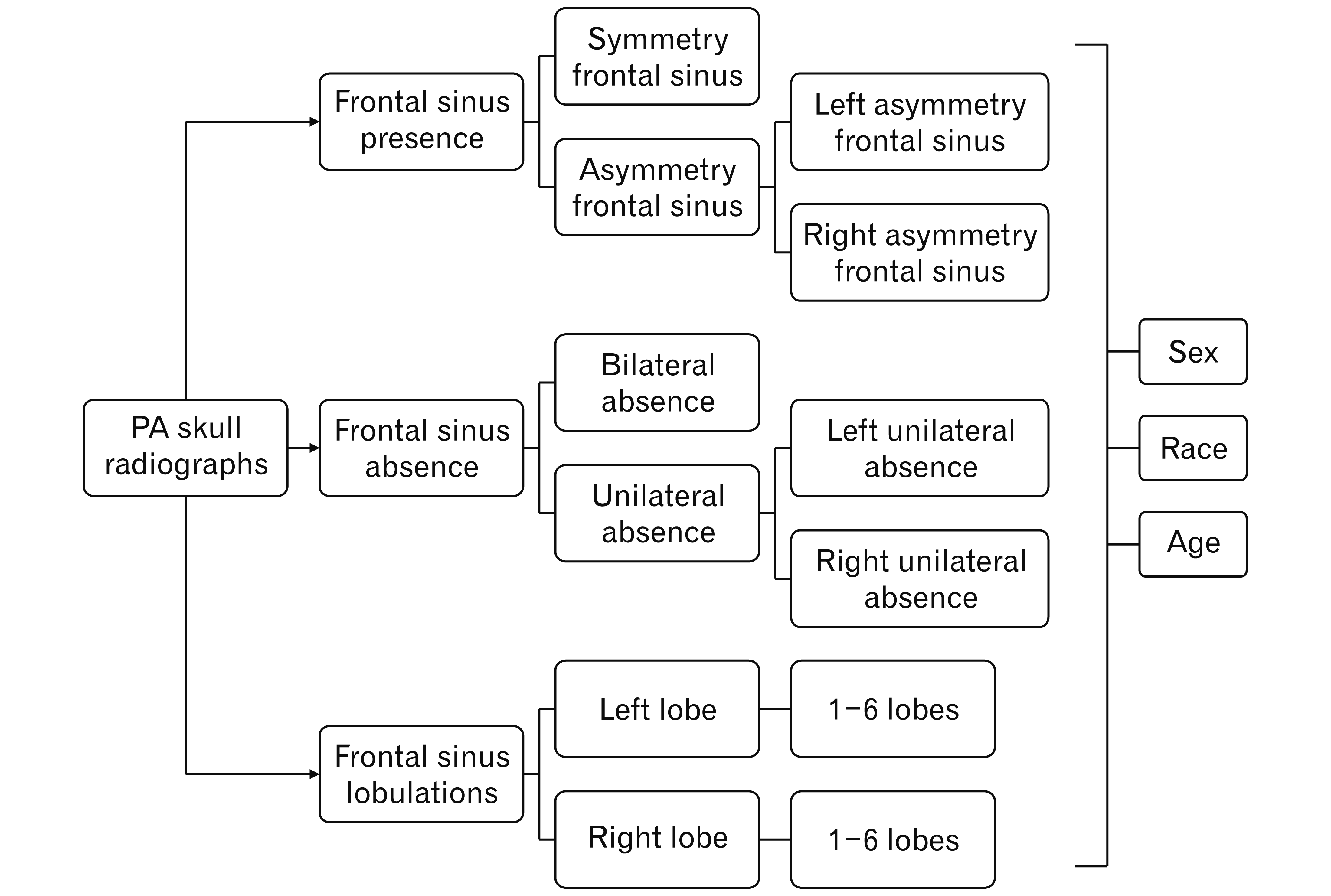
7]. The present study highlighted the findings on the morphological variation of frontal sinus patterns in relation to sex, race, and age groups of Malaysians. The right and left frontal sinuses develop separately, and one or more cells are formed on each side, separated by partial septa [
16]. Consequently, the frontal sinuses may appear asymmetrical due to the independent development or not develop at all. It is not uncommon to find an absent frontal sinus [
7]. The percentage of bilateral absence of frontal sinus was 2.7% in the Malaysian population, which is relatively similar to other Asian populations, like Verma et al. [
4], which reported 5.4% non-existence of frontal sinus in the South Indian population. Similarly, Patil et al. [
11] had found bilateral aplasia of only 1.0% among the North Indian population. There were 2.0% unilateral absence cases in our study, which is similarly low compared to Indian and Ireland populations, with 4.3% and 2.0% of total unilateral absence, respectively [
1]. Among 2.0% of total unilateral cases, the left frontal sinus was absent in two cases, and the right was absent in six cases. Overall, the absence of frontal sinus occurred in only 19 cases.
This current study demonstrated that the presence of frontal sinus was found in 95.4% of the subjects, and symmetrical frontal sinus comprised 40.8% of the total cases. In contrast, Verma et al. [
4] showed that there was 78.5% symmetry in the Indian population, and in a study by Shireen et al. [
15], symmetrical frontal sinus was noted in 83.2% of the Saudi population samples, suggesting that the majority of the Indian and Saudi population have more symmetrical frontal sinus compared to Malaysians. In our study, asymmetry cases were seen in 54.5% of samples. The results were consistent with those studies by Taniguchi et al. [
17], who obtained 43.1% in the Japanese population. David and Saxena [
14] reported symmetry frontal sinus in 58.0% of subjects in the Indian population. The discrepancy between percentage frequency in this study and other preceding studies may be due to environmental factors (
i.e., climates of different countries) and individual health that might affect the variability of the frontal sinus pattern among different populations [
4]. The climatic factor on the frontal sinus morphology is speculated to be associated with heat retention and insulation that occurs in cold environments that contribute to the smaller size and absence of the frontal sinus [
18].
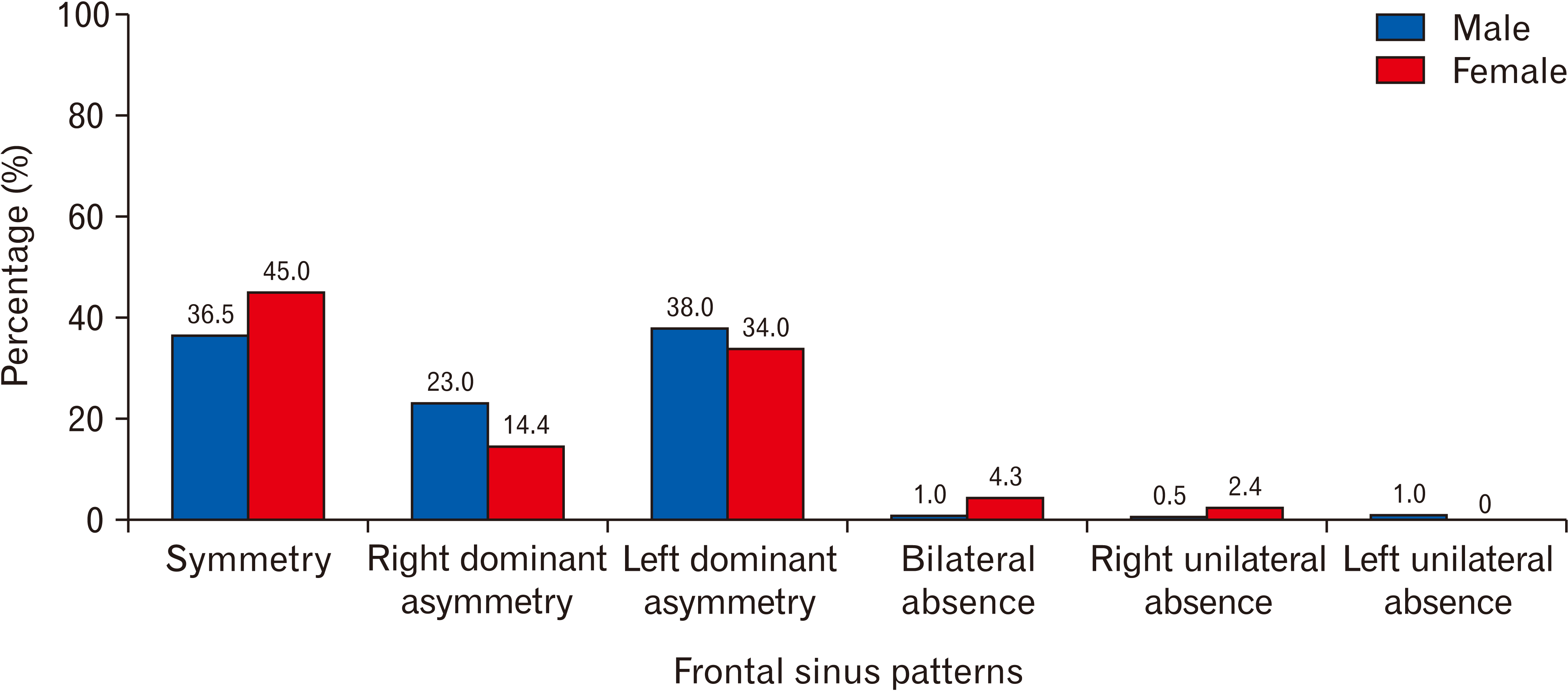
Our study demonstrated that the frontal sinus exhibits morphological variability between both sexes. The absence of frontal sinus is greater in females (6.7%) compared to males (2.5%), and this finding is in accordance with the findings of the Indian population (male: 4% and female: 12%), Japanese population (male: 13% and female: 23%), and Turkish population (male: 1.3% and female: 5.0%) [
1].
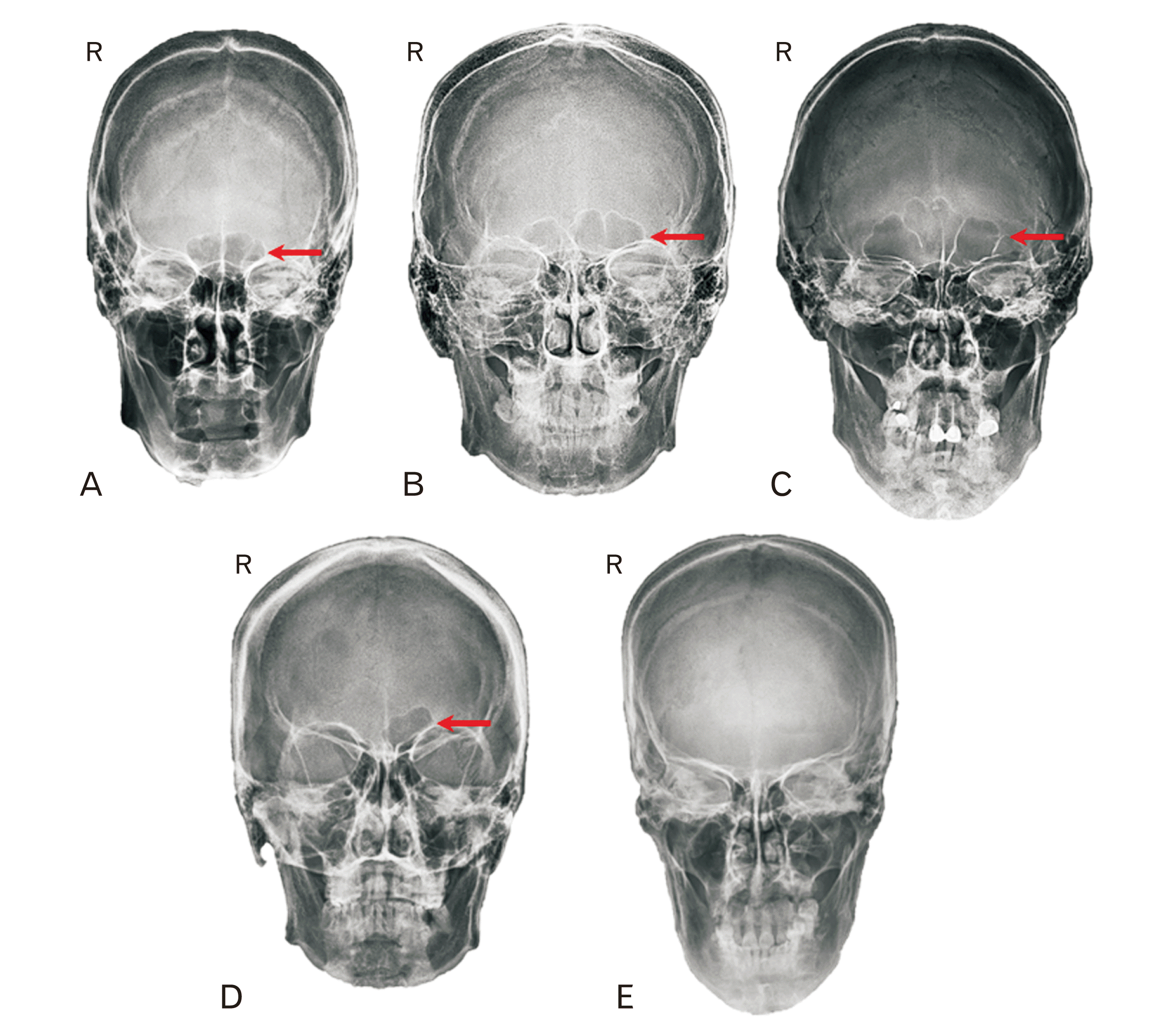
The central septa of the frontal sinus in this current study presented that majority of males had central septa sloping toward the right (38.0%) and left (23.0%), indicating the left and right asymmetry. The result is similar to Verma et al. [
4], who reported that males mostly appeared with asymmetry frontal sinus (left asymmetry: 2.7%, and right asymmetry: 0.9%). The sloping of the central septum is due to the independent development of both sides of the frontal sinus, leading to the existence of one side being larger and crossing the midline [
1,
15]. The symmetry of the frontal sinus is commonly seen amongst females, 45.0% out of the total samples. This is consistent with what has been found in previous studies among the South Indian population [
4,
14], with 39% and 32.0% of the females, respectively. Data on lobulations shows frontal sinus with 3 to 5 lobes often appears among males compared to females. A similar presentation was seen in the Indian population [
4].
The morphological variation of frontal sinus was evaluated among three main races of the Malaysian population (
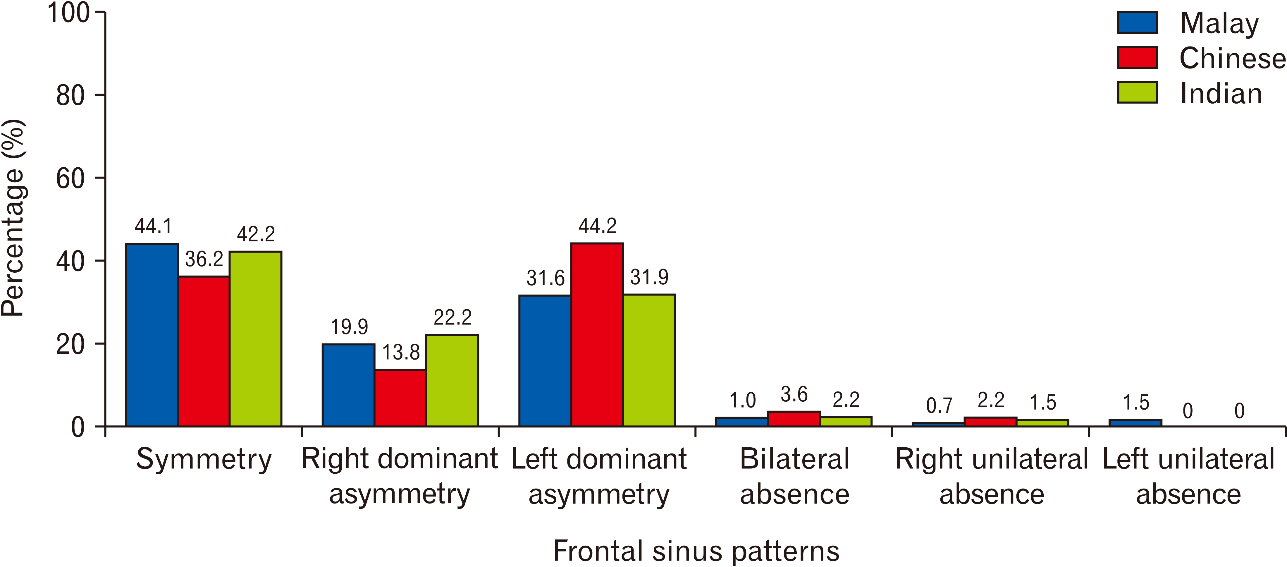
i.e., Malay, Chinese, and Indian) in the present study. The frontal sinus was absent in the majority of the Chinese race, with 5.8%. Malay individuals demonstrated symmetry patterns of 44.1%, and Indian and Chinese showed 42.2% and 36.2% symmetry patterns, respectively. This suggested that Malay typically have symmetrical frontal sinus more often than other races. Chinese (44.2%) most commonly have a central septum sloping to the right side, whereas Indians (22.2%) often have a central septum sloping to the left side, suggesting right asymmetry of the frontal sinus. In other populations, the pattern’s variation among the New Mexican population demonstrates that Black individuals often showed right asymmetry than White individuals [
19]. The inconsistency of the distribution of frontal sinus among races may be explained by genetic factors [
4]. The distribution of lobulations among these three races is random. Though, all races commonly have one to three lobes on both sides of the frontal sinus. To date, the discussion among race groups is limited due to the limitation of literature regarding the relationship between frontal sinus morphology and ancestry [
20]. The frontal sinus patterns variation of the three main races among Malaysian have not been analysed and compared against one another in this manner.
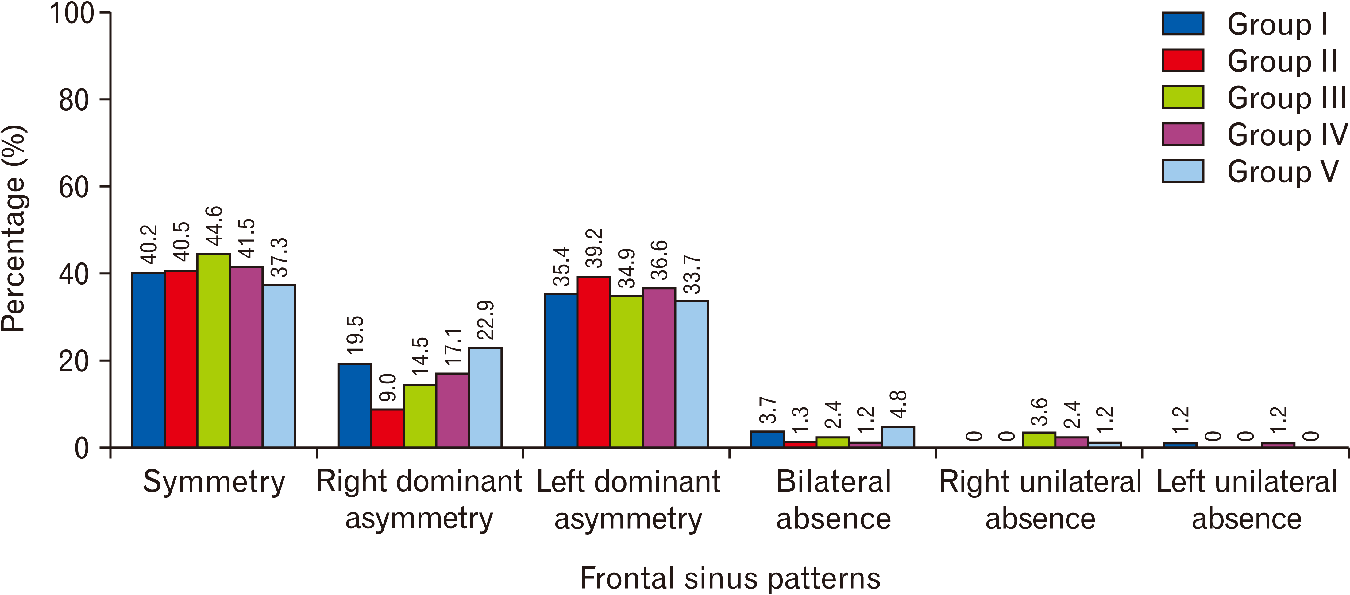
This study revealed the variation of frontal sinus morphology in different age groups. The cases of absent frontal sinus are typically seen among those in the age group III and V, with 6.0% of the subjects. Symmetry cases were seen as the highest in age group III (44.6%). Whilst percentage of central septum slops to the left and right side is often found in age groups V (22.9%) and II (39.2%), respectively. Despite the unpredictable distribution of lobulations, all the age groups typically appeared with one to three lobes on both sides of the frontal sinus. There is still limited published literature related to the frontal sinus patterns and age group done in this comprehensive manner.
This study includes the limitation of unidentified frontal sinus morphological distribution among minority races in Malaysia, such as Iban and Kadazan Dusun, which consist of 1.0 % population in Malaysia [
21]. Future studies could be done to explore the frontal sinus pattern distribution specifically to all the minority races in Malaysia. Nevertheless, this study provided a novel insight into the frontal sinus patterns among its major races of the Malaysian population.
In conclusion, the study revealed that the distribution of frontal sinus morphology varies according to sex, race and age to some degree. As the frontal sinus morphology is population-specific and data specific to the Malaysian population is limited, this study helps to build a database of the frontal sinus morphology, thus enhancing the potential use of frontal sinus in forensic investigation and assisting neurosurgeons in surgical planning involving frontal bone among Malaysian.
Go to :



 PDF
PDF Citation
 Print
Print


 XML Download