Journal List > Anat Cell Biol > v.55(3) > 1516079993

Shetty, Patil, Mobin, Gowda, Puttamallappa, Vamadevaiah, and Kunjappagounder: Study of course and termination of brachial artery by dissection and computed tomography angiography methods with clinical importance

Abstract

The Brachial artery is a continuation of the axillary artery, from the inferior border of the tendon of teres major to the neck of the radius, terminating into radial and ulnar arteries just a cm distal to the elbow joint. Unlike veins, variations in the arteries are comparatively less common. Anatomical variations of the brachial artery occur in almost 20% of the cases and are commonly found during routine dissection or clinical practice. To observe the variations in the course and termination of brachial artery by dissection and computed tomography (CT) angiography methods. The present study was conducted on 40 upper limbs each in the department of Anatomy & Radiology of JSS Medical College and Hospital, Mysuru. The brachial artery was traced from origin to termination and variations were noted and photographed. Patients who were undergoing CT angiography of the upper limbs in JSS Hospital were included in the study. Variations noted and compared with the dissection method. In the present study, normal patterns of the brachial arterial course and termination were observed in 31 specimens. The remaining 9 specimens showed variant course and termination in the brachial artery like an unusually tortuous superficial brachial artery, superficial brachio-ulnar artery and brachio-radial artery. CT angiography showed 6 variations and a tortuous brachial artery. A detailed description of the vascular pattern of upper limbs especially variations in their origin and termination is of extreme importance in clinical practice. The knowledge of these variations is important for catheterization, graft harvesting, arteriovenous fistula creation, shunt application and astrup examination.

Introduction

The brachial artery is the main artery of the upper limb. It begins as a continuation of the axillary artery at the distal border of the tendon of teres major muscle and ends at the level of the neck of the radius by dividing into radial and ulnar arteries.
It gives profunda brachii, superior and inferior ulnar collateral, nutrient, and muscular arteries in the arm. The branches of the brachial artery anastomose with the branches of the radial and ulnar arteries around the elbow joint. Because of this extensive collateral circulation, blood flow around the elbow joint and to the distal part of the limb is not discontinued in any position of the elbow joint [1].
Rodriguez-Niedenfuhr [2] classified the different arterial variations of the major arteries of the upper limb into seven types after a detailed study of arterial variations in 192 specimens:
  1. Superficial brachial: Brachial artery crosses anterior to the median nerve, instead of posterior to the median nerve, and at the level of the elbow joint it divides into the radial and ulnar arteries.

  2. Accessory brachial: Originates above the elbow joint from the upper third of the brachial artery. It crosses the median nerve in front to unite the brachial artery proximal to the elbow before it divides into ulnar and radial arteries.

  3. Brachioradial: This is the high origin of the radial artery, and runs superficial to the median nerve in the arm.

  4. Superficial radial: radial artery with a normal origin, but crosses over the tendons of anatomical snuffbox at the wrist.

  5. Superficial brachio-ulnar: This is the high origin of the ulnar artery and courses over the superficial forearm flexors muscles.

  6. Brachio-ulnar: the high origin of the ulnar artery but normal course along forearm and hand.

  7. Superficial brachioulnoradial artery: superficial brachial artery dividing into radial and ulnar arteries at the elbow and the normal brachial artery continues as the common interosseous artery.

Development of brachial artery

The axis artery of the upper limb is derived from the lateral branch of the 7th cervical intersegmental artery. The main arterial trunk grows outwards along the ventral axial line and terminates in the capillary plexus of the developing limb bud. Digital arteries arise from the capillary plexus.
The proximal part of the main arterial trunk forms the axillary and brachial arteries and its distal part persists as the interosseous artery. The median artery which arises later from the interosseous artery runs along the median nerve and joins the capillary plexus in the palm. The ulnar artery develops as a branch from the original brachial artery trunk and extends down the ulnar side of the forearm. Initially, the radial artery arises more proximally than the ulnar artery from the brachial artery trunk. Later, the radial artery is connected with the main trunk next to the origin of the ulnar artery. The upper portion of its origin usually undergoes regression. Thus radial and ulnar arteries arise at the same level [2, 3].
Our present study is aimed to observe the variations in the course and termination of the brachial artery by dissection and computed tomography (CT) angiography methods. The need for this study was as this type of study was not reported in literature and CT angiography is one of the commonest procedure done routinely nowadays in some clinical conditions and the knowledge of variations in brachial artery is essential to perform clinical interventions.
Objectives: (i) to study the course and termination of the brachial artery by dissection method and to study the course and termination of the brachial artery by CT Angiography method of upper limb; (ii) to correlate the variations of brachial artery (course and termination) by dissection method with CT Angiography method.

Materials and Methods

The present study was conducted on 40 upper limbs each in the Department of Anatomy and Radiology of JSS Medical College and Hospital, Mysuru. For the present study we have utilized 16 males (32 limbs) and 4 females (8 limbs) cadavers. Out of 40 limbs, 20 belonged to right side and 20 belonged to left side in both cadaveric study and CT angiographic study. All the cadavers belonged to the voluntary body donars who had registered under Body Donation Association (BDA) program in our institute and belonged to the age group between 65–70 years.The Donated bodies and patients belong to Mysuru district of Karnataka state, South India. It took two years (two MBBS batches 1919–1920 and 2020–2021 dissection practicals) to conduct this study. During our regular dissection practical classes for MBBS students, we observed some interesting variations in the brachial artery and incorporated the CT Angiographic study to make our research study more interesting and knowledgeable.
The brachial artery was traced from origin to termination, variations noted and photographed. Patients who are undergoing CT angiography of upper limb in JSS Hospital were included in the study. Variations noted and compared with dissection method (IEC No. JSSMC/IEC/210421/15NCT/2021-22).

Inclusion and exclusion criteria

Inclusion criteria: Donated bodies to JSSMC, BDA, and patients undergoing CT angiography of upper limb for renal dialysis, pain in the upper limb/reduced pulse in JSS Hospital.
Exclusion criteria: damaged limbs and trauma to upper limb arteries.

Results

During routine dissection of the upper limb for undergraduate students, we observed a few very rare variations in the course and termination of the brachial artery. The textbook description of the brachial artery was found in 31 limbs out of 40 specimens (Table 1).
Variations observed in 9 limbs were as follows:
Variation in the course of the brachial artery tortuous superficial brachial artery.

Variation in the course of the brachial artery

Tortuous superficial brachial artery

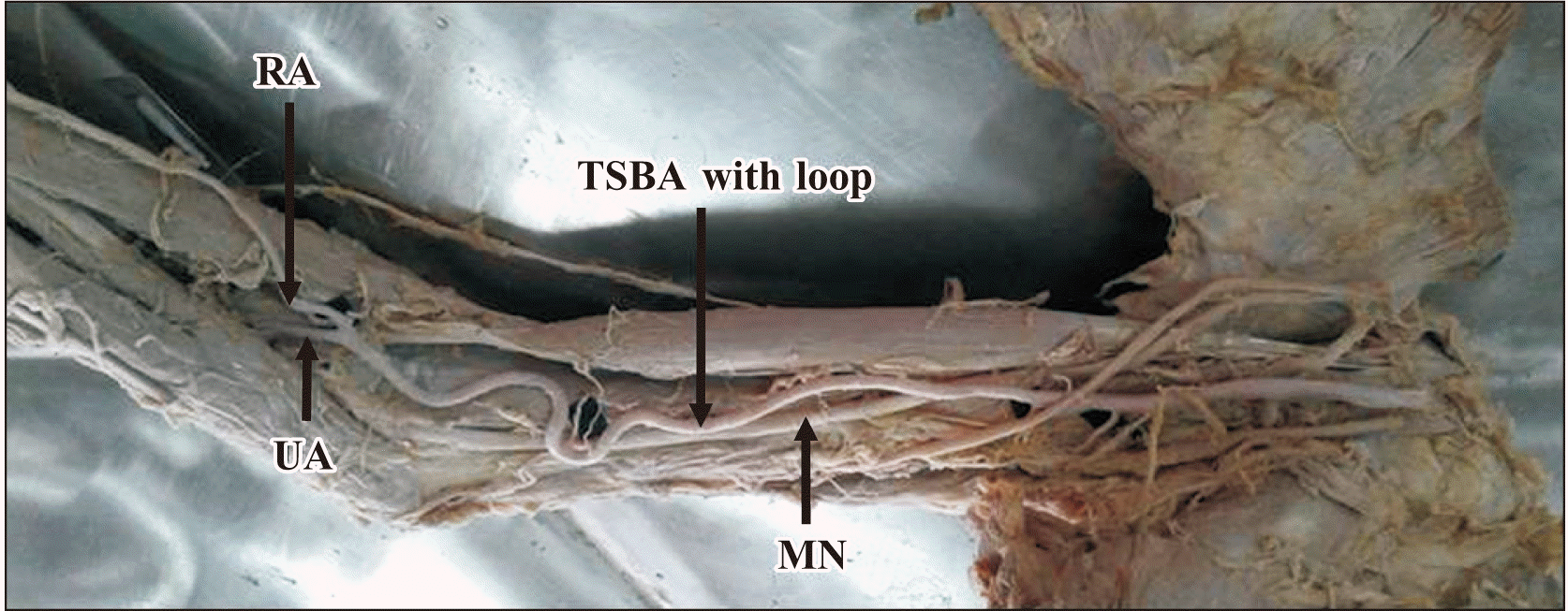
In one limb, the brachial artery had an unusually tortuous course with a loop. The loop was just above the elbow joint. The loop gave two thin muscular branches, one to the biceps brachii and another one to the brachialis. The median nerve was passing posterior to the brachial artery instead of anterior. It was dividing into ulnar and radial arteries in the cubital fossa and they had a normal course in the forearm (Fig. 1). This type of variation in the brachial artery course has not been reported in the literature so far.
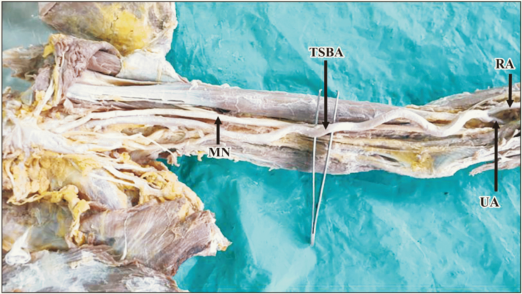
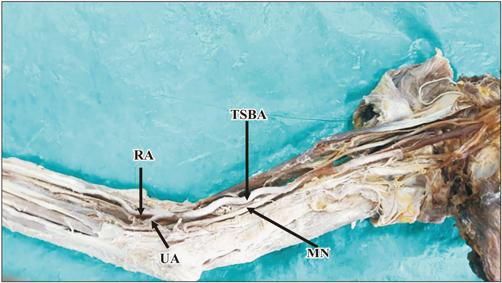
In one cadaver there was a bilateral tortuous superficial brachial artery. In both, the limbs’ brachial artery was highly tortuous and was passing superficially to the median nerve in the arm and entered the cubital fossa, and divided into radial and ulnar arteries. Radial and ulnar arteries were also slightly tortuous in the distal forearm (Figs. 2, 3).
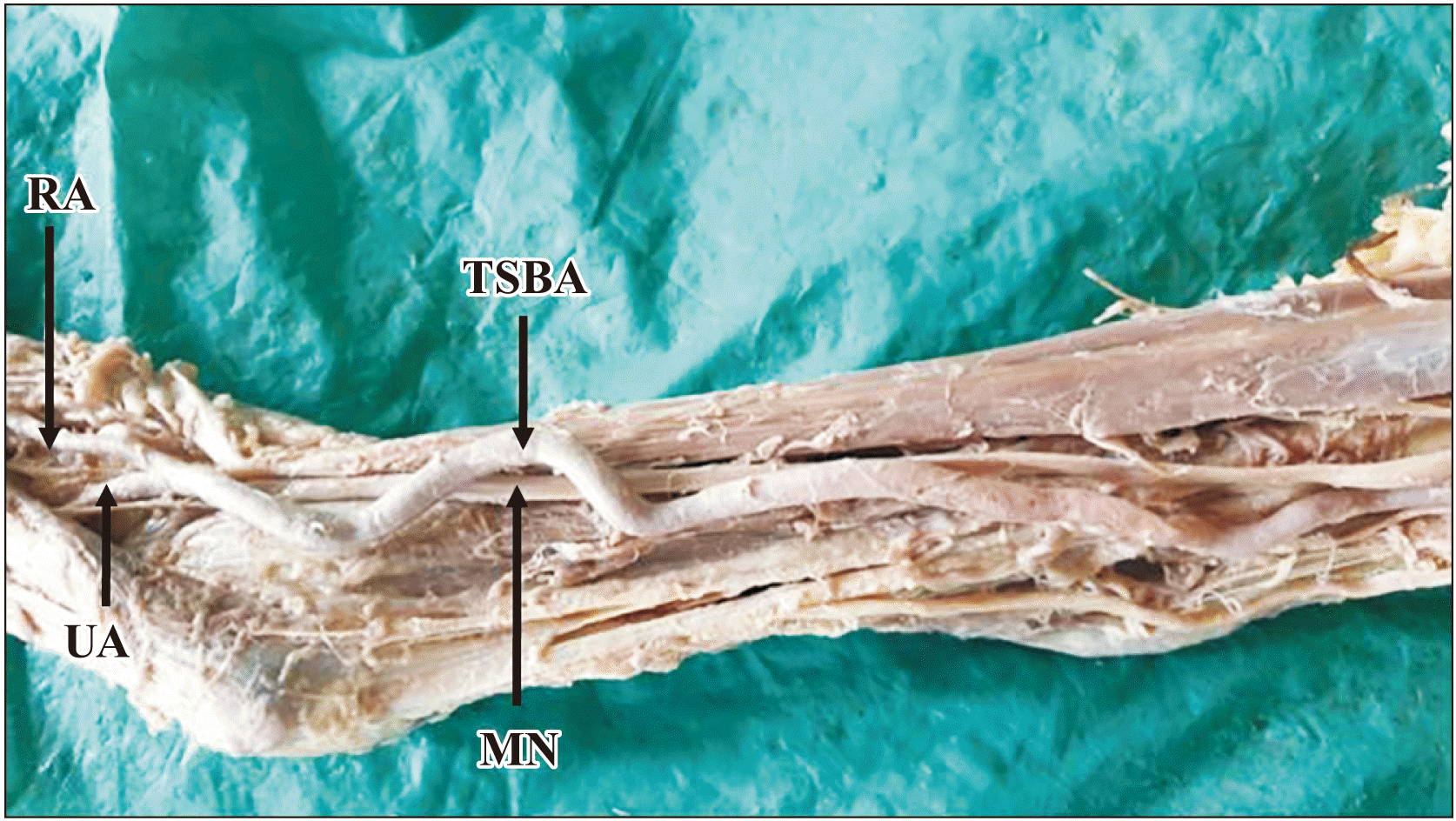
In another specimen, the right limb had a tortuous superficial brachial artery, passing superficial to the median nerve in the arm and entering the cubital fossa and divided into radial and ulnar arteries (Fig. 4).

Variation in the termination

Superficial brachio-ulnar artery

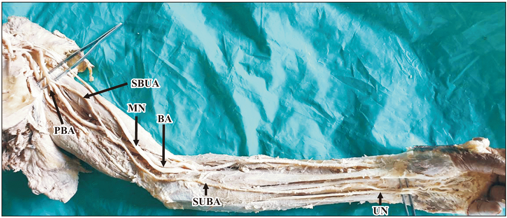
Superficial brachio-ulnar artery—also called the high origin of the ulnar artery. In one left limb, there was a high origin of ulnar artery from the brachial artery almost at the same level where profunda brachii artery was arising from the brachial artery. Superficial brachio-ulnar artery passed along the medial side of the arm and medial to the median nerve. In the cubital fossa, the median nerve was in between brachial and superficial brachio-ulnar arteries. It entered the forearm, superficial to flexor muscles of the forearm. It was not accompanying the ulnar nerve in the forearm only at the distal third of the forearm, superficial to the flexor retinaculum it was lateral to the ulnar nerve and entered the palm to form the superficial palmar arch—type B supplying all the digits. The superficial brachio-ulnar artery didn’t give any branches in its course. The brachial artery was crossed by the median nerve and later the median nerve was lying in between brachial and superficial brachio-ulnar arteries in the cubital fossa. The brachial artery trifurcated in the cubital fossa and gave radial recurrent, radial, and common interosseous arteries.
Radial recurrent artery—after passing posterior to the tendon of biceps brachii; it is divided into ascending and descending branches.
Radial artery—was larger in caliber than the superficial brachio-ulnar artery but showed normal course and distribution.
Common interosseous artery—gave anterior and posterior ulnar recurrent arteries, muscular branches, and divided into anterior and posterior interosseous arteries (Fig. 5).
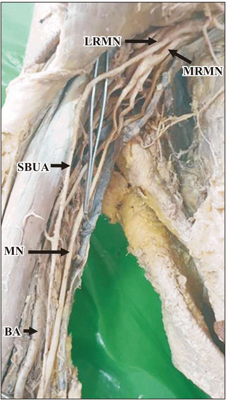
In another limb, a superficial brachio-ulnar artery was taking origin from the axillary artery. It crossed the lateral root of the median nerve and passed lateral to the median nerve in the upper part of the arm. Later it crossed the brachial artery and the median nerve to reach the cubital fossa. It was passing superficial to flexor muscles in the forearm and entered the palm superficial to flexor retinaculum to form the superficial palmar arch. Throughout its course, it didn't give any branches. The brachial artery was seen dividing into a larger radial artery and a common interosseous artery in the cubital fossa. The radial artery was wider in caliber to compensate for the thin ulnar artery (Fig. 6).

Brachio-radial artery

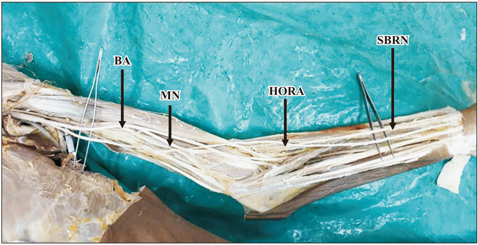
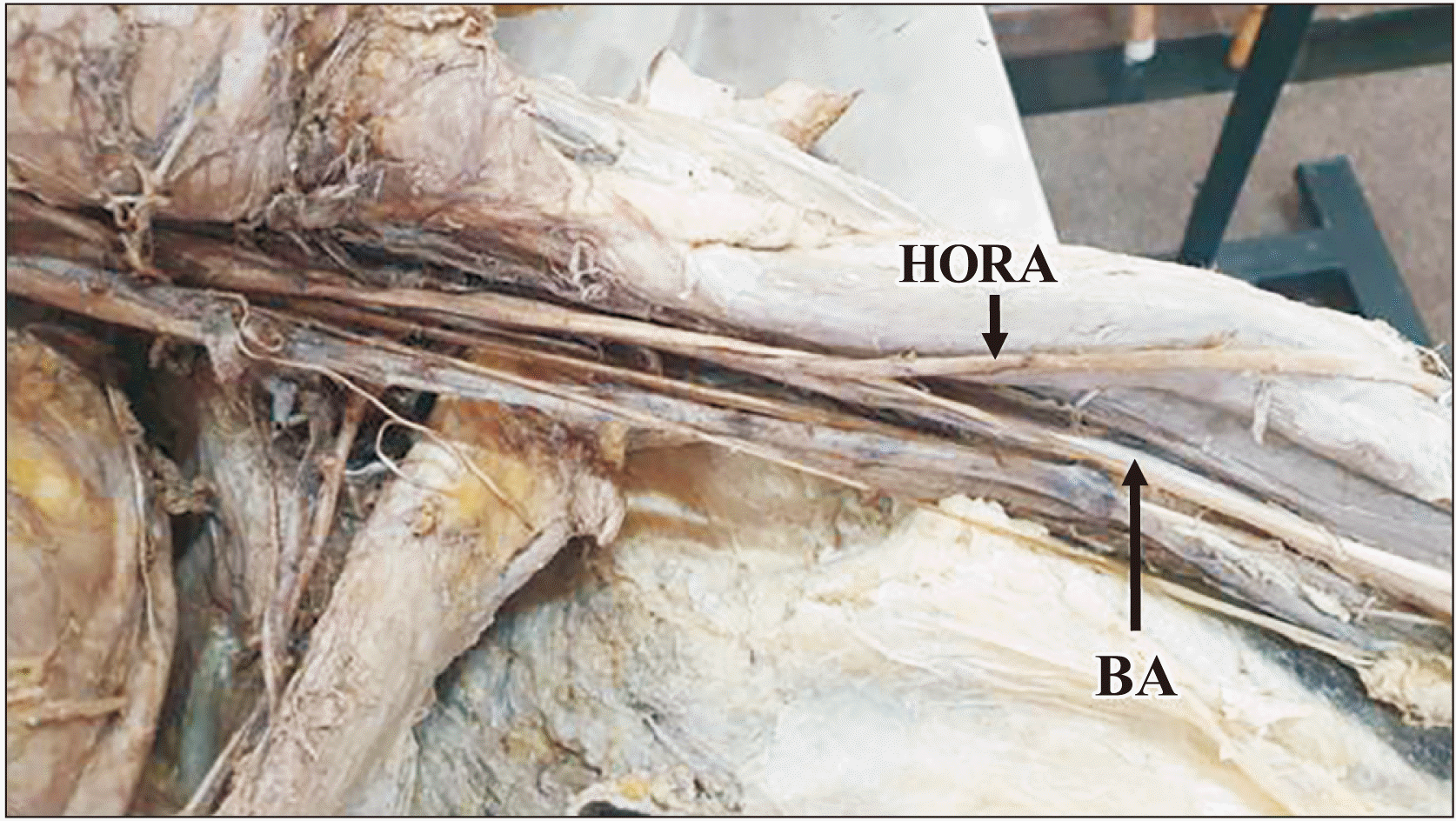
Brachio-radial artery also called the high origin of radial artery (HORA). Three specimens showed HORA but the termination of the brachial artery in all the three specimens was different. In the first specimen—HORA crossed the median nerve and brachial artery and reached the lateral side of the arm. In the cubital fossa, it was lateral to the tendon of the biceps brachii and continued on the lateral side of the forearm. Here it was accompanied by the lateral cutaneous nerve of the forearm and then entered the anatomical snuff box. Throughout its course, it didn't give any branches. The brachial artery was seen passing superficially to the median nerve and then entering the cubital fossa. In the cubital fossa, the brachial artery was the medial most structure. The median nerve was lying in between the brachial artery and tendon of the biceps brachii. Brachial artery gave radial recurrent, ulnar recurrent arteries and divided into ulnar and common interosseous arteries (Fig. 7).
The second and third HORA was in one specimen that is bilateral.
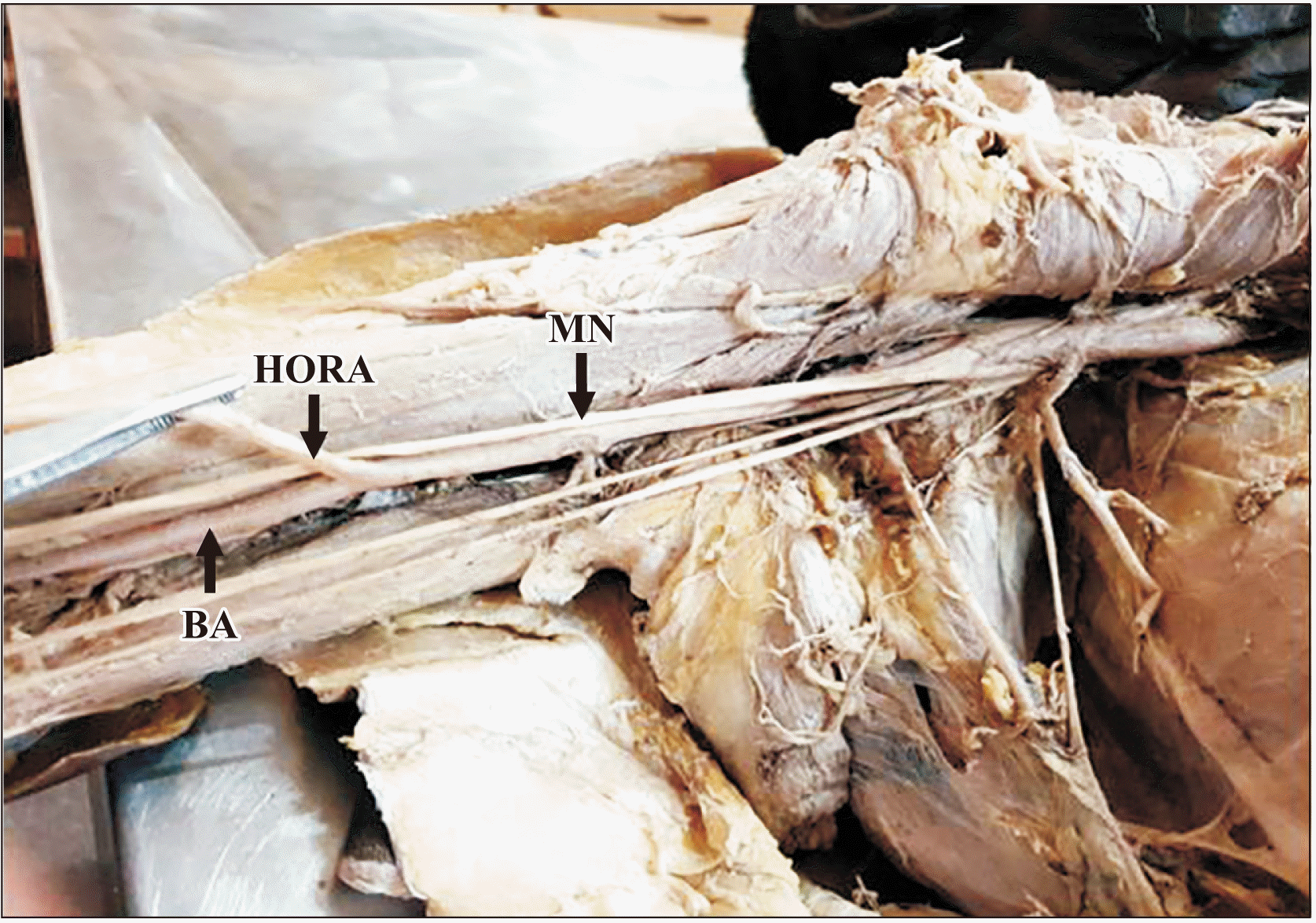
In the right limb—HORA was arising almost in the middle of the arm from the brachial artery. It crossed the median nerve in the arm and lateral to the tendon of biceps brachii it entered the forearm. The brachial artery in the cubital fossa gave the ulnar artery, median artery, and common interosseous artery. The ulnar artery was passing in between the fibers of the median nerve. The persistent median artery with the median nerve entered the palm through the carpal tunnel. It didn't join the ulnar artery to complete the superficial palmar arch. Type H incomplete superficial palmar arch was seen where the median artery and ulnar artery supplied 21/2 digits each separately (Fig. 8).
In the left limb—HORA was arising almost in the middle of the arm from the brachial artery. After crossing the median nerve in the arm and lateral to the tendon of biceps brachii in the cubital fossa it entered the lateral side of the forearm. Brachial artery gave ulnar artery and common interosseous artery (Fig. 9).
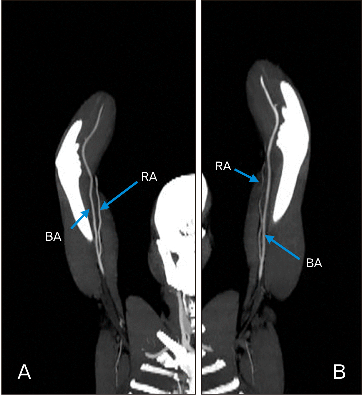
CT angiography showed six variations out of 40 samples. The identified upper limb CT angiograms were evaluated and analysed by 2 independent trained radiologists, who were blinded to the clinical records. The analysts subjectively evaluated the overall technical quality and anatomical variations. Six limbs were showing a high origin of the radial artery and in one individual brachial artery was slightly tortuous (Fig. 10). In one individual HORA was bilateral. Results were expressed in terms of frequency and percentages.

Discussion

Arteries are normally straight tubes that effectively carry blood to the distal organs. Sometimes, arteries may take a tortuous course with unusual twists and turns either due to abnormal embryological development or due to vascular diseases. A mild tortuous course of arteries is asymptomatic without clinical symptoms, a severe tortuous course can result in ischemic shock of the distal parts. Clinical studies associate tortuous arteries with aging, atherosclerosis, hypertension, diabetes mellitus, and genetic defects [4].
Tortuous arteries and veins are common angiographic findings in many studies and clinical investigations [5]. With the advance in imaging technology, more tortuous vessels are being detected. Various types of tortuosity have been reported in clinical investigations like looping, twisting, angulation, and kinking vessels [5]. Tortuosity has been reported in the aorta, vertebral, iliac, femoral, coronary, cerebral, and internal carotid arteries by Ertugrul [6].
Ashwini and Vasantha [7], in their study, reported unusual tortuous brachial, ulnar, and radial arteries with loops and bends. But brachial artery had prominent loops when compared to the radial and ulnar arteries. In our present study, the brachial artery had a loop that was passing superficial to the median nerve, this has not been reported in the literature so far. According to Dobrin et al. [8], degradation of elastin in the wall of the vessel results in aneurysm and elongation of the vessel resulting in tortuosity. The tortuous brachial artery may be mistaken for a vein and may complicate intravenous drug administration and percutaneous brachial catheterization [9, 10]. Median nerve passing posterior to the brachial artery is also very rare, seen only in 2% of cases [11]. Since the median nerve is passing posterior to the tortuous brachial artery it can compress the nerve resulting in carpal tunnel syndrome-like symptoms [12, 13].
The brachial artery is accessed for coronary angiography, aortic and peripheral interventions like angioplasty, stenting, thrombolysis, and complex endovascular repair of abdominal aortic and thoracic aortic aneurysms [14, 15]. An unusual tortuous course of the brachial artery may cause difficulties or failure of the procedure during clinical interventions.
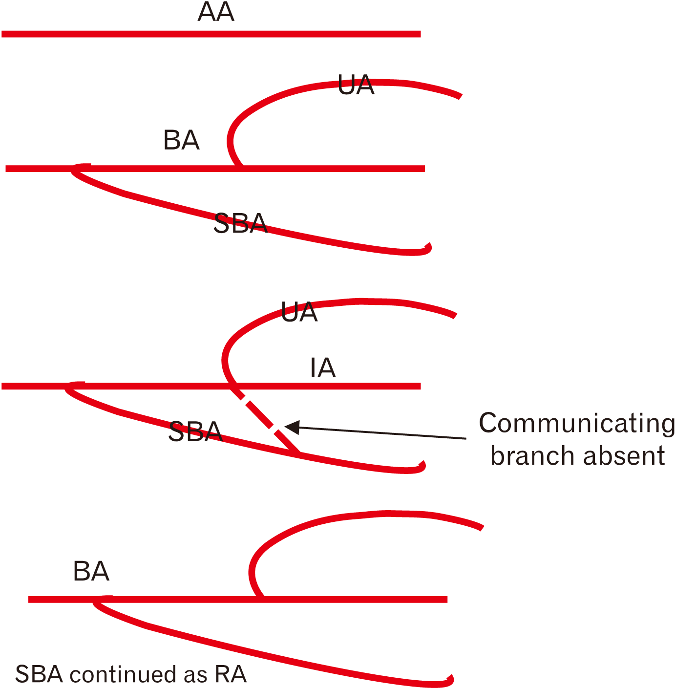
According to Rodríguez-Niedenführ et al. [2], the superficial brachial artery crosses anterior to the median nerve, instead of posterior to the median nerve, and at the level of the elbow joint, it divides into the radial and ulnar arteries. The incidence of superficial brachial artery varies from 3.6% to 9.6% [2]. This type of variation of the brachial artery is different from the normal anatomy and maybe one of the reasons for the unknown cause of median nerve entrapment neuropathy [13]. Another variation seen quite often is the division of the brachial artery into radial and ulnar arteries at a higher level than the usual level with an incidence of 8% of cases [16-18]. Such a high breakup is called “high bifurcation of the brachial artery” and may occur in various forms. The high origin of the radial artery is the most common variation when the radial artery arises first, the common trunk later divides into the ulnar artery, and the common interosseous artery [1, 2, 16]. According to Yang et al. [19], superficial brachial artery gave rise to radial and ulnar arteries in the cubital fossa in 8.9% of cases, continued in the forearm as the radial artery in 2.3% of cases, or ended in the upper arm in 1.0% of cases. The superficial brachial artery never continued as ulnar artery in their study (Fig 11; schematic representation of developmental basis for superficial radial artery continuing as radial artery).
High bifurcation of the brachial artery may result in major clinical complications like high failure rate and reduced patency of an arteriovenous (AV) fistula. Lioupis et al. [20] reported that an increased chance of failure of AV shunt is 45% in patients having high bifurcation of the brachial artery when compared to 23% with patients having a normal branching pattern. Variations in the brachial artery pattern may cause difficulties in conducting flap harvesting during reconstructive surgeries and in arteriography [21]. We have followed the Rodriguez- Niedenfuhr classification of upper limb arteries and have reported superficial brachial as superficial brachial artery and not as high bifurcation of the brachial artery.
The superficial brachioulnar artery is an ulnar artery of high origin and runs superficial to the flexors in the forearm. This is a very rare variation of the brachial artery and the reported incidence of the high origin of ulnar artery taking origin from the axillary artery varies between 0.17%–2% [22].
When the high origin of the ulnar artery is present, the brachial artery commonly terminates as the radial and common interosseous arteries [2]. High origin of ulnar artery from the axillary artery has been reported by Jacquemin et al. [23] and T Nakatani et al. [24].
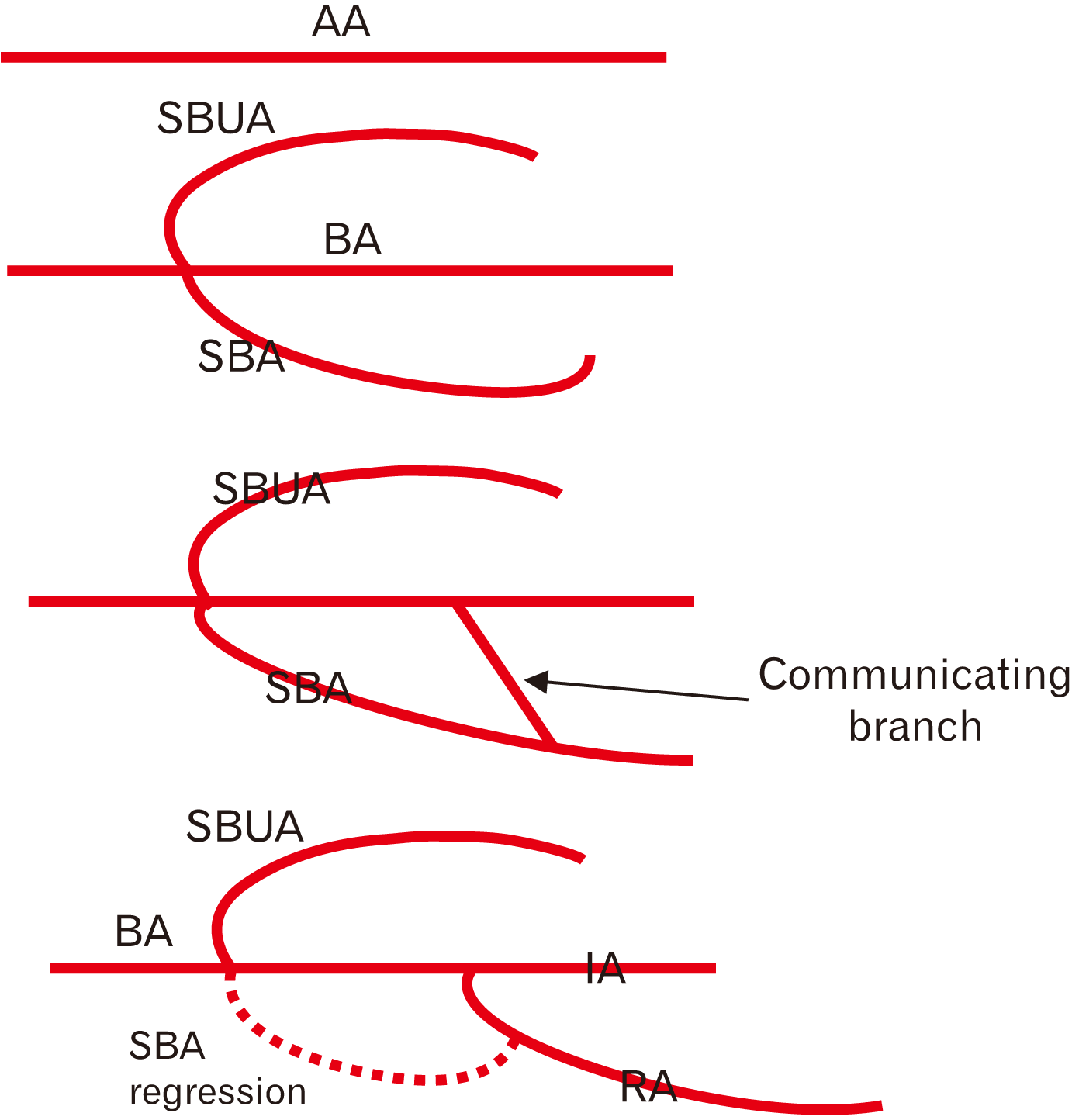
The reported incidence of the high origin of ulnar artery taking origin from the brachial artery is 0.7% according to Adachi [25] and 1.5% according to Rodríguez-Niedenführ et al. [2]. In our study, it is observed in 5% of cases. High origin of ulnar artery from the brachial artery has been reported by Bozer et al. [26] and Nakatani et al. [27]. Since it passes superficial to the bicipital aponeurosis and in clinical practice it could be mistaken for a vein leading to intra-arterial injection and difficulties in angiographic interventions [28] and also has a higher risk of getting damaged during forearm flap surgery [29]. The high origin of the ulnar artery should always be kept in mind during forearm flaps surgery or when the radial artery is used as a conduit in CABG [30, 31]. McWilliams and Sodha [32] reported that the high origin of the ulnar artery was clinically mistaken for vein inflammation - phlebitis but with Doppler ultrasound, it can be diagnosed correctly (Fig 12; Schematic representation of developmental basis for superficial brachioulnar artery). Every variation in the peripheral vascular anatomy can be related to either regression or persistence of the artery proximally in the upper limb. The type of variation presented in this case is due to persistence of ulnar artery in the arm and failure of formation of communication between ulnar and axial arteries in cubital fossa. The superficial brachioulnar artery in upper part of forearm can be explained on the basis of haemodynamic mechanism between deep and superficial arteries in the forearm. Change in the embryonic haemodynamics during angiogenic sprouting may result in primitive vasculature which persists or regresses in unusual patterns [33].
The reported incidence of HORA varies from 4.67% to 15.6% in cadaveric studies [34, 35] and 7% to 8.54% in angiographic studies [36, 37] and it is the most frequent variation observed in the brachial artery. In our study, it was observed in 7.5% of cases in cadavers and 15% of cases in CT angiographic study.
The radial artery has been used in coronary artery bypass grafting, cosmetic surgeries such as forearm flaps, and renal dialysis by making an autogenous AV fistula [38, 39].
The transradial approach becomes more difficult in the presence of brachioradial artery [40] and also results in the development of tortuosity, which can increase the risk of failure of trans- radial catheterization [41]. The radial artery is used in AV fistula creation in the forearm as preferred access for hemodialysis in end-stage renal disease. The most common complication of AV fistula is ischemic steal syndrome, which can occur in 70% of radiocephalic fistula and 90% of brachiocephalic fistula [42]. However, symptoms of hand ischemia due to postoperative steal syndrome are more probably to occur in 1%–2% of radiocephalic fistula and 5%–10% of brachiocephalic fistula [43, 44]. But according to Morsy et al. [45] and Zibari et al. [46], postoperative steal syndrome occurs in less than 1% in radiocephalic fistula and 6% in brachiocephalic fistulae. Duplex ultrasound and arteriography should aim to cross-examine the arteries proximal and distal to the fistula in addition to fistula flow [47]. The high bifurcation of the brachial artery is not rare. An unusually small brachial artery should remind clinicians to investigate the possibility of high bifurcation of the brachial artery. A simple ultrasound examination can detect the anomaly and allow the diameter assessment for clinical decision-making regarding access creation [47]. According to Rodríguez-Niedenführ et al. [2] and Singer [3], this type of anomaly is due to the failure of formation of the communicating branch between the superficial brachial artery and axial artery at the level of the elbow and thus superficial brachial artery did not disappear but continues as radial artery (Table 2) [37, 48-56].
In conclusions, CT angiography has become an important diagnostic imaging method for the evaluation of upper limb vascular diseased abnormalities. The advantage of CT angiography is that it is available 24 hours and has minimum interference. In our study, HORA is commonest variation seen in both cadaveric and in CT angiographic methods and the percentage is high in CT angiographic method. Therefore CT angiography is essential before designing suitable invasive or surgical procedures for successful surgery or interventions.

Notes

Author Contributions

Conceptualization: HS. Data acquisition: HS, MHNG, VP. Data analysis or interpretation: HS, VP. Drafting of the manuscript: HS, VP. Critical revision of the manuscript: VP, NM, VSP, RMV, PK. Approval of the final version of the manuscript: all authors.

Conflicts of Interest

No potential conflict of interest relevant to this article was reported.

References

1. Standring S. 2008. Gray's anatomy: the anatomical basis of clinical practice. 40th ed. Churchill Livingstone;Edinburgh: p. 905–6.
2. Rodríguez-Niedenführ M, Vázquez T, Nearn L, Ferreira B, Parkin I, Sañudo JR. 2001; Variations of the arterial pattern in the upper limb revisited: a morphological and statistical study, with a review of the literature. J Anat. 199(Pt 5):547–66. DOI: 10.1046/j.1469-7580.2001.19950547.x. PMID: 11760886. PMCID: PMC1468366.
crossref
3. Singer E. 1933; Embryological pattern persisting in the arteries of the arm. Anat Rec. 55:403–9. DOI: 10.1002/ar.1090550407.
crossref
4. Han HC. 2012; Twisted blood vessels: symptoms, etiology and biomechanical mechanisms. J Vasc Res. 49:185–97. DOI: 10.1159/000335123. PMID: 22433458. PMCID: PMC3369246.
crossref
5. Weibel J, Fields WS. 1965; Tortuosity, coiling, and kinking of the internal carotid artery. I. Etiology and radiographic anatomy. Neurology. 15:7–18. DOI: 10.1212/WNL.15.1.7. PMID: 14257832.
crossref
6. Ertugrul A. 1967; Diffuse tortuosity and lengthening of the arteries. Circulation. 36:400–7. DOI: 10.1161/01.CIR.36.3.400. PMID: 6033167.
crossref
7. Ashwini C, Vasantha K. 2014; An unusual tortuous brachial artery and its branches: histological basis and its clinical perspective. Int J Life Sci Biotechnol Pharm Res. 3:121–4.
8. Dobrin PB, Schwarcz TH, Baker WH. 1988; Mechanisms of arterial and aneurysmal tortuosity. Surgery. 104:568–71. PMID: 3413685.
9. Du Toit DF, Sunshine M, Knott-Craig C, Laker L. 1985; Gangrene of the hand and forearm after inadvertent intra-arterial injection of pyrazole. A case report. S Afr Med J. 68:491–2. PMID: 4049165.
10. Handlogten KS, Wilson GA, Clifford L, Nuttall GA, Kor DJ. 2014; Brachial artery catheterization: an assessment of use patterns and associated complications. Anesth Analg. 118:288–95. DOI: 10.1213/ANE.0000000000000082. PMID: 24445630.
11. Tubbs R, Shoja MM, Loukas M. 2016. Bergman's comprehensive encyclopedia of human anatomic variation. John Wiley & Sons;New York: DOI: 10.1002/9781118430309.
12. Segal R, Machiraju U, Larkins M. 1992; Tortuous peripheral arteries: a cause of focal neuropathy. Case report. J Neurosurg. 76:701–4. DOI: 10.3171/jns.1992.76.4.0701. PMID: 1545266.
13. Nkomozepi P, Xhakaza N, Swanepoel E. 2017; Superficial brachial artery: a possible cause for idiopathic median nerve entrapment neuropathy. Folia Morphol (Warsz). 76:527–31. DOI: 10.5603/FM.a2017.0013. PMID: 28198531.
crossref
14. Gan HW, Yip HK, Wu CJ. 2010; Brachial approach for coronary angiography and intervention: totally obsolete, or a feasible alternative when radial access is not possible? Ann Acad Med Singap. 39:368–73. PMID: 20535426.
15. Alvarez-Tostado JA, Moise MA, Bena JF, Pavkov ML, Greenberg RK, Clair DG, Kashyap VS. 2009; The brachial artery: a critical access for endovascular procedures. J Vasc Surg. 49:378–85. discussion 385DOI: 10.1016/j.jvs.2008.09.017. PMID: 19028057.
crossref
16. Panagouli E, Anagnostopoulou S, Venieratos D. 2014; Bilateral asymmetry of the highly bifurcated brachial artery variation. Rom J Morphol Embryol. 55:469–72. PMID: 24970004.
17. Cherukupalli C, Dwivedi A, Dayal R. 2007; High bifurcation of brachial artery with acute arterial insufficiency: a case report. Vasc Endovascular Surg. 41:572–4. DOI: 10.1177/1538574407305798. PMID: 18166644.
crossref
18. Tsoucalas G, Eleftheriou A, Panagouli E. 2020; High bifurcation of the brachial artery: an embryological overview. Cureus. 12:e7097. DOI: 10.7759/cureus.7097. PMID: 32231893. PMCID: PMC7098415.
crossref
19. Yang HJ, Gil YC, Jung WS, Lee HY. 2008; Variations of the superficial brachial artery in Korean cadavers. J Korean Med Sci. 23:884–7. DOI: 10.3346/jkms.2008.23.5.884. PMID: 18955798. PMCID: PMC2580020.
crossref
20. Lioupis C, Mistry H, Junghans C, Haughey N, Freedman B, Tyrrell M, Valenti D. 2010; High brachial artery bifurcation is associated with failure of brachio-cephalic autologous arteriovenous fistulae. J Vasc Access. 11:132–7. DOI: 10.1177/112972981001100209. PMID: 20155716.
crossref
21. Hansdak R, Arora J, Sharma M, Mehta V, Suri RK, Das S. 2015; Unusual branching pattern of brachial artery - embryological basis and clinicoanatomical insight. Clin Ter. 166:65–7. DOI: 10.7417/CT.2015.1817. PMID: 25945432.
22. Natsis K, Papadopoulou AL, Paraskevas G, Totlis T, Tsikaras P. 2006; High origin of a superficial ulnar artery arising from the axillary artery: anatomy, embryology, clinical significance and a review of the literature. Folia Morphol (Warsz). 65:400–5. PMID: 17171623.
23. Jacquemin G, Lemaire V, Medot M, Fissette J. 2001; Bilateral case of superficial ulnar artery originating from axillary artery. Surg Radiol Anat. 23:139–43. DOI: 10.1007/s00276-001-0139-2. PMID: 11462864.
crossref
24. Nakatani T, Tanaka S, Mizukami S, Shiraishi Y, Nakamura T. 1996; The superficial ulnar artery originating from the axillary artery. Ann Anat. 178:277–9. DOI: 10.1016/S0940-9602(96)80068-9. PMID: 8712378.
crossref
25. Adachi B. 1928. Das Arteriensystem der Japaner. Volume 1:Maruzen;Kyoto: p. 285–356. German. DOI: 10.1016/s0940-9602(96)80068-9.
26. Bozer C, Ulucam E, Kutoglu T. 2004; A case of originated high superficial ulnar artery. Trakia J Sci. 2:70–3.
27. Nakatani T, Tanaka S, Mizukami S. 1998; Superficial ulnar artery originating from the brachial artery and its clinical importance. Surg Radiol Anat. 20:383–5. DOI: 10.1007/BF01630626. PMID: 9894322.
crossref
28. Chin KJ, Singh K. 2005; The superficial ulnar artery--a potential hazard in patients with difficult venous access. Br J Anaesth. 94:692–3. DOI: 10.1093/bja/aei548. PMID: 15814810.
crossref
29. Latika A, Rima Dada R. 2005; Superficial ulnar artery- a case report. Indian J Pract Dr. 2:7–8. DOI: 10.18410/jebmh/2017/289. PMID: 26715898. PMCID: PMC4681723.
30. Jacob J, Deshpande R, Desai J. 2005; Superficial ulnar artery. Eur J Cardio Thorac Surg. 28:494. DOI: 10.1016/j.ejcts.2005.02.048. PMID: 16111612.
crossref
31. Porter CJ, Mellow CG. 2001; Anatomically aberrant forearm arteries: an absent radial artery with co-dominant median and ulnar arteries. Br J Plast Surg. 54:727–8. DOI: 10.1054/bjps.2001.3706. PMID: 11728121.
crossref
32. McWilliams RG, Sodha I. 2000; Doppler ultrasound diagnosis of a superficial ulnar artery. Eur J Ultrasound. 12:155–7. DOI: 10.1016/S0929-8266(00)00106-3. PMID: 11118923.
crossref
33. Sieger J, Patel L, Sheikh K, Parker E, Sheng M, Sakthi-Velavan S. 2019; Superficial brachioulnar artery and its clinical significance. Anat Cell Biol. 52:333–6. DOI: 10.5115/acb.19.008. PMID: 31598363. PMCID: PMC6773909.
crossref
34. Rodríguez-Baeza A, Nebot J, Ferreira B, Reina F, Pérez J, Sañudo JR, Roig M. 1995; An anatomical study and ontogenetic explanation of 23 cases with variations in the main pattern of the human brachio-antebrachial arteries. J Anat. 187(Pt 2):473–9. PMID: 7592009. PMCID: PMC1167441.
35. Weathersby HT. 1956; Anomalies of brachial and antebrachial arteries of surgical significance. South Med J. 49:46–9. DOI: 10.1097/00007611-195601000-00010. PMID: 13281685.
36. Karlsson S, Niechajev IA. 1982; Arterial anatomy of the upper extremity. Acta Radiol Diagn (Stockh). 23:115–21. DOI: 10.1177/028418518202300206. PMID: 7090847.
crossref
37. Uglietta JP, Kadir S. 1989; Arteriographic study of variant arterial anatomy of the upper extremities. Cardiovasc Intervent Radiol. 12:145–8. DOI: 10.1007/BF02577379. PMID: 2507150.
crossref
38. Jennings WC, Mallios A, Mushtaq N. 2018; Proximal radial artery arteriovenous fistula for hemodialysis vascular access. J Vasc Surg. 67:244–53. DOI: 10.1016/j.jvs.2017.06.114. PMID: 28912005.
crossref
39. Keen JA. 1961; A study of the arterial variations in the limbs, with special reference to symmetry of vascular patterns. Am J Anat. 108:245–61. DOI: 10.1002/aja.1001080303. PMID: 14454801.
crossref
40. Patel T, Shah S, Pancholy S, Rao S, Bertrand OF, Kwan T. 2014; Balloon-assisted tracking: a must-know technique to overcome difficult anatomy during transradial approach. Catheter Cardiovasc Interv. 83:211–20. DOI: 10.1002/ccd.24959. PMID: 23592578.
crossref
41. Ostojić Z, Bulum J, Ernst A, Strozzi M, Marić-Bešić K. 2015; Frequency of radial artery anatomic variations in patients undergoing transradial heart catheterization. Acta Clin Croat. 54:65–72. PMID: 26058245.
42. Anderson CB, Etheredge EE, Harter HR, Codd JE, Graff RJ, Newton WT. 1977; Blood flow measurements in arteriovenous dialysis fistulas. Surgery. 81:459–61. PMID: 847655.
43. Berman SS, Gentile AT, Glickman MH, Mills JL, Hurwitz RL, Westerband A, Marek JM, Hunter GC, McEnroe CS, Fogle MA, Stokes GK. 1997; Distal revascularization-interval ligation for limb salvage and maintenance of dialysis access in ischemic steal syndrome. J Vasc Surg. 26:393–402. discussion 402–4. DOI: 10.1016/S0741-5214(97)70032-6. PMID: 9308585.
crossref
44. Odland MD, Kelly PH, Ney AL, Andersen RC, Bubrick MP. 1991; Management of dialysis-associated steal syndrome complicating upper extremity arteriovenous fistulas: use of intraoperative digital photoplethysmography. Surgery. 110:664–9. discussion 669–70. PMID: 1925955.
45. Morsy AH, Kulbaski M, Chen C, Isiklar H, Lumsden AB. 1998; Incidence and characteristics of patients with hand ischemia after a hemodialysis access procedure. J Surg Res. 74:8–10. DOI: 10.1006/jsre.1997.5206. PMID: 9536965.
crossref
46. Zibari GB, Rohr MS, Landreneau MD, Bridges RM, DeVault GA, Petty FH, Costley KJ, Brown ST, McDonald JC. 1988; Complications from permanent hemodialysis vascular access. Surgery. 104:681–6.
47. Zamani P, Kaufman J, Kinlay S. 2009; Ischemic steal syndrome following arm arteriovenous fistula for hemodialysis. Vasc Med. 14:371–6. DOI: 10.1177/1358863X09102293. PMID: 19808723.
crossref
48. Bidarkotimath S, Avadhani R, Arunachalam K. 2011; Primary pattern of arteries of upper limb with relevance to their variations. Int J Morphol. 29:1422–8. DOI: 10.4067/S0717-95022011000400059.
crossref
49. Vatsala AR, Rajashekar HV, Angadi AV, Sangam . 2013; Variation in the branching pattern of Brachial artery: a morphological and statistical study. Int J Biol Med Res. 4:2920–3.
50. Sonje P, Kanaskar N, Arole V, Shevade S, Awari P, Vatsalaswamy . 2014; Study of variations in the branching pattern of brachial artery. Int J Cur Res Rev. 6:55–60.
51. Deepa TK, John Martin K. 2016; An anatomical study of variations in termination of brachial artery, with its embryological basis and clinical significance. Int J Med Res Health Sci. 5:85–9.
52. Kaur A, Sharma A, Sharma M. 2017; Variation in branching pattern of brachial artery. Int J Sci Stud. 5:213–7.
53. Ojha P, Prakash S. 2019; Variations in branching pattern of brachial artery - a study in cadavers. Int J Sci Res. 8:33–6. DOI: 10.36106/ijsr/1300946.
crossref
54. Balakrishnan R, Kavya , Sharmada KL. 2020; Arterial variation in the brachial artery and its clinical implications. MedPulse Int J Anat. 13:5–8. DOI: 10.26611/10011312.
55. Konarik M, Musil V, Baca V, Kachlik D. 2020; Upper limb principal arteries variations: a cadaveric study with terminological implication. Bosn J Basic Med Sci. 20:502–13. DOI: 10.17305/bjbms.2020.4643. PMID: 32343941. PMCID: PMC7664784. PMID: 2ff53371eadb4f6ebb70390d1b74ab85.
crossref
56. Celik HH, Görmüs G, Aldur MM, Ozçelik M. 2001; Origin of the radial and ulnar arteries: variations in 81 arteriograms. Morphologie. 85:25–7. PMID: 11534414.

Fig. 1
Right upper limb showing unusually TSBA. TSBA, tortuous superficial brachial artery; MN, median nerve; RA, radial artery; UA, ulnar artery.
acb-55-3-284-f1.tif
Fig. 2
Left upper limb showing TSBA. TSBA, tortuous superficial brachial artery; RA, radial artery; UA, ulnar artery; MN, median nerve.
acb-55-3-284-f2.tif
Fig. 3
Right upper limb showing TSBA. TSBA, tortuous superficial brachial artery; RA, radial artery, UA, ulnar artery; MN, median nerve.
acb-55-3-284-f3.tif
Fig. 4
Right upper limb showing TSBA. TSBA, tortuous superficial brachial artery; RA, radial artery; UA, ulnar artery; MN, median nerve.
acb-55-3-284-f4.tif
Fig. 5
Left upper limb showing SBUA or high origin of ulnar artery. SBUA, superficial brachio-radial artery; BA, brachial artery; MN, median nerve; PBA, profunda brachii artery, UN, ulnar nerve.
acb-55-3-284-f5.tif
Fig. 6
Right upper limb showing high origin of ulnar artery from axillary artery. LRMN, lateral root of the median nerve; MRMN, medial root of the median nerve; SBUA, superficial brachio-radial artery; MN, median nerve; BA, brachial artery.
acb-55-3-284-f6.tif
Fig. 7
Left upper limb showing brachio-radial artery or high origin of radial artery. SBRN, superficial branch of radial nerve; HORA, high origin of radial artery; MN, median nerve; BA, brachial artery.
acb-55-3-284-f7.tif
Fig. 8
Right upper limb showing brachio-radial artery or HORA. HORA, high origin of radial artery; MN, median nerve; BA, brachial artery.
acb-55-3-284-f8.tif
Fig. 9
Left upper limb showing brachio-radial artery or HORA. HORA, high origin of radial artery; BA, brachial artery.
acb-55-3-284-f9.tif
Fig. 10
Computed tomography angiography images showing brachio-radial artery and tortuous brachial artery. (A) High origin of radial artery, (B) high origin of radial artery. RA, radial artery; BA, brachial artery.
acb-55-3-284-f10.tif
Fig. 11
Schematic representation of developmental basis for SBA that continues as radial artery. AA, axis artery; UA, ulnar artery; BA, brachial artery; SBA, superficial brachial artery; IA, interosseous artery; RA, radial artery.
acb-55-3-284-f11.tif
Fig. 12
Schematic representation of developmental basis for SBUA. AA, axis artery; SBUA, superficial brachio-ulnar artery; BA, brachial artery; SBA, superficial brachial artery; IA, interosseous artery; RA, radial artery.
acb-55-3-284-f12.tif
Table 1
Showing comparison of results between cadaveric samples and angiography patients
Cadaveric sample CT Angiography patient
Number 20 cadavers, 40 limbs 20 patients
Sex 32 limbs males
08 limbs females
32 limbs males
08 limbs females
Side Variations observed on R, L, R, R, L, R, L, R , L Variations observed on R, R, R, R, R, L
Outcome Variations observed more on the right side and in females Variations observed more on the right side and in males
Table 2
Showing compilation of the data available on the variations in the branching pattern of brachial artery published in last 10 years and comparing them with the present study
Author name and year No. of cases Tortuous Brachial artery High bifurcation of brachial artery or superficial brachial artery High origin of radial artery High origin of ulnar artery
Bidarkotimath and Avadhani (2011) [48] 100 limbs 2 cases (2.0%) 1 case (1.0%)
Vatsala et al. (2013) [49] 54 limbs 1 case (1.8%) 2 cases (3.7%) 2 cases (3.7%)
Sonje et al. (2014) [50] 50 limbs - 1 case (1.9%) 2 cases (3.7%)
Deepa and John Martin (2016) [51] 102 limbs 2 cases (2.0%) -
Kaur et al. (2017) [52] 40 limbs 4 cases (10.0%) -
Ojha et al. (2019) [53] 80 limbs 3 cases (3.8%) 1 case (1.3%)
Balakrishnan et al. (2020) [54] 78 limbs 12 cases (15.4%) 1 case (1.2%)
Konarik et al. (2020) [55] 423 limbs 40 cases (9.5%) 12 cases (2.8%) 2 cases (0.5%)
Uglietta et al. (1989) (angiographic study) [37] 100 cases - 7 cases (7.0%) 2cases (2.0%)
Celik et al. (2001) (angiographic study) [56] 81 cases - 7 cases (8.6%) 1 case (1.2%)
Present study (cadaveric study) (CT Angiographic study) 40 limbs 4 cases* 4 cases (10.0%)* 3 cases (7.5%)/6 cases (15.0%) 2 cases (5.0%)

*Tortuous brachial artery and Superficial brachial artery in same specimens.

TOOLS
Similar articles