Abstract
To determine the morphology of the lacrimal sac fossa and bony nasolacrimal duct using computed tomography for obtaining detailed anatomical understanding of the drainage system and utilizing these measurements in planning for dacryocystorhinostomy (DCR) and nasolacrimal duct (NLD) obstruction in normal southwest (SW) population of Iran. One-hundred-sixty-five cases referred for the diagnosis of neuro-ophthalmic conditions were retrospectively studied. Measurements of lacrimal sac fossa were taken on three anatomical sections (upper, middle, and lower planes) utilizing a digital caliper/protractor instrument. Lacrimal thickness and two measurements of maxillary bone thickness were taken at each plane—namely, the “midpoint thickness” and the “maximum thickness.” The anterior extent of the nasal mucosa and NLD width was also evaluated. The mean maximum thickness of the maxillary bone at the three anatomical planes of the lacrimal sac fossa was 4.07 mm, 4.78 mm, and 5.60 mm, respectively. The midpoint thickness of the maxillary bone at each plane was 2.38 mm, 1.99 mm, and 1.68 mm, respectively, in both sexs. The lacrimal bone thickness at each level was 0.76 mm, 0.69 mm, and 0.67 mm, respectively. The proportion of the lacrimal sac fossa comprising the lacrimal bone at lower plane was 43.57% and showed a positive correlation with age (P=0.01). The mean anteroposterior bony nasolacrimal diameter was 5.94 mm with no significant difference between patient sex and age. According to the results, its indicate that performing an osteotomy during DCR could be easier in the Iranian SW population compared to other ethnics.
The lacrimal sac fossa (LSF) is a shallow depression just within medial wall of the orbital socket, comprising a thick maxillary bone and a thin lacrimal bone in variable ratios [1]. The lacrimal bone rests anterior to the ethmoid bone and contains a fossa for the nasolacrimal sac. LSF is bounded in front by the anterior lacrimal crest on the frontal process of the maxilla, and behind by the posterior lacrimal crest of the lacrimal bone [2]. The LSF is joined to the lacrimal duct where tears flow from the upper lacrimal sac of the orbital margin to the inferior meatus of nasal cavity [3]. The lacrimo-maxillary suture is situated between the anterior and posterior lacrimal crest, indicating the anastomosis of the frontal process of the maxilla to the lacrimal bone [2]. The variation in lacrimo-maxillary suture position is responsible for variable bone proportions of lacrimal fossa [2]. Therefore, an appropriate anatomical knowledge of the LSF is essential for lacrimal drainage surgeries, such as dacryocystorhinostomy (DCR) and conjunctivodacryocystorhinostomy (CDRC) [4]. During DCR procedure, a bone window (ostium) is formed at the LSF to make a bypass from the lacrimal sac to the nasal cavity [4]. To increase DCR success chance, it is crucial to detach the nasal mucosa and provide an ostium of sufficient width to expose the lacrimal sac. The creation of an appropriately sized and properly positioned bony ostium will minimize the risk of complications and damage to adjoining anatomy such as the orbit and skull base [5, 6]. Different studies showing that there is a variability in the LSF morphology and intranasal space between the different ethnic groups age and sex [7-10]. Generally, the lacrimal bone proportion of the lacrimal fossa appears to be higher in Indians, compared to other racial groups such as the Caucasians, Koreans, or black African [10-13].
The objective of this research is to measure the distribution and thickness of the bones that comprise the LSF, bony nasolacrimal duct (NLD) diameter, using computed tomography for obtaining precise anatomical knowledge of the drainage system, to evaluate how these values are affected by patient sex and age, and to exploit these in interventions of lacrimal drainage surgery in normal southwest population of Iran.
This research was a noninterventional retrospective case series of normal southwest Iranian populations who have settled in this area for three generations. The study was approved by the Education and Research center ethics committee of the Ahvaz Jundishapur University of Medical Sciences under number IR. AJUMS. HGOLESTAN. REC.1400.075. The Ethics committee assigned a waiver of informed consent for this study based on the ethical guidelines for medical and health research involving human subjects established by the Iranian Ministry of Health. The waiver was granted since the research was a retrospective chart review, and not an interventional study.
All the patients referred for the diagnosis or management of neuro-ophthalmic conditions between January 2019 and March 2021 were included in this study. Patient exclusion criteria are: subjects with bilateral blowout fracture or multiple facial fractures, patients with a history of orbital, lacrimal or facial bone surgery, severely deviated nasal septum and patients who had axial computed tomography (CT) scans with the thickness of >2 mm. All examinations were performed with a 16-slice CT scanner (Siemens SOMATOM Sensation 16; Siemens, Erlangen, Germany). Spiral 2.0 mm or less axial 1-mm–thick sections parallel to the infraorbitomeatal line and coronal as well as sagittal scans perpendicular to this plane were acquired with a field of view of 180 mm, kV 120, Rotation Time 1.0, Pitch 0.8, matrix size of 512×512, and a bone window algorithm (window width: 2,500; window level: 500) [8].
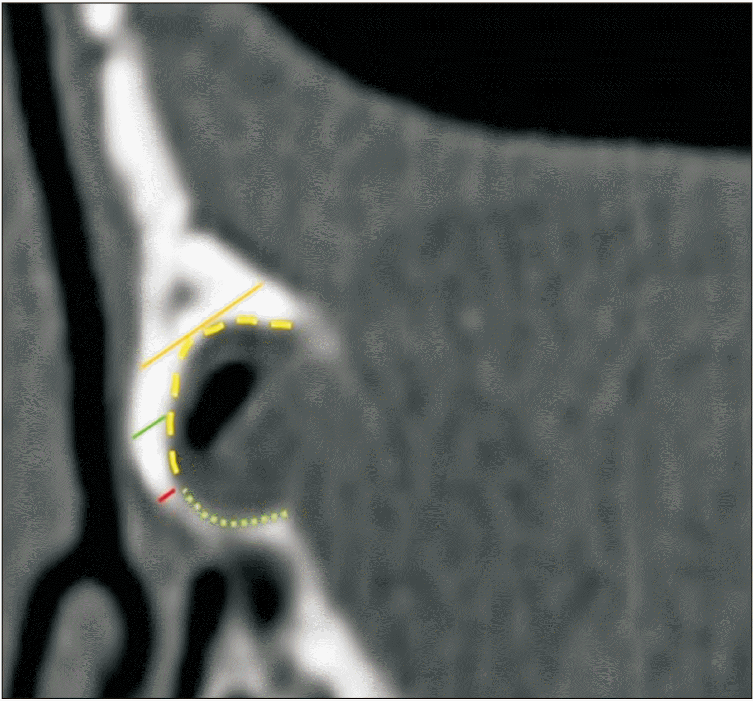
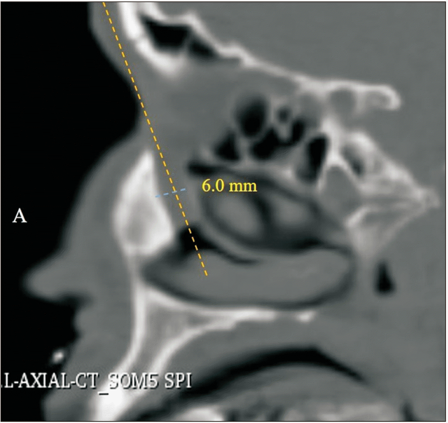
The measurement method was conducted regarding a previous research by Sarbajna et al. [7] (Figs. 1, 2), and only one orbit from each case was included in the study. The first axial cut below the fronto-lacrimal-maxillary suture that indicated the concavity of LSF was determined as the “upper section” (the most cranial cut), and the “lower section” (the most caudal cut) was the last axial cut before a complete nasolacrimal duct ring appeared. The “middle section” was identified as the cut midway between the upper and lower sections [10]. All measurements were done using the digital caliper/protractor tool of the viewing workstation (Syngo CT workstation; Siemens) by two trained radiologists. As identified in Fig. 1A and B, the dimensions of the LSF were calculated on the three sections after magnification of the images. For each of the 3 axial sections, the maximum and midpoint thickness of the maxillary bone (orange and green dotted line in Fig. 2), as well as the thickness of the lacrimal bone (red line in Fig. 2) just posterior to the lacrimo-maxillary suture were taken at 30° from the coronal plane. A digital freehand caliper was used to measure the curvilinear length of the maxillary and lacrimal bone in the LSF (Fig. 2). The ratio of the length of the lacrimal bone with relation to the entire length of the LSF was determined as follows: length of the lacrimal bone/total length of the LSF ×100 [7]. The same freehand caliper was also utilized to measure the nasal mucosal length from the level of the anterior aspect of the fossa to the internal contour of the anterior curvature of nasal mucosa at the lower axial sections (Fig. 2) [10]. In another part of present research, we also measured antero-posterior NLD inner diameter by same caliper tool [9]. The axial and coronal sections were aligned along the center of the NLD; consequently, providing sagittal measurements to be selected from reconstructed images lying in the plane perpendicular to the long axis of the NLD (Fig. 3).
Data statistical evaluation was performed using the SPSS version 24 (IBM Corp., Armonk, NY, USA). Patient age, thickness and proportion were compared between sexes using the Student’s t-test, and revealed as means±standard deviations. Pearson’s correlation coefficient was used to evaluate correlation between the thickness and patient age. To determine factors affecting the proportion such as patient age, thickness and length, multivariate linear regression analysis with stepwise variable selection was done. A P-value of less than 0.05 was considered statistically significant.
Computed Tomography axial and sagittal images of 117 males (72.7%) and 44 females (27.3%) were studied, and results of measurements and statistical analyses are shown in Tables 1–4 and Figs. 1–3. One-hundred-sixty-five left sides from 161 cases (age: 40.59±17.4 years) were included in this research. In both sexes, the maximum thickness of the maxillary bone increased from the upper to the lower plane (P=0.004). Interestingly, the maximum thickness on the middle plane has a positive correlation with age (P=0.02). In contrast, the midpoint thickness reduced from the upper to the lower plane. The thickness of lacrimal bone was relatively equal throughout the lacrimal fossa. The thickness on each plane did not show sex-related difference (Pearson’s correlation coefficient), but had a significant relationship with age in middle (P=0.03) and lower plane (P=0.01), respectively. The proportion of lacrimal fossa comprising lacrimal bone for lower plane did not show sex-related difference (males: 46.63%±9.45% and females: 43.41%±9.46%), but had positive correlation with age (P=0.01). The mean antero-posterior diameters of left bony nasolacrimal duct was 5.94±0.89 mm for both sexes. This study found no significant difference between patient sex, age, and lacrimal duct diameter (LDD). Nasal mucosa length (NML), for lower plane, showed significant sex-related difference (P=0.001), but had no correlation with patient age. Using a stepwise process, all items were removed from the regression model for the proportion on the upper plane (P>0.05). Predictors for the proportion on each plane were as follows: patient age at the level of the middle plane (adjusted r2=0.032; P=0.02) and patient age and maximum thickness at the level of the lower plane (adjusted r2=0.0001; P=0.05). However, the correlation was very weak.
In the present research, using orbital CT scan, we have determined the ratio, thickness of the maxillary and lacrimal bone on the LSF, intranasal space, and nasolacrimal duct diameter of south-west population of Iran. This is the first time that such an investigation has been conducted in this geographic area. The anatomical structure of LSF is made anteriorly by a combination of the frontal process of the maxillary bone and the lacrimal bone at the posterior. The depth of the fossa gradually increased toward the lower level and joined to the nasolacrimal duct [14]. An adequately sized and accurately placed osteotomy is a prerequisite for a DCR with a successful outcome [15]. Many studies have reported the racial differences in the anatomy of the intranasal space and LSF [10, 11]. Thus, a comprehensive knowledge of the anatomical variation of the LSF is essential in planning the surgeon for a convenient surgical procedure during DCR.
In the study ahead, the maxillary bone on an average revealed an increased thickness from upper to the lower plane with an average maximum thickness of 4.07 mm on the upper plane and 5.6 mm on the lowermost plane. The results shown that there is a positive correlation between patient age and maximum thickness on the middle plane. On the contrary, the midpoint thickness was greater on the upper plane, with an average size of 2.38 mm.
The lacrimal bone thickness was approximately uniform throughout the lacrimal fossa with a maximum measurement of 0.76 and 0.69 mm on the upper and middle plane, and 0.67 mm on the lowermost plane. Interestingly, the correlation between patient age and lacrimal thickness on lower plane shown a sex related difference. A previous cadaveric study in United Kingdom [16], and a research performed by Gore et al. [10] exhibited a clearly thinner lacrimal bone (Black African, 0.09–0.12 mm; Caucasians, 0.09–0.10 mm) compared to present research. Despite the anatomic racial characteristics, we cannot rule out the assumption that diverse measurement techniques and tools may have been produced different reports.
The results shown that on an average, at mid-fossa level 43.57% of lacrimal bone constituting the lacrimal fossa wall in both sexs. Subsequently, an inverse correlation was recognized between patient age and percentage of lacrimal fossa forming lacrimal bone. This could mean that DCR is more difficult in older patients than in younger subjects. These findings are consistent with the study conducted by Sarbajna et al. [7], which measured the morphology of the LSF in the Japanese population. In a similar study on black Africans and Caucasians which conducted by Gore et al. [10], they explained that the proportion of lacrimal bone of both races was less in almost all the planes compared to the Iranian southwest population in present research. Agarwal and Kumar's research [17] showed that the proportion of lacrimal and maxillary bone in the LSF to be 50%. These results demonstrate a higher proportion of lacrimal bone percentage compared to present inquiry. However, the calculations in their investigation have been based on an overall average in relation to the entire fossa rather than specific anatomical sections. The lacrimal bone proportion was considerably less in the inquiry conducted by Woo et al. [11] in Asian population (upper, 21.0%; middle, 31.0%; lower, 37.6%). However, this study presented results almost had similar findings to that of Kang et al. [12] (upper, 39%; middle 39%; lower, 43%). This study results show that the average length of nasal mucosa anterior to lacrimal fossa is 25.6 mm and there is a meaningful correlation between both sexs. Gore et al. [10] disclosed that, length of nasal mucosa in Black African (in lower plane) is 16.0 mm and 24.2 mm in Caucasians, respectively. The present findings seem to be consistent with Gore et al. [10] observations, because anthropological evidence shows that the population living in southwestern Iran is Caucasian [18]. With reference to these findings, this might be even more important in patients of different ethnic groups, where there is only limited nasal mucosa available for soft-tissue anastomosis.
Another part of this study was to investigate the differences in normal NLD anatomy between Iranian southwest (SW) community, to add to the existing knowledge considering the aetiology and possibility of primary acquired nasolacrimal duct obstruction (PANDO). Janssen et al. [19] study in Europeans found a narrower NLD diameter in PANDO cases as compared to normal subjects. To our knowledge, the present research is the first in the Middle East to measure the antero-posterior (AP) diameter of the normal bony nasolacrimal duct. In the present inquiry, some scans were excluded due to septal deviation and sinusitis, as chronic sinusitis can lead to bone remodeling and therefore could affect the size of the NLD. A few investigations have been published on sex variations correlated with the bony nasolacrimal canal [19]. A survey by Groessl et al. [20] declared that the lower nasolacrimal fossa and the middle bony lacrimal duct are significantly smaller in females than in males. In contrast to previous studies [21-23], our results did not show any significant correlation between patient age, sex and AP diameter of nasolacrimal duct. Lin et al. [9] performed a similar study in four ethnic groups. They showed that bony nasolacrimal duct is greater in caliber (inner diameter) if the patient is of Afro-Caribbean or Oriental origin compared to European or South Asian. In the present research, the mean AP diameter of the nasolacrimal duct was 5.94 mm in both sexes, almost similar to Afro-Caribbean (5.5 mm) and Oriental ethnicity (5.7 mm) [9].
Taken together, the maxillary bone was thicker in Iranian SW population, compared to the other ethnics mentioned in this survey (with except for Black African). However, because of the larger proportion of lacrimal bone forming the LSF in Iranian population, the following conclusions can be drawn that performing osteotomy could be relatively easier in the Iranian SW population compared to other races. According to findings and since the maximum thickness of maxillary bone at the lower plane, the authors propose that the rhinostomy is easier to begin and continue at the upper or middle planes in the lacrimal fossa.
A recent systematic review article disclosed that the general agreement was that NLD anatomy is not an important cause in PANDO. However, the etiopathogenesis of PANDO remains poorly recognized [24]. The evidence from this study indicates European and South Asian patients have narrower NLDs as compared to Iranian SW population. So, NLD obstruction in Iranian subjects may be more likely due to secondary acquired causes.
Although the suggested investigation is the first inquiry in this demographic area, but has some limitations. First, the retrospective design structure of study, and second, no possibility of direct compare our results with those studied in other ethnics using statistical methods. Third, we obtained the data from the left side (unaffected side) subjects. Fourth, its include theoretical relation to clinical application. Fifth, further comparative investigations are required to give the anatomic differences, which would provide better instructions. In this regard, patients do not generally need orbital CT scan prior to DCR procedure and it would be challenging to justify radiation hazard to perform an examination that provides a comparison of anatomical measurements with the ease of DCR.
The study ahead to be of good clinical value, because the key to being a successful ophthalmic surgeon lies in the awareness and ability to change surgical technique when anatomic variation is experienced.
The bony anatomy of the LSF differs according to ethnic. Present research evaluated the bony constituents of the LSF in the Iranian SW population. Our results reveal that maximal lacrimal to maxillary bone proportion and a higher NLD diameter are both found in this community. These results show that performing an osteotomy during DRC could be easier in the Iranian SW population compared to other races.
Acknowledgements
The results reported in this paper were part of the residency research thesis project of Samad Shahryari. The authors thank to head of medical imaging center of Golestan Hospital for its cooperation. The Research Deputy of the Ahvaz Jundishapur University of Medical Sciences, Ahvaz, Iran (grant number U-00031), funded this study.
Notes
References
1. Hartikainen J, Aho HJ, Seppä H, Grenman R. 1996; Lacrimal bone thickness at the lacrimal sac fossa. Ophthalmic Surg Lasers. 27:679–84. DOI: 10.3928/1542-8877-19960801-07. PMID: 8858634.

2. Shams PN, Abed SF, Shen S, Adds PJ, Uddin JM. 2012; A cadaveric study of the morphometric relationships and bony composition of the caucasian nasolacrimal fossa. Orbit. 31:159–61. DOI: 10.3109/01676830.2011.648809. PMID: 22551366.

3. Valencia MRP, Takahashi Y, Naito M, Nakano T, Ikeda H, Kakizaki H. 2019; Lacrimal drainage anatomy in the Japanese population. Ann Anat. 223:90–9. DOI: 10.1016/j.aanat.2019.01.013. PMID: 30797973.

4. Rajak SN, Psaltis AJ. 2019; Anatomical considerations in endoscopic lacrimal surgery. Ann Anat. 224:28–32. DOI: 10.1016/j.aanat.2019.03.010. PMID: 30953809.

5. Yang JW, Oh HN. 2012; Success rate and complications of endonasal dacryocystorhinostomy with unciformectomy. Graefes Arch Clin Exp Ophthalmol. 250:1509–13. DOI: 10.1007/s00417-012-1992-x. PMID: 22623114. PMCID: PMC3460168.

6. Gupta N. Gupta N, editor. 2021. Complications of endoscopic dacryocystorhinostomy. Endoscopic Dacryocystorhinostomy. Springer;Singapore: p. 167–75. DOI: 10.1007/978-981-15-8112-0_12. PMCID: PMC9128314.

7. Sarbajna T, Takahashi Y, Valencia MRP, Ito M, Nishimura K, Kakizaki H. 2019; Computed tomographic assessment of the lacrimal sac fossa in the Japanese population. Ann Anat. 224:23–7. DOI: 10.1016/j.aanat.2019.03.008. PMID: 30953810.

8. Yong AM, Zhao DB, Siew SC, Goh PS, Liao J, Amrith S. 2014; Assessment of bony nasolacrimal parameters among Asians. Ophthalmic Plast Reconstr Surg. 30:322–7. DOI: 10.1097/IOP.0000000000000101. PMID: 25069069.

9. Lin Z, Kamath N, Malik A. 2021; Morphometric differences in normal bony nasolacrimal anatomy: comparison between four ethnic groups. Surg Radiol Anat. 43:179–85. DOI: 10.1007/s00276-020-02614-4. PMID: 33184673.

10. Gore SK, Naveed H, Hamilton J, Rene C, Rose GE, Davagnanam I. 2015; Radiological comparison of the lacrimal sac fossa anatomy between black Africans and Caucasians. Ophthalmic Plast Reconstr Surg. 31:328–31. DOI: 10.1097/IOP.0000000000000457. PMID: 26039331.

11. Woo KI, Maeng HS, Kim YD. 2011; Characteristics of intranasal structures for endonasal dacryocystorhinostomy in asians. Am J Ophthalmol. 152:491–8.e1. DOI: 10.1016/j.ajo.2011.02.019. PMID: 21669403.

12. Kang D, Park J, Na J, Lee H, Baek S. 2017; Measurement of lacrimal sac fossa using orbital computed tomography. J Craniofac Surg. 28:125–8. DOI: 10.1097/SCS.0000000000003262. PMID: 27930466.

13. Purevdorj B, Dugarsuren U, Tuvaan B, Jamiyanjav B. 2021; Anatomy of lacrimal sac fossa affecting success rate in endoscopic and external dacryocystorhinostomy surgery in Mongolians. Anat Cell Biol. 54:441–7. DOI: 10.5115/acb.21.081. PMID: 34620735. PMCID: PMC8693133.

14. Örge FH, Boente CS. 2014; The lacrimal system. Pediatr Clin North Am. 61:529–39. DOI: 10.1016/j.pcl.2014.03.002. PMID: 24852150.

15. Tao H, Ma ZZ, Wu HY, Wang P, Han C. 2014; Anatomic study of the lacrimal fossa and lacrimal pathway for bypass surgery with autogenous tissue grafting. Indian J Ophthalmol. 62:419–23. DOI: 10.4103/0301-4738.121137. PMID: 24817745. PMCID: PMC4064215.

16. Yung MW, Logan BM. 1999; The anatomy of the lacrimal bone at the lateral wall of the nose: its significance to the lacrimal surgeon. Clin Otolaryngol Allied Sci. 24:262–5. DOI: 10.1046/j.1365-2273.1999.00235.x. PMID: 10472456.

17. Agarwal M, Kumar V. 2012; Morphological study of fossa for lacrimal sac: contributions by lacrimal and maxilla. J Anat Soc India. 61:234–41. DOI: 10.1016/S0003-2778(12)80037-2.

18. Amanolahi S. 2005; A note on ethnicity and ethnic groups in Iran. Iran Cauc. 9:37–41. DOI: 10.1163/1573384054068105.

19. Janssen AG, Mansour K, Bos JJ, Castelijns JA. 2001; Diameter of the bony lacrimal canal: normal values and values related to nasolacrimal duct obstruction: assessment with CT. AJNR Am J Neuroradiol. 22:845–50. PMID: 11337326. PMCID: PMC8174956.
20. Groessl SA, Sires BS, Lemke BN. 1997; An anatomical basis for primary acquired nasolacrimal duct obstruction. Arch Ophthalmol. 115:71–4. Erratum in: Arch Ophthalmol 1997;115:655. DOI: 10.1001/archopht.1997.01100150073012. PMID: 9006428.

21. Bulbul E, Yazici A, Yanik B, Yazici H, Demirpolat G. 2016; Morphometric evaluation of bony nasolacrimal canal in a caucasian population with primary acquired nasolacrimal duct obstruction: a multidetector computed tomography study. Korean J Radiol. 17:271–6. DOI: 10.3348/kjr.2016.17.2.271. PMID: 26957913. PMCID: PMC4781767.

22. Park JH, Huh JA, Piao JF, Lee H, Baek SH. 2019; Measuring nasolacrimal duct volume using computed tomography images in nasolacrimal duct obstruction patients in Korean. Int J Ophthalmol. 12:100–5. DOI: 10.18240/ijo.2019.01.16. PMID: 30662848. PMCID: PMC6326919.

23. McCormick A, Sloan B. 2009; The diameter of the nasolacrimal canal measured by computed tomography: gender and racial differences. Clin Exp Ophthalmol. 37:357–61. DOI: 10.1111/j.1442-9071.2009.02042.x. PMID: 19594561.

24. Ali MJ, Paulsen F. 2019; Etiopathogenesis of primary acquired nasolacrimal duct obstruction: what we know and what we need to know. Ophthalmic Plast Reconstr Surg. 35:426–33. DOI: 10.1097/IOP.0000000000001310. PMID: 30730434.

Fig. 1
The upper (A), middle (B), and lower axial planes (C) through the left maxillary and lacrimal bones.

Fig. 2
The thickness and length of the right maxillary and lacrimal bones on the lower plane. The maximum (orange line) and midpoint thickness of the maxillary bone (green line), and the thickness of the lacrimal bone near the lacrimo-maxillary suture (red line) were measured. The length of the maxillary (yellow broken line) and lacrimal bone in the lacrimal fossa (green dotted line) was also measured.

Fig. 3
Sagittal section showing the nasolacrimal duct measurement. Yellow and blue dotted line indicate the longitudinal axis and anteroposterior diameter of bony nasolacrimal duct, respectively. A: anterior.

Table 1
Correlation between patient sex and thickness on each plane (upper, middle, and lower axial plane)
Table 2
Correlation between patient age and thickness on each plane (upper, middle, and lower axial plane)
| Lacrimal fossa parameter | Correlation coefficient | P-value |
|---|---|---|
| Maximum thickness | ||
| Upper | 0.147 | 0.06 |
| Middle | 0.178* | 0.02 |
| Lower | 0.034 | 0.66 |
| Midpoint thickness | ||
| Upper | 0.068 | 0.39 |
| Middle | –0.022 | 0.78 |
| Lower | –0.042 | 0.59 |
| Lacrimal bone thickness | ||
| Upper | –0.057 | 0.47 |
| Middle | –0.169* | 0.03 |
| Lower | –0.192* | 0.01 |
Table 3
Correlation between patient age and maxillary length (ML), lacrimal length (LL), nasal mucosa length (NML), and lacrimal duct diameter (LDD) and proportion of lacrimal fossa comprising lacrimal bone proportion (LP)
| Lacrimal fossa parameter | Correlation coefficient | P-value |
|---|---|---|
| ML | 0.18 | 0.01 |
| LL | –0.15 | 0.04 |
| NML | 0.11 | 0.14 |
| LDD | 0.07 | 0.37 |
| LP | –0.2 | 0.01 |
Table 4
Correlation between patient sex and maxillary length (ML), lacrimal length (LL), nasal mucosa length (NML), lacrimal duct diameter (LDD), and proportion of lacrimal fossa comprising lacrimal bone proportion (%) (LP)


 PDF
PDF Citation
 Print
Print


 XML Download