This article has been corrected. See "Erratum " in Volume 57 on page 297.
Abstract
Background
Gaucher disease (GD) is an autosomal recessive disorder characterized by excessive accumulation of glucosylceramide in multiple organs. This study was performed to determine the detection rate of GD in a selected patient population with unexplained splenomegaly in Korea.
Methods
This was a multicenter, observational study conducted at 18 sites in Korea between December 2016 and February 2020. Adult patients with unexplained splenomegaly were enrolled and tested for β-glucosidase enzyme activity on dried blood spots (DBS) and in peripheral blood leukocytes. Mutation analysis was performed if the test was positive or indeterminate for the enzyme assay. The primary endpoint was the percentage of patients with GD in patients with unexplained splenomegaly.
Results
A total of 352 patients were enrolled in this study (male patients, 199; mean age, 48.42 yr). Amongst them, 14.77% of patients had concomitant hepatomegaly. The most common sign related to GD was splenomegaly (100%), followed by thrombocytopenia (44.32%) and, anemia (40.91%). The β-glucosidase activity assay on DBS and peripheral leukocytes showed abnormal results in sixteen and six patients, respectively. Eight patients were tested for the mutation, seven of whom were negative and one patient showed a positive mutation analysis result. One female patient who presented with splenomegaly and thrombocytopenia was diagnosed with type 1 GD. The detection rate of GD was 0.2841% (exact 95% CI, 0.0072‒1.5726).
Gaucher disease (GD) is an autosomal recessive lysosomal storage disorder caused by congenital deficiency of lysosomal enzyme acid β-glucosidase (glucocerebrosidase) [1, 2]. The excess accumulation of glucosylceramide in macrophages in multiple organs causes various manifestations of GD such as hepatosplenomegaly, anemia, thrombocytopenia, bone pain, etc. [1]. GD has been traditionally categorized into three classic variants based on the presence or absence of neurologic manifestation. Type 1 GD (GD1), the non-neuronopathic form, is the most prevalent subtype, accounting for up to 94% of patients in Europe, Canada, and the United States [3]. The incidence of GD1 is variable with an estimated prevalence of 1–2 in 100,000 worldwide and as high as 1 in 850 in the Ashkenazi Jewish population [2]. In contrast, Type 2 (acute neuronopathic) and type 3 (chronic neuronopathic) GD are relatively more common in East Asian countries, including South Korea [4, 5].
Gaucher disease is diagnosed by measuring acid β-glucosidase enzyme activity levels in nucleated cells or on dried blood spots (DBS). However, defining β-glucocerebrosidase (GBA) mutation through gene analysis is needed for the confirmative diagnosis [3]. Currently, there are two types of treatment modalities; enzyme replacement and substrate reduction therapy, which can reverse many of the non-neurological manifestations [6].
Gaucher disease is frequently associated with non-specific symptoms that mimic other hematological malignancies [7]. It has been reported that 86% of patients with GD in the United States and 73% in Australia/New Zealand consulted hemato-oncologists for the evaluation of their symptoms [8]. In a survey conducted among 406 hemato-oncologists, only 20% of physicians considered GD in the differential diagnosis even in the presence of all the classic symptoms of GD [8]. This low percentage is attributable to the rarity, frequent non-specific and mild to moderate initial symptoms, and low level of awareness about GD.
Although the prevalence rate of GD has not yet been evaluated in Asian countries, according to a report from Yoo’s group, approximately 60 Korean patients from 50 families have been identified until 2013, demonstrating an enormous gap from the pan-ethnic prevalence of 1 in 40,000–100,000 live births [9, 10]. Therefore, determining the prevalence rate of GD will provide important insights into the ethnical and clinical features of GD in a Korean population.
The aim of this study was to determine the detection rate of GD in a selected population of patients with unexplained splenomegaly, who were referred to hemato-oncology departments in Korea. The design of the study mirrored real-life clinical practice.
This was a national, multicenter, observational study conducted at 18 sites (hemato-oncologists) in Korea between December 2016 and February 2020. The study included a maximum of three visits, a screening or baseline visit (visit 1), collection of first blood sample (visit 2) for enzyme assay, and collection of second blood sample (visit 3; within 12 wk after visit 2) for mutation analysis. Based on the patient’s discretion, procedures scheduled at visit 2 were also conducted at visit 1 or at a later date (within 12 wk after visit 1). Visit 3 was conducted if the patient tested positive or indeterminate for the enzyme assay.
This study was conducted in accordance with the Declaration of Helsinki, International Conference on Harmonization Good Clinical Practice guidelines, and applicable local regulatory requirements. The study protocol was approved by an independent ethics committee or institutional review board at each study site. Written informed consent was obtained from each participant prior to any study-related procedure.
Patients of either sex aged >19 years with unexplained splenomegaly detected by physical examination and/or imaging techniques including ultrasound, computed tomography (CT), or magnetic resonance imaging (MRI) and patients who had a history of splenectomy were eligible for participation in this study. Patients with chronic B or C viral hepatitis and patients with alcoholic or viral liver cirrhosis were excluded from the study; however, patients whose splenomegaly was suspected due to GD upon physician’s discretion were eligible, even if they met the exclusion criteria.
On visit 1, demographics and baseline characteristics (age, sex, date of diagnosis of splenomegaly, method of diagnosis, size of splenomegaly, presence of hepatomegaly, past medical/surgical history, present medical history, family history of GD), history of bone marrow biopsy, signs and symptoms, and details of laboratory investigations were collected. On visit 2, a sample for enzyme assay (acid β-glucosidase activity) was collected. This whole blood sample was divided into 2 heparin tubes of 2 cc and 9 cc and sent for analysis to a designated central lab (LabGenomics Co., Ltd, South Korea) where acid β-glucosidase enzyme activity was measured on DBS and in peripheral leukocytes. The parallel testing of acid sphingomyelinase deficiency (ASMD) was performed on DBS upon protocol amendment to address the clinical similarities between Gaucher disease and Niemann Pick Type A/B caused by deficiency of acid sphingomyelinase. On visit 3, if the result of the enzyme assay was positive or indeterminate (visit 2), mutation analysis was performed to confirm the diagnosis. The sample (5 cc of whole blood) was collected in an ethylenediamine tetraacetic acid or heparin tube. The cut-off value used for DBS was 5.0 µmol/L/h and the normal range for peripheral leukocytes was 6.0–9.0 nmol/h/mg protein.
The primary endpoint was the percentage of patients with GD in patients with unexplained splenomegaly in Korea. The secondary endpoints included the identification of clinical features which were highly predictive of GD in the Korean patients who visited the hemato-oncology department and the proportion of patients with abnormal DBS results of acid sphingomyelinase activity in patients with unexplained splenomegaly. The exploratory endpoint is the percentage of patients with an abnormal result of acid sphingomyelinase on parallel DBS testing.
Gaucher disease is an ultra-rare disease that occurs in about 1 of 40,000 people [5] and there is a lack of data on the detection rate in high-risk patients in Asia. A study from Italy reported an interim detection rate of 3.6% (seven patients) after screening 196 high-risk patients (those with splenomegaly and/or thrombocytopenia) at the time of developing the study design for this study [11]. Considering the primary objective of this study, 600 patients with unexplained splenomegaly were needed to detect 20 patients with GD. The detection rate of GD was expressed as number and percentage. Clinical, hematological, and biochemical parameters of patients affected by GD were compared to those of unaffected patients using the Wilcoxon rank-sum (Mann–Whitney U-test) test for continuous variables or Fisher’s exact test for categorical variables.
The present study was terminated early due to difficulty with enrolment. A total of 395 patients were screened; of which, 352 patients completed the study (Fig. 1). The most common reason for exclusion was missing the primary endpoint (N=19) due to the incompletion of all required testing. Of the 352 patients, the mean age was 48.42±15.81 SD years and 56.53% (N=199) were men. Demographic and clinical characteristics are summarized in Table 1. The most common method of diagnosis of splenomegaly was using CT (71.02%), followed by abdominal ultrasound (Abd U/S) (23.01%) and physical examination (6.53%).
The median (range) duration of splenomegaly was 90 (1–15629) days and the mean size of splenomegaly was 14.57±3.40 SD cm. A total of 14.77% (N=52) patients had hepatomegaly. Of 352 patients, 161 patients had a history of bone marrow biopsy, of which 73 (20.74%) patients had a specific abnormality. The most common sign related to GD was splenomegaly (N=352, 100%), followed by thrombocytopenia (N=156, 44.32%) and anemia (N=144, 40.91%). One patient had a family history of GD: this patient was a sibling of another patient.
The mean hemoglobin was 12.21±2.73 SD g/dL and it was abnormal in 52.01% (N=181) of patients (Table 2). The mean serum iron was 85.89±62.24 SD µg/dL, serum ferritin was 541.61±1099.10 SD µg/dL, and transferrin was 144.94±123.09 SD mg/dL. Abnormal serum iron, serum ferritin, and transferrin were observed in 38.83%, 41.18%, and 46.67% of patients, respectively.
A total of 16 patients had abnormal results in the assay of acid β-glucosidase activity by DBS testing. The mean value of acid β-glucosidase activity in 352 patients was 8.06±4.88 SD nmol/h/mg protein by peripheral leukocyte testing. The acid β-glucosidase activity assay in peripheral leukocyte test showed that 86.93% (N=306) of patients had normal acid β-glucosidase activity, 11.36% (N=40) had slightly decreased acid β-glucosidase activity at which level GD is not suspected (heterozygous, etc.), and 1.70% (N=6) had abnormal acid β-glucosidase activity and were suspected of having GD. Of 188 patients who were tested for ASMD by DBS testing, 1.06% (N=2) had abnormal results.
A total of eight patients were tested for the mutation, of which seven were negative and one patient tested positive. None of the patients who underwent GBA mutation analysis was diagnosed as a carrier. One patient who had an abnormal result in both DBS and peripheral leukocyte tests was positive for the GBA gene mutation test (Fig. 2). The detection rate of GD was 0.2841% (exact 95% CI, 0.0072–1.5726).
As there was only one patient who was diagnosed with GD1, it was challenging to statistically compare hematological and biochemical data between those with and without GD. The data are summarized in Supplementary Table 1.
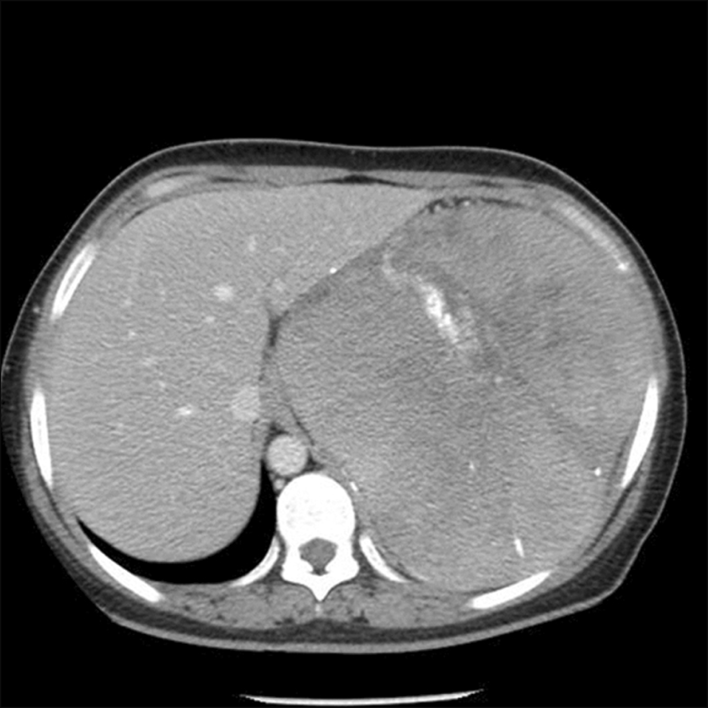
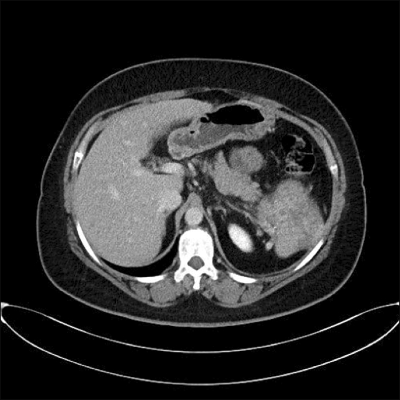
The patient was a 34-year-old Korean woman with unexplained splenomegaly that was diagnosed in 1991. This patient had a near-total splenectomy approximately 27 years prior, but the remnant spleen had enlarged, which prompted suspicion of GD. Splenomegaly and thrombocytopenia were observed without a family history of GD. The size of the splenomegaly was 18.4 cm detected by CT (Fig. 3, 4). The patient had no hepatomegaly but had a specific abnormality on bone marrow biopsy that showed Gaucher cells. There was no evidence of short stature, osteopenia, or osteonecrosis; however, the patient had a history of chronic osteomyelitis in the left distal femur at the age of ten. There was evidence of operation scar and iliotibial band defect. The level of hemoglobin was 11.00 g/dL; white blood cells (WBC), 4.12×10³/L; red blood cells (RBC), 3.55×10⁶/L, and platelet count, 82.00×10³/L. The biochemical parameters were as follows: serum iron, 58.50 µg/dL; serum ferritin, 1673.90 µg/dL; blood urea nitrogen (BUN), 13.00 mg/dL; creatinine, 0.53 mg/dL, serum glutamic-pyruvic transaminase, 15.00 U/L; serum glutamic-oxaloacetic transaminase, 36.00 U/L; total bilirubin, 1.55 mg/dL; total protein, 8.20 g/dL; and albumin, 3.50 g/dL. The result for acid β-glucosidase enzyme activity was abnormal in DBS and 0.20 nmol/h/mg in peripheral leukocytes. The mutation analysis confirmed the diagnosis of GD. GBA gene sequencing detected c.754T>A (p.Phe252Ile, aka F213I) and c.1448T>C (p.Leu483Pro, aka L444P) mutation. This patient was immediately started on enzyme replacement therapy upon diagnosis.
Gaucher disease, albeit rare, is not a hard-to-diagnose disorder with the availability of a relatively simple enzyme assay for β-glucosidase activity and confirmatory molecular analysis for the GBA gene [3]. The difficulty arises from recognizing nonspecific symptoms due to low awareness and the perception that GD is too rare to be seen at the hemato-oncology clinic.
This study found a low detection rate of 0.2841% in a selected Korean population presenting unexplained splenomegaly. Type 1 GD accounts for more than 94% of all patients diagnosed with GD worldwide; however, epidemiologic surveys in Korea indicate that more than half of Korean patients with GD have either GD2 or GD3 [3-5, 12]. This study was conceptualized with the fundamental question of whether such a small proportion of patients with GD1 is a true representation of Korean patients or is it skewed due to low awareness among hemato-oncologists that may have led to under-diagnosis in patients presenting without apparent neurologic involvement.
It is important to note that this study enrolled only adult patients whose symptoms may not have been severe or specific enough to raise suspicion during childhood. Also, considering previous reports describing a high proportion of neuronopathic forms of the disease [4, 5, 12], the vast majority of patients in Korea may have CNS manifestations that could have already been detected in childhood or managed by other specialties, including pediatricians and neurologists.
The present study demonstrated a lower detection rate of GD compared to that of other countries. A study conducted in Italy reported that 3.3% of patients with splenomegaly and/or thrombocytopenia were diagnosed with GD [11]. Our study enrolled patients with unexplained splenomegaly without the requirement for additional symptoms such as thrombocytopenia to make the screening available for a wider population; additionally, it relied on interim data from the Cappellini study in which all (100%) identified patients had splenomegaly and none had thrombocytopenia alone [13].
The patient who was diagnosed with GD in this study showed classical features of splenomegaly, thrombocytopenia, anemia, bone lesion, and hyperferritinemia. Hyperferritinaemia is commonly observed in patients with GD1 and recent studies have proposed GD-focused diagnostic flow charts starting from hyperferritinemia [11, 14]. It would be reasonable to propose more targeted screening using splenomegaly as an overarching symptom with additional consideration of other key hematologic manifestations such as thrombocytopenia and hyperferritinemia in hemato-oncology clinics.
The GBA mutations in Korean patients were different from those of patients from other countries, including Italy, but were similar to those in patients from Japan and China [5, 13]. N370S, the most common mutation in the Ashkenazi Jewish population that is known to be associated with GD1 has never been reported in Korean patients, including those enrolled in previous studies [5, 15]. According to a recent report from the International Collaborative Gaucher Group (ICGG) registry, the diverse global population has two GBA mutations in patients with GD3; L444P (77%) and D409H (7%) [16]. In Korea, L444P (20.8%) is the most prevalent mutation and is often associated with a neuronopathic form of the disease, followed by G46E (13.9%), which is suspected to be associated with the non-neuronopathic form, and F213I (12.5%) [13]. The most frequent allele found in Japan is F213I, which is associated with neurological manifestations of GD and is identified in all three types of GD in the Korean population [13, 17]. The GD patient identified in this study also had L444P and F213I mutations which align with these reported data. This wide allelic heterogeneity in GBA mutation may have resulted in different clinical phenotypes presented in Korean patients.
The results of bone marrow biopsy in this study showed abnormality not only for the patient identified with GD but also for those without GD (45.74%). Bone marrow biopsy is frequently used to confirm the presence of Gaucher cells [18]. However, occasionally, pseudo-Gaucher cells are observed in non-Gaucher patients [19-21]. Moreover, there are less invasive diagnostic methods to confirm GD such as enzyme assay and molecular analysis of GBA mutation [3]. Patients presenting with thrombocytopenia and without GD symptoms (44.16%) in the present study were at high risk of a bleeding complication during a bone marrow biopsy. Therefore, bone marrow biopsy should be judiciously conducted and should probably be discouraged as a routine examination for diagnosing GD [2]. In the acid β-glucosidase enzyme activity assay, 1.42% of those without GD showed abnormal results and were suspected of GD.
Although the diagnosis of ASMD was not confirmed due to the unavailability of peripheral leukocyte and sphingomyelin phosphodiesterase 1 (SMPD1) gene analysis, it is reasonable to conduct parallel testing of GD and ASMD considering that 1.06% (N=2) of patients showed abnormality in ASMD testing and similarities in clinical manifestations such as hepatosplenomegaly, thrombocytopenia, and possible skeletal involvement. The deficiency of acid sphingomyelinase activity is associated with Niemann-Pick disease A/B, another lysosomal storage disorder that may have overlapping symptoms with Gaucher disease [22]. These results indicate that hemato-oncologists may need to consider the potential diagnosis of Niemann-Pick A/B disease in patients with unexplained splenomegaly. Since GD1 is generally underdiagnosed, alternate approaches such as newborn screening are being explored to increase the diagnostic rate [23]. Moreover, the high proportion of patients with an undetermined underlying diagnosis for their splenomegaly warrants further research delving into the causes of unknown splenomegaly.
Real-world data related to GD will contribute to a better understanding of this disease in the Korean population. As this was an observational study, it was impossible to mandate all patients to take required testing and, thus, it was left to the treating physician to decide whether the patient needs a mutation analysis, especially when the result was indeterminate or borderline low at a heterozygous level for enzyme testing. Therefore, all the patients with abnormalities in either DBS or peripheral leukocyte did not undergo mutation analysis rendering incomplete or missing data. In the present study, it was impossible to compare the clinical features of GD and non-GD patients because of an insufficient number of patients with GD for statistical analysis. Further studies are warranted to validate the robust criteria for at-risk or high-risk patients and determine generalized clinical features of GD in Korea.
In conclusion, the detection rate of GD in probable high-risk patients in Korea was lower than expected. This may be attributed to allelic heterogeneity of the GBA gene and less stringent screening criteria focusing on unexplained splenomegaly only. However, it is still important that hemato-oncologists consider the potential diagnosis of GD for patients with unexplained splenomegaly to reduce the misdiagnosis or delayed diagnosis and reduce the risk of complications.
ACKNOWLEDGMENTS
The authors would like to thank the study participants, their families, and caregivers who were involved in this study. The authors also thank Sapna Patil of Sqarona Medical Communications LLP, Mumbai for medical writing assistance which was paid for by Sanofi. Editorial support was also provided by Anahita Gouri and Rohan Mitra from Sanofi India.
REFERENCES
1. Grabowski GA. 2008; Phenotype, diagnosis, and treatment of Gaucher's disease. Lancet. 372:1263–71. DOI: 10.1016/S0140-6736(08)61522-6. PMID: 19094956.

2. Mistry PK, Cappellini MD, Lukina E, et al. 2011; A reappraisal of Gaucher disease-diagnosis and disease management algorithms. Am J Hematol. 86:110–5. DOI: 10.1002/ajh.21888. PMID: 21080341. PMCID: PMC3058841.

3. Kaplan P, Baris H, De Meirleir L, et al. 2013; Revised recommendations for the management of Gaucher disease in children. Eur J Pediatr. 172:447–58. DOI: 10.1007/s00431-012-1771-z. PMID: 22772880.

4. Lee JY, Lee BH, Kim GH, et al. 2012; Clinical and genetic characteristics of Gaucher disease according to phenotypic subgroups. Korean J Pediatr. 55:48–53. DOI: 10.3345/kjp.2012.55.2.48. PMID: 22375149. PMCID: PMC3286762.

5. Jeong SY, Park SJ, Kim HJ. 2011; Clinical and genetic characteristics of Korean patients with Gaucher disease. Blood Cells Mol Dis. 46:11–4. DOI: 10.1016/j.bcmd.2010.07.010. PMID: 20729108.

6. Gary SE, Ryan E, Steward AM, Sidransky E. 2018; Recent advances in the diagnosis and management of Gaucher disease. Expert Rev Endocrinol Metab. 13:107–18. DOI: 10.1080/17446651.2018.1445524. PMID: 30058864. PMCID: PMC6129380.

7. Thomas AS, Mehta AB, Hughes DA. 2013; Diagnosing Gaucher disease: an on-going need for increased awareness amongst haematologists. Blood Cells Mol Dis. 50:212–7. DOI: 10.1016/j.bcmd.2012.11.004. PMID: 23219328.

8. Mistry PK, Sadan S, Yang R, Yee J, Yang M. 2007; Consequences of diagnostic delays in type 1 Gaucher disease: the need for greater awareness among hematologists-oncologists and an opportunity for early diagnosis and intervention. Am J Hematol. 82:697–701. DOI: 10.1002/ajh.20908. PMID: 17492645.

9. Andrade-Campos MM, de Frutos LL, Cebolla JJ, et al. 2020; Identification of risk features for complication in Gaucher's disease patients: a machine learning analysis of the Spanish registry of Gaucher disease. Orphanet J Rare Dis. 15:256. DOI: 10.1186/s13023-020-01520-7. PMID: 32962737. PMCID: PMC7507684. PMID: 29234a24352744b8afb0e10a2ead564d.

10. Yoo HW, Lee HJ, Kim DS, et al. 2014. Disease Management Guideline on LSD - 2. Gaucher disease. The Korean Society of Inherited Metabolic Disease & Medical Review;Seoul, Korea:
11. Motta I, Consonni D, Stroppiano M, et al. 2021; Predicting the probability of Gaucher disease in subjects with splenomegaly and thrombocytopenia. Sci Rep. 11:2594. DOI: 10.1038/s41598-021-82296-z. PMID: 33510429. PMCID: PMC7843616. PMID: b22675ddb7324d238338d5c54148d366.

12. Charrow J, Andersson HC, Kaplan P, et al. 2000; The Gaucher registry: demographics and disease characteristics of 1698 patients with Gaucher disease. Arch Intern Med. 160:2835–43. DOI: 10.1001/archinte.160.18.2835. PMID: 11025794.
13. Motta I, Filocamo M, Poggiali E, et al. 2016; A multicentre observational study for early diagnosis of Gaucher disease in patients with splenomegaly and/or thrombocytopenia. Eur J Haematol. 96:352–9. DOI: 10.1111/ejh.12596. PMID: 26033455.

14. Marchi G, Nascimbeni F, Motta I, et al. 2020; Hyperferritinemia and diagnosis of type 1 Gaucher disease. Am J Hematol. 95:570–6. DOI: 10.1002/ajh.25752. PMID: 32031266.

15. Diaz GA, Gelb BD, Risch N, et al. 2000; Gaucher disease: the origins of the Ashkenazi Jewish N370S and 84GG acid beta-glucosidase mutations. Am J Hum Genet. 66:1821–32. DOI: 10.1086/302946. PMID: 10777718. PMCID: PMC1378046.
16. El-Beshlawy A, Tylki-Szymanska A, Vellodi A, et al. 2017; Long-term hematological, visceral, and growth outcomes in children with Gaucher disease type 3 treated with imiglucerase in the International Collaborative Gaucher Group Gaucher Registry. Mol Genet Metab. 120:47–56. DOI: 10.1016/j.ymgme.2016.12.001. PMID: 28040394.

17. Kawame H, Eto Y. 1991; A new glucocerebrosidase-gene missense mutation responsible for neuronopathic Gaucher disease in Japanese patients. Am J Hum Genet. 49:1378–80. PMID: 1840477. PMCID: PMC1686467.
18. Stirnemann J, Vigan M, Hamroun D, et al. 2012; The French Gaucher's disease registry: clinical characteristics, complications and treatment of 562 patients. Orphanet J Rare Dis. 7:77. DOI: 10.1186/1750-1172-7-77. PMID: 23046562. PMCID: PMC3526516.

19. Cozzolino I, Picardi M, Pagliuca S, et al. 2016; B-cell non-Hodgkin lymphoma and pseudo-Gaucher cells in a lymph node fine needle aspiration. Cytopathology. 27:134–6. DOI: 10.1111/cyt.12254. PMID: 26033037.

20. Sharma P, Das R, Bansal D, Trehan A. 2015; Congenital dyserythropoietic anemia, type II with SEC23B exon 12 c.1385 A → G mutation, and pseudo-Gaucher cells in two siblings. Hematology. 20:104–7. DOI: 10.1179/1607845414Y.0000000166. PMID: 24801240.

21. Chatterjee T, Dewan K, Ganguli P, et al. 2013; A rare case of hemoglobin E hemoglobinopathy with Gaucher's disease. Indian J Hematol Blood Transfus. 29:110–2. DOI: 10.1007/s12288-012-0153-z. PMID: 24426351. PMCID: PMC3636348.

22. Ferreira CR, Gahl WA. 2017; Lysosomal storage diseases. Transl Sci Rare Dis. 2:1–71. DOI: 10.3233/TRD-160005. PMID: 29152458. PMCID: PMC5685203.

23. Burton BK, Charrow J, Hoganson GE, et al. 2017; Newborn screening for lysosomal storage disorders in Illinois: the initial 15-month experience. J Pediatr. 190:130–5. DOI: 10.1016/j.jpeds.2017.06.048. PMID: 28728811.

Table 1
Demographic and clinical characteristics.
| Parameter | N=352 |
|---|---|
| Age (yr) | 48.42±15.81 SD |
| Gender, N (%) | |
| Male | 199 (56.53) |
| Female | 153 (43.47) |
| Weight (kg) | 66.95±13.51 SD |
| Height (cm) | 166.38±9.35 SD |
| Nationality, N (%) | |
| Korean | 351 (99.72) |
| Non-Korean | 1 (0.28) |
| Method of diagnosis of splenomegaly*, N (%) | |
| Magnetic resonance imaging | 250 (71.02) |
| Abdominal ultrasound | 81 (23.01) |
| Physical examination | 23 (6.53) |
| Computed tomography | 2 (0.57) |
| Others | 6 (1.70) |
| Duration of splenomegaly (days), median (range) | 90 (1–15629) |
| Size of splenomegaly, cm (N=223) | 14.57±3.40 SD |
| Hepatomegaly, N (%) | 52 (14.77) |
| Bone marrow biopsy result, N (%) [N=161] | |
| No specific abnormality | 88 (25.00) |
| Specific abnormality | 73 (20.74) |
| Confirmed | 1 (0.28) |
| Unconfirmed | 160 (45.45) |
| Signs and symptoms related to Gaucher, N (%) | |
| Splenomegaly | 352 (100.00) |
| Thrombocytopenia | 156 (44.32) |
| Anemia | 144 (40.91) |
| Hepatomegaly | 52 (14.77) |
| Bone pain | 14 (3.98) |
| Pathologic fracture | 1 (0.28) |
| Osteonecrosis | 1 (0.28) |
| Others | 41 (11.65) |
| Duration of signs and symptoms (days), median (range) | 338 (1–15629) |
Table 2
Summary of laboratory parameters.


 PDF
PDF Citation
 Print
Print


 XML Download