Abstract
Lateral compartment syndrome of the lower leg is rarely observed. Hence, there may be difficulty in diagnosis as its clinical patterns are different and more complicated than usual. We report two rare cases of a 20-year-old and a 28-year-old diagnosed with isolated lateral compartment syndrome who had either a surgical or conservative treatment. The comparison was done by analyzing the progression of neurological manifestation, electromyography, and nerve conduction study for two years. In the final follow-up, the patient who underwent the surgical treatment showed a shorter recovery time. However, both patients showed a full recovery from neurologic deficits.
Acute compartment syndrome is one of the orthopedic emergency diseases and is defined as a state of causing blood circulatory disturbance into the tissues in the muscle compartment due to increased tissue pressure in the closed compartment or state of capillary circulation being decreased to less than the degree required for tissue survival.1) Once pressure increases in the closed compartment, the pressure is difficult to be normalize by itself and if it is left untreated for several hours, muscle and nerve function get gradually depressed.2) This causes irreversible, permanent functional disturbance and eventually causes structural deformity. Therefore, capillary circulation must be recovered as soon as possible with early diagnosis and decompression in order to prevent fatal complications.2)
Lateral compartment syndrome of a lower leg, although rarely observed, may have difficulty in diagnosing as its clinical patterns are different from and more complicated than other usual ones. This study was aimed to see the changing patterns of each patient with isolated lateral compartment syndrome who had either surgical or conservative treatment.
This study was approved by the Institutional Review Board in our institution (Inje University Busan Paik Hospital, IRB No, 2021-09-015).
Level of Evidence: Level 4, case series study.
A 20-year-old man developed pain in right lower leg 3 days ago after playing soccer for 3 hours. He had no specific medical history. The symptom was watched over because he thought himself to have a muscle sprain then. From the next morning, ankle dorsiflexion and greater toe extension power started to get lost, and the patient visited the emergency room.
In physical examination, direct tenderness and soft tissue swelling were observed in the lateral side of the affected lower leg; motor grade of ankle dorsiflexion and greater toe extension was measured at 0/5, and hypoesthesia in the outer region of the right foot and 2nd and 3rd metatarsal bones region was found. Pulses from dorsalis pedis artery and posterior tibial artery were well-palpitated. Intracompartmental pressure (ICP) measurement was performed with a slit catheter and the ICP within the lateral compartment of the lower leg was 40 mmHg. Magnetic resonance imaging (MRI) was performed for the right lower leg and signal increase limited to peroneus longus muscle and peroneus brevis muscle was observed in T1 and T2 images (Fig. 1).
In the light of such clinical features and imaging examination results, isolated lateral compartment syndrome in the lower leg was diagnosed and fasciotomy was performed along the axis of the fibula. The aseptic dressing was done postoperatively with the surgical site opened and surgical loop tightening (Fig. 2). After 4∼5 days from the initial surgery, additional wound debridement and sequential wound suture were done. From 15 days after the first procedure, the surgical site came out to be clean and a secondary suture was conducted (Fig. 3).
In the electromyography conducted 20 days after injury, signal loss in deep peroneal nerve was observed and in physical examination, right ankle dorsiflexion and greater toe extension exercise were both measured at 1/5 and right ankle plantarflexion observed at 3/5, deep and superficial peroneal nerve sensation was measured up to 50% (Table 1).
Four months after the date of initial surgery, ankle dorsiflexion and greater toe extension motor improved to 4/5, and active ankle dorsiflexion and plantarflexion range of motion were measured to be 10° and 40°, each. The peroneal nerve sensation was still measured at 50%. At 6 months, peroneal nerve sensation recovered up to 60%, and range-of-motion was recovered as much as the same level compared with the unaffected side (Fig. 4, Table 2). At 1 year after initial surgery, superficial peroneal nerve sensation recovered up to 100% and deep peroneal nerve sensation remained 60%. In electromyography, improved patterns were observed in the lower leg muscles except for extensor digitorum brevis muscle (Table 3). In the telephone call done in twenty-one months postoperatively, all of his sensations were improved.
A 28-year-old man had pain in the right lower leg after having a motorcycle accident 4 weeks before hospitalization. He had no specific medical history. When he got injured, he had severe pain of visual analog scale 8 points, but the pain subsided and eventually disappeared so that he watched over the symptom at home. But starting from 1 week after the initial trauma, right foot dorsiflexion power started to weaken. The weakness showed timely aggravation and in 4 weeks after the initial trauma, the patient eventually showed right foot drop. At this point, the patient visited our center as an outpatient.
In the physical examination, direct tenderness or soft tissue swelling in right lower leg was not prominent; he complained of sensory loss in the entire region of the top of the foot in the front part; motor grade of foot invertor muscle was measured at 5/5 and foot evertor muscle at 4/5, but tibialis anterior muscle 2/5, extensor hallucis longus muscle 0/5, and extensor digitorum longus muscle 0/5. The pulses in dorsalis pedis artery and posterior tibial artery were well-felt. ICP measurement was performed with a slit catheter and the ICP within the lateral compartment of the lower leg was 30 mmHg. In the MRI taken right after the first visit, a signal increase in T2 images limited to peroneus longus muscle and peroneus brevis muscle was observed (Fig. 5). In electromyography, signal depression in the right deep and superficial peroneal nerve and posterior tibial nerve was observed (Table 4).
In light of such findings, isolated lateral compartment syndrome in a lower leg was diagnosed, but at the time when he was hospitalized, pain or edema in the lateral compartment of a right lower leg was mild. It was thought that he was under the improving phase and determined to perform conservative treatment which included initial non-weight bearing, anti-inflammatory drug medication, and gradual physical therapy.
Three months after the initial trauma, motor grade of foot invertor muscle 5/5, foot evertor muscle 5/5, tibialis anterior muscle 3/5, extensor hallucis longus muscle 0/5, and extensor digitorum longus muscle 0/5 were observed, and deep and superficial peroneal nerve sensation was measured at 50% or so. Six months after injury, motor grade of foot invertor muscle was improved to 5/5, foot evertor muscle 5/5, tibialis anterior muscle 4/5, extensor hallucis longus muscle 3/5, and extensor digitorum longus muscle 4/5 with deep and superficial peroneal nerve sensation at 60%. Nine months after injury, which was a final follow-up, all motor grade of foot invertor muscle, foot evertor muscle, tibialis anterior muscle, and extensor hallucis longus muscle improved to motor grade 4/5 (Table 4). In terms of range of motion, right ankle dorsiflexion was measured to be 20° and greater toe extension to be 70°. Superficial and deep peroneal nerve sensation improved up to 80%. In follow-up electromyography, posterior tibialis nerve was normal and the signal of deep and superficial peroneal nerve improved compared to the previous tests. In telephone call done in 2 years after injury, sensory and motor both were fully recovered.
The lower leg is comprised of four compartments and in the case of compartment syndrome, it occurs most frequently in the anterior compartment. But, in the case of lateral compartment syndrome, it is rare and known to be occurred mostly by low-energy trauma. Isolated lateral compartment syndrome may be caused by internal and external injury of ankle,3) riding,4) continuous lithotomy position5) in urological or gynecological surgery, and peroneus longus muscle damage.6,7) In the case of isolated lateral compartment syndrome, symptoms may not appear prominently, which makes early diagnosis and treatment difficult,4,8) and secondarily, paralysis in peroneal nerve may occur.
In general, isolated lateral compartment syndrome of the lower leg accompanies degradation in the superficial peroneal nerve. But in this study, patients showed prominent motor weakness, and signal depression in the deep peroneal nerve was observed in the electromyography. Common peroneal nerve is forming the bottom of the fibular tunnel, penetrating into the deep fascia of peroneus longus. And it passes by the lateral compartment while passing by the posterior side of peroneus longus muscle.9) Due to this distinguishing anatomical structure, deep peroneal nerve might be captured by increased lateral compartment pressure and hematoma between the fibular tunnel and peroneus longus muscle, which is considered to have the potential to cause paralysis.10)
This study reports two patients diagnosed with isolated lateral compartment syndrome of the lower leg which does not occur commonly. Both patients did not accompany large fractures nor severe damage and pain was mild or in improving status. Because of these clinical features, diagnosis was not made immediately in both patients; 1 week after initial trauma in a 20-year-old patient and 4 weeks after initial trauma in a 28-year-old patient. Although patients fully recovered from initial deficits in a telephone call, earlier diagnosis and subsequent treatment is still an important factor to manage the compartment syndrome. Therefore, even with the relatively mild symptom, thorough physical examination, imaging examination including MRI, and electromyography should be considered if isolated lateral compartment syndrome in the lower leg is clinically suspected.
Although there are many case reports regarding isolated lateral compartment syndrome of the lower leg, there is no definite guidelines about treatment so far. This case report differentiates from others in two points. First, in similar settings (age, low energy trauma, delayed diagnosis and treatment, presentation of both sensory and motor deficit), surgery was performed in one case and conservative treatment was done in another. Second, serial follow-up with electromyography was done in both cases.
In this study, despite completely different types of treatments were used, motor and sensory deficits of the lower leg completely recovered in the last follow-up. This may implicate both surgical and careful conservative treatment have a good prognosis in isolated lateral compartment syndrome of a lower leg. The only difference between the two patients was recovery time. In a 20-year-old patient who underwent surgical treatment, motor grade of ankle dorsiflexion and greater toe extension recovered up to nearly normal level in 4 months postoperatively, and up to completely normal level in 6 months postoperatively. On the other hand, in a 28-year-old patient who underwent conservative treatment fully recovered the motor deficit in 9 months after the initial trauma. According to these findings, the surgical treatment seems to have some advantages on shorter recovery time than conservative treatment.
There are some limitations of this study. First, this is a case report. Therefore, further study on a larger scale or meta-analysis should be done in order to compare the clinical outcomes between surgical and conservative treatment in isolated lateral compartment syndrome of a lower leg. If a further case report is going to be made, serial follow-up of electromyography may be useful. Second, this study compared the clinical outcome by serial follow-up of physical examination and electromyography. Further study including a questionnaire which can effectively represent the patient’s functional outcome may be useful to compare the outcome.
REFERENCES
1. Mubarak SJ, Hargens AR, Owen CA, Garetto LP, Akeson WH. 1976; The wick catheter technique for measurement of intramuscular pressure. A new research and clinical tool. J Bone Joint Surg Am. 58:1016–20. DOI: 10.2106/00004623-197658070-00022. PMID: 977611.

2. Whitesides TE, Heckman MM. 1996; Acute compartment syndrome: update on diagnosis and treatment. J Am Acad Orthop Surg. 4:209–18. doi: 10.5435/00124635-199607000-00005. DOI: 10.5435/00124635-199607000-00005. PMID: 10795056.

3. Gabisan GG, Gentile DR. 2004; Acute peroneal compartment syndrome following ankle inversion injury: a case report. Am J Sports Med. 32:1059–61. doi: 10.1177/0363546503261726. DOI: 10.1177/0363546503261726. PMID: 15150059.
4. Ebenezer S, Dust W. 2002; Missed acute isolated peroneal compartment syndrome. CJEM. 4:355–8. doi: 10.1017/S1481803500007776. DOI: 10.1017/S1481803500007776. PMID: 17608982.

5. Nicholson P, Devitt A, Stevens M, Mahalingum K. 1998; Acute exertional peroneal compartmental syndrome following prolonged horse riding. Injury. 29:643–4. doi: 10.1016/S0020-1383(98)00143-0. DOI: 10.1016/S0020-1383(98)00143-0.

6. Lee RY, Colville JM, Schuberth JM. 2009; Acute compartment syndrome of the leg with avulsion of the peroneus longus muscle: a case report. J Foot Ankle Surg. 48:365–7. doi: 10.1053/j.jfas.2009.01.009. DOI: 10.1053/j.jfas.2009.01.009. PMID: 19423039.

7. Naidu KS, Chin T, Harris C, Talbot S. 2009; Bilateral peroneal compartment syndrome after horse riding. Am J Emerg Med. 27:901.e3–5. doi: 10.1016/j.ajem.2008.11.009. DOI: 10.1016/j.ajem.2008.11.009. PMID: 19683135.

8. Davies JA. 1979; Peroneal compartment syndrome secondary to rupture of the peroneus longus. A case report. J Bone Joint Surg Am. 61:783–4. DOI: 10.2106/00004623-197961050-00027.

9. Gloobe H, Chain D. 1973; Fibular fibrous arch. Anatomical considerations in fibular tunnel syndrome. Acta Anat (Basel). 85:84–7. doi: 10.1159/000143983. DOI: 10.1159/000143983. PMID: 4713100.
10. Ryan W, Mahony N, Delaney M, O'Brien M, Murray P. 2003; Relationship of the common peroneal nerve and its branches to the head and neck of the fibula. Clin Anat. 16:501–5. doi: 10.1002/ca.10155. DOI: 10.1002/ca.10155. PMID: 14566896.

Figure 1
Right side lower leg magnetic resonance imaging taken for 20-year-old man. Signal increase is shown limited to peroneus longus muscle and peroneus brevis muscle. (A) T2-weighted axial image with fat suppression. (B) T1-weighted axial image with gadolinium enhancement.
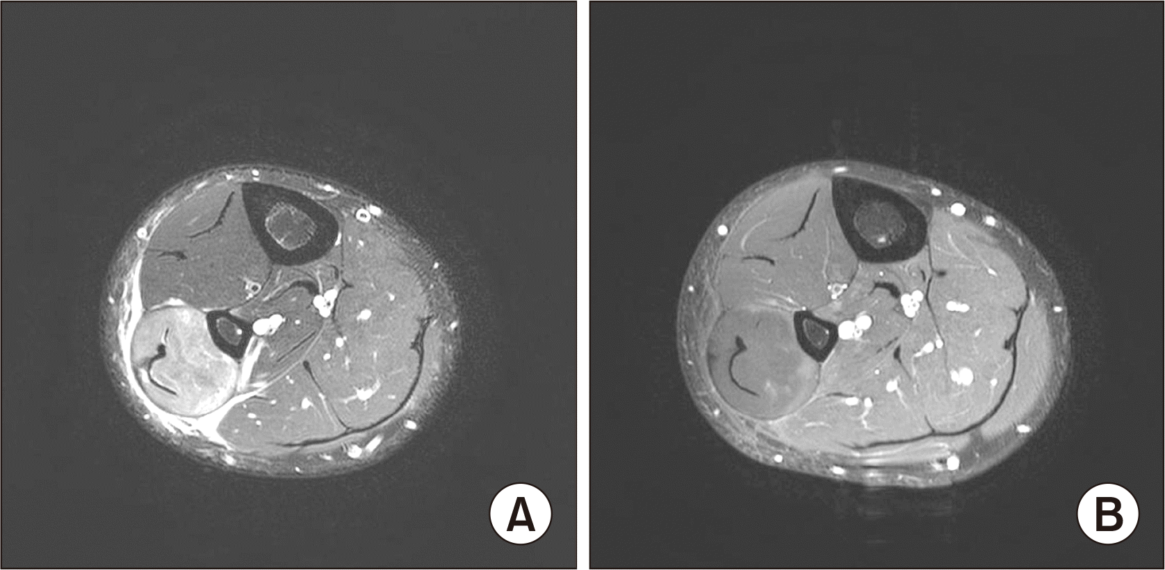
Figure 2
Surgical treatment was done for 20-year-old man who was diagnosed with isolated lateral compartment syndrome in the lower leg. After fasciotomy and debridement, aseptic dressing was done with the surgical site opened and tightening was done with surgical loop. Figures are taken in 1 day postoperatively.
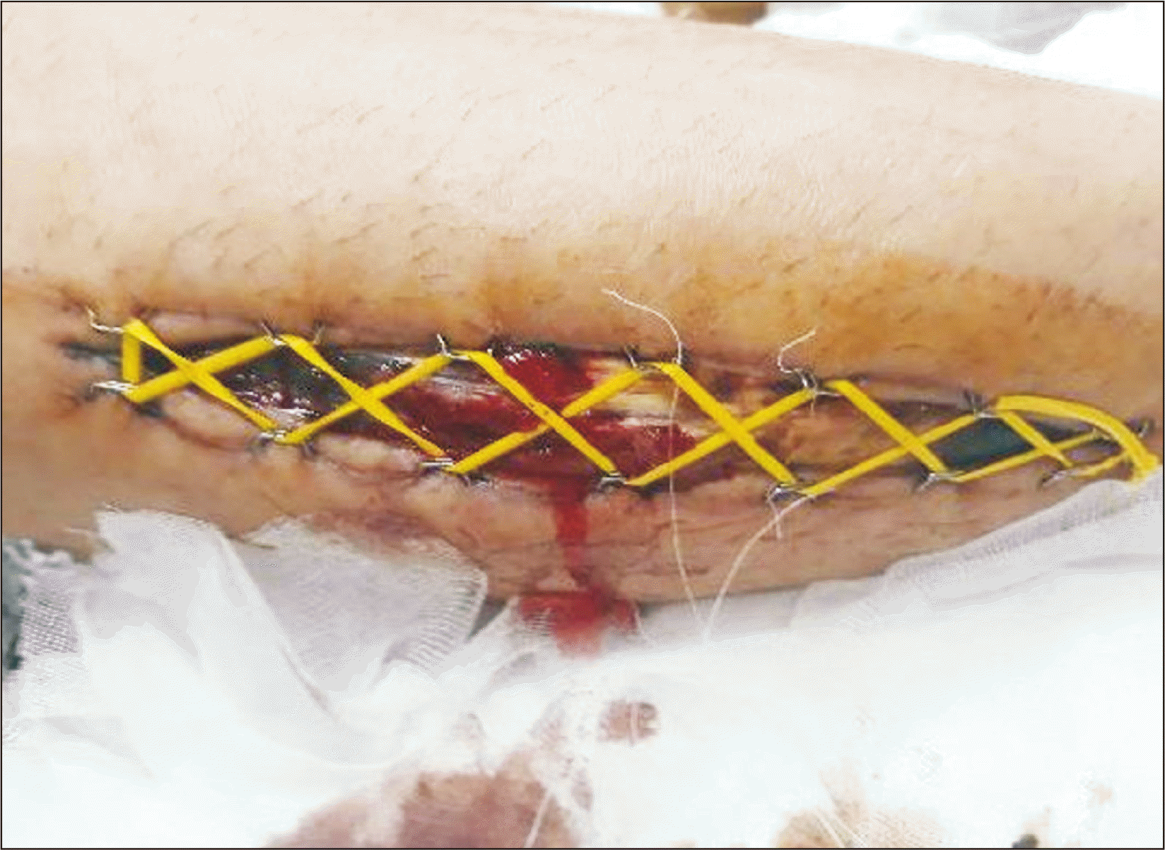
Figure 3
After initial surgery, surgical site came out to be clean in 20-yearold man with isolated lateral compartment syndrome. Secondary suture was done in 15 days postoperatively. Drainage tube was also inserted.
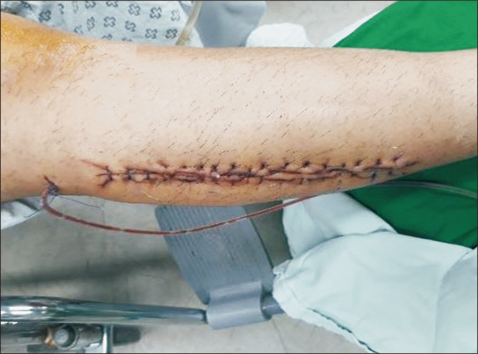
Figure 4
At 6 months postoperatively, rangeof-motion of ankle dorsiflexion, plantarflexion, and greater toe extension recovered almost same level as unaffected side.
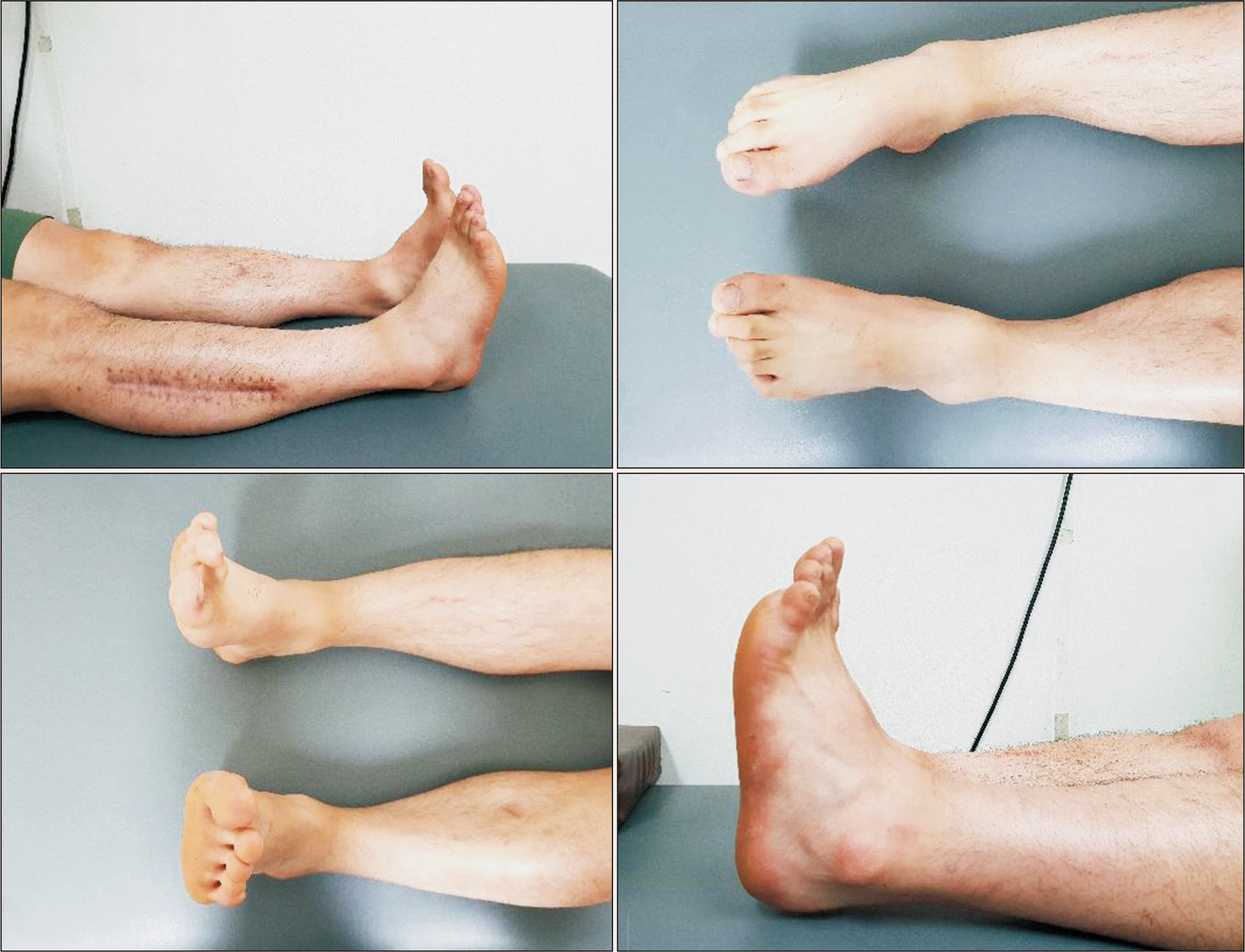
Figure 5
Right side lower leg magnetic resonance imaging taken for 28-year-old man. Signal increase is shown in peroneus longus muscle and peroneus brevis muscle in T2-weighted images.
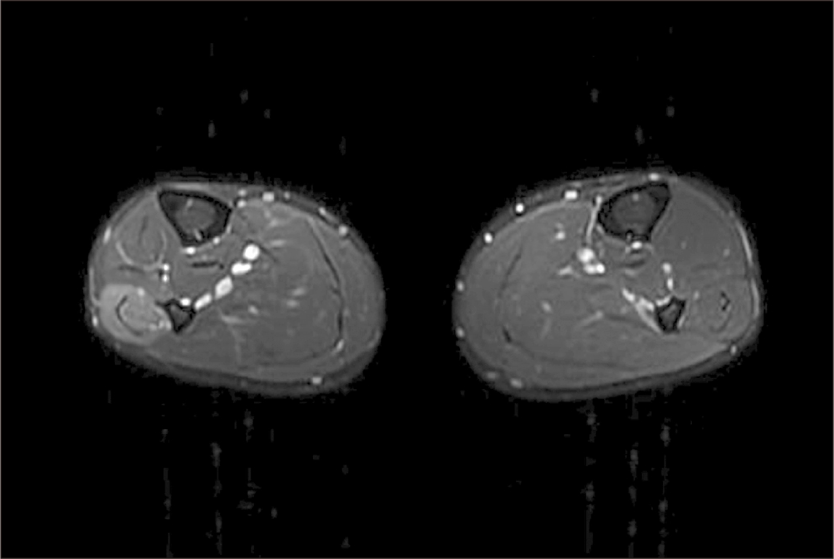
Table 1
Electromyography, 20-Year-Old Patient
Table 2
Nerve Conduction Studies 20-Year-Old Patient
Table 3
Electromyography, 28-Year-Old Patient
Table 4
Nerve Conduction Studies 28-Year-Old Patient


 PDF
PDF Citation
 Print
Print


 XML Download