

 PDF
PDF Citation
 Print
Print


INTRODUCTION
Atrial fibrillation (AF) is the most common arrhythmia, and its occurrence is associated with aging, high blood pressure, hyperthyroidism, and various cardiovascular conditions [1]. While exercise can have cardiovascular benefits, it ironically is also a risk factor for AF [2]. Exercise-induced AF correlates highly with exercise intensity, and in particular, high-intensity exercise has induced AF even in individuals without underlying cardiovascular disease [3,4]. However, the relationship between exercise intensity and AF generation is not yet clearly understood.
AF features structural and electrical remodeling of the atrium that can both cause the disease and be caused by it [5]. Atrial enlargement [6], hypertrophy [7] and fibrosis [8] reflect structural remodeling, and functional changes of ion channels reflect electrical remodeling [9]. Normal atrial activity results from the normal functioning of ion channels associated with action potential (AP) and repolarization [10], but ion channel dysfunction by electrical remodeling can cause abnormal changes in AP and consequent AF [11]. Atrial AP duration is a major determinant of heart rate and refractory phase, and is one of the most sensitive factor affecting AF. The shape and frequency of AP are controlled by the functions of various ion channels such as voltage-gated Na+, Ca2+, and K+ channels [12-14].
Short AP duration and related changes in heart rate and refractory period are believed to be involved in AF pathogenesis. Researchers observed decreased Ca2+ influx into atrial myocytes through L-type Ca2+ channels (ICaL) after AF or tachypacing [15,16]. Another important factor that decreases AP duration in AF is increased K+ channel functioning [10,17]. K+ channel upregulation contributed to tachypacing or AF in animal models of cardiac remodeling [18]. Potential targets include the diverse K+ channels expressed in the atrium, including G-protein activated inwardly rectifying K+ (GIRK) [19], ATP-sensitive K+ [20], and voltage-gated K+ (KV) channels [21].
Molecular mechanisms, including changes in ion channels that induce or are induced by AF, have shown promise, but there are little data on the specific protection or risk associated with AF induction. Therefore, with this study, we examined the effects of exercise intensity on AF induction. Specifically, we induced AF in mice using acetylcholine-calcium chloride (ACh + CaCl2) and focused on the ion channel activity after either no exercise or regular treadmill exercise at low, moderate, or high intensity for 4 weeks.
METHODS
Animals and preparation of atrial myocytes
We used 8-week-old male ICR (CD-1) mice (body weight 35 ± 5 g). All animal experiments followed the ethical guidelines of ARRIVE and all experimental protocols were approved by Konkuk University Animal Experimental Ethics Committee (approval no. KUB181007). To obtain hearts and prepare atrial myocytes, mice were injected intraperitoneally using a 4:1 mixture of alfaxan (alfaxalone 10 mg/ml; Jurox Inc., Rutherford, NSW, Australia) and rumpun (xylazine hydrochloride 23.32 mg/ml; Bayer, Leverkusen, Germany) and then completely anesthetized. The heart was dissociated, and the experiment was conducted.
After connecting the cannula to the aorta of the removed heart, normal Tyrode’s (NT) solution (143 mM NaCl, 5.4 mM KCl, 0.33 mM NaH2PO4, 1.8 mM CaCl2, 0.5 mM MgCl2, 11 mM glucose, 5 mM 4-(2-hydroxyethyl)-1-piperazineethanesulfonic acid (HEPES), (pH adjusted to 7.4 using NaOH) was flowed at a rate of 8 ml/min for 3–5 min at 37°C. Then Ca2+-free NT was perfused until the heart stopped beating.
Ca2+-free NT supplemented with collagenase type II (0.14 mg/ml; Worthington Biochemical Corp., Lakewood, NJ, USA) was flowed for 10–15 min for enzymatic treatment, and then the treated enzyme was washed out with Ca2+-free NT for 5 min. The tissues were then washed again with Kraft-Brühe (KB) solution (50 mM KCl, 50 mM K-glutamate, 20 mM KH2PO4, 3 mM MgCl2, 20 mM taurine, 20 mM glucose, 10 mM HEPES, and 0.5 mM N,N,Nl,Nl,-tetraacetic acid, pH 7.4 adjust with KOH). The atrial myocytes were then dispersed with gentile agitation using a dropper and stored in a refrigerator at 4°C.
Exercise protocol
We randomly assigned the mice into no, low, moderate, or high intensity treadmill exercise group. Before start of the exercise protocol, the mice ran at 10 m/min with no incline for 30 min a day for 5 days to adapt to the exercise protocol. During the 4-week exercise protocol after the adaptation, treadmill duration (30 min) and incline (5%) remained the same for all intensities: low, 12 m/min; moderate, 15 m/min; and high, 18 m/min [22]. To make the mice continue running according to the exercise protocol, we provided an electric shock at 1–2 Hz (1.22 mA current with 200 msec width per pulse) [23]. After the exercise intervention, we induced AF with ACh-CaCl2 (injection for 7 days) and observed the related variables.
Atrial fibrillation by acetylcholine and calcium chloride injection
We first randomly assigned the mice to one of five experimental groups: 1) control (n = 25): mice injected with physiological salt solvent without ACh + CaCl2, no exercise; 2) non-ex AF (n = 25): mice injected with ACh + CaCl2 to induce AF, no exercise; 3) low-ex AF (n = 25): mice injected with ACh + CaCl2 to induce AF, low-intensity exercise; 4) mid-ex AF (n = 25): mice injected with ACh + CaCl2 to induce AF, moderate intensity exercise; and 5) high-ex AF (n = 25): mice injected with ACh + CaCl2 to induce AF, high intensity exercise. After the mice exercised for 4 weeks, we induced AF by injecting a solution of ACh (25 µg/ml) and CaCl2 (6 mg/ml) into the tail vein at 10 ml/kg body weight for 7 days [24]. We recorded lead II electrocardiograms (ECG, PowerLab data acquisition system; AD Instruments, Colorado Springs, CO, USA) on the day before and after the ‘7-day ACh-CaCl2 treatment’ to examine the changes of ECG by the ACh-CaCl2 treatment in each group. After recording the ECG after the AF induction, we rapidly removed the heart using surgical scissors under deep anesthesia.
Patch-clamp experiment
We used a conventional whole-cell configuration of patch-clamp technique to record the cell membrane ion currents and AP with an Axopatch 200B amplifier (Molecular Devices, Sunnyvale, CA, USA) or EPC-8 amplifier (HEKA Instruments, Reutlingen, Germany). Data were digitized using custom-built software (R-clamp; provided by Dr. S.Y. Ryu) or with the Digidata 1440A at a sampling rate of 5 kHz after being low-pass filtered at 1 kHz. We also controlled voltage pulse generation by R-clamp or pClamp 10.1 software (Molecular Devices) running on an IBM-compatible Pentium computer. The patch pipettes were pulled from borosilicate capillaries (Clark Electromedical Instruments, Pangbourne, UK) using a puller (PP-83; Narishige, Tokyo, Japan); we used patch pipettes that featured a resistance of 1.5–2.5 MΩ when filled with the pipette solution.
After fixing the membrane voltage to –70 mV (holding potential), we elicited the outward and inward K+ currents using ramp or step pulses under voltage-clamp mode, and recorded action potentials with current injection (250–400 nA for 1 ms) under the current clamp mode. The resting membrane potential was recorded under I0 mode. We recorded the GIRK currents using the nystatin-perforated whole-cell patch method and considered the currents activated by acetylcholine (100 µM, for 2 min) at the holding potential of –40 mV as GIRK currents. The current-voltage (I–V) relationship of GIRK was briefly investigated using ramp pulses. ICaL currents were measured using a 10 mV incremented step-pulse protocol after the voltage was maintained at –70 mV (holding potential) and depolarized to –50 mV to exclude Na+ currents.
Solution
We used NT as the bath solution to record the currents. For outward K+ currents and AP, the composition of the pipette solution was 135 mM KCl, 5 mM NaCl2, 5 mM magnesium adenosine trisphophate (Mg-ATP), 10 mM HEPES, and 5 mM EGTA. The pH was adjusted to 7.35 using KOH. For ICaL currents, the pipette solution contained 120 mM CsCl, 12 mM NaCl, 10 mM HEPES, 20 mM tetraethyl ammonium chloride, 0.1 mM EGTA, 5 mM MgCl2, and 10 mM K2ATP, with pH adjusted to 7.25 with CsOH. For recording GIRK currents, NT was used as the perfusion solution, and the pipette solution was 140 mM KCl, 5 mM NaCl, 10 mM HEPES, 5 mM EGTA (KOH-adjusted to pH 7.2). 200 µg/ml of nystatin was added to the pipette solution just before seal formation.
Immunohistological study
Immunohistochemistry was performed to confirm the level of fibrosis, an index of morphological remodeling following AF. After the heart was excised, it was fixed in 4% paraformaldehyde in PBS for one day. A paraffin block (with 5 mm thickness) was used and stained with 0.1% toluidine blue 1% NaCl. Representative tissues stained in the fibrous area were observed under 400× magnification with an Olympus DP 70 optical microscope (Olympus, Shinjuku, Japan).
Statistical analysis
We analyzed all data using Origin 8.0 (OriginLab, Northampton, MA, USA) or SPSS 22.0 (IBM, Armonk, NY, USA) and calculated average and standard error (SE) for all data. We used one-way ANOVA to confirm between-group differences and Tukey’s post-hoc test for significant factors. We set significance at α = 0.05.
RESULTS
Arrhythmia induction by acetylcholine and calcium chloride
First, we confirmed that we had induced AF-like arrhythmia after the ACh + CaCl2 treatment because it was present in the AF groups but not in the saline-injected control group (Fig. 1A).
There were no differences in heart rate between the five groups before the ACh + CaCl2 treatment: rates for control, non-ex AF, low-ex AF, mid-ex AF, and high-ex AF group were, respectively, 545 ± 17.07 beats/min, 547.5.77 ± 14.36 beats/min, 547.5 ± 18.87 beats/min, 555 ± 19.36 beats/min, and 550 ± 7.07 beats/min. After the ACh + CaCl2 treatment for 7 days, however, heart rate increased significantly in all the AF groups (Fig. 1A, B; control, 547.5 ± 14.36 beats/min; non-ex AF, 750 ± 24.49 beats/min; low-ex AF, 681.25 ± 46.97 beats/min; mid-ex AF, 772.5 ± 37.74 beats/min; high-ex AF, 826.25 ± 40.69 beats/min). There were also irregular RR intervals apparent in non-ex, mid-ex, and high-ex AF (Fig. 1A). Notably, both heart rate and % increase of heart rate after AF induction were significantly lower in low-ex and significantly higher in high-ex than in the other AF groups (Fig. 1B, C). That is, high-intensity treadmill exercise promoted AF-like increase in heart rate that was induced by ACh + CaCl2, whereas low-intensity exercise suppressed the heart rate increase.
Morphological changes
Immunohistochemistry was also performed to examine morphological changes such as fibrosis of atrial muscle. Fig. 2A shows representative toluidine blue-stained atrial tissues from each group, with fibrotic tissues in the blue stain; the figure shows greater fibrosis in the AF groups than in the control group and the least fibrosis in low-ex (Fig. 2A). Regarding heart hypertrophy, heart weight/body weight ratio was significantly higher (p = 0.008) in high-ex AF than in the control group (Fig. 2B), but there were no differences in atrial myocyte size measured by membrane capacitance (Fig. 2C). In short, heart fibrosis and weight increased following AF induction after exercise dependent on exercise intensities, but cell size was not affected.
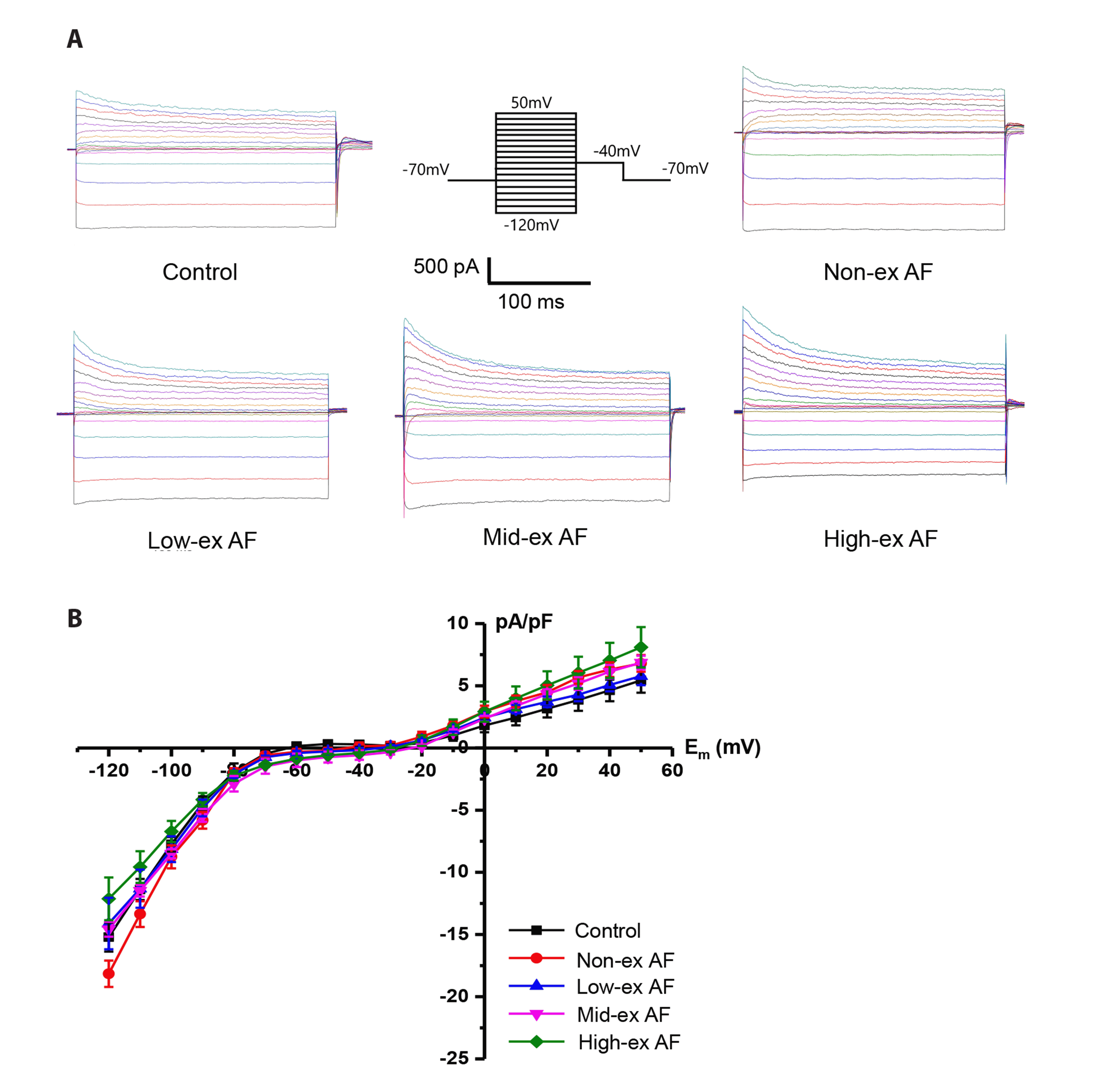
Inward rectifier and voltage-gated K+ currents
Next, we used patch clamp to examine the inward rectifier and outward voltage-gated K+ currents, eliciting the currents using voltage-step pulses from –120 mv to +50 mV at 10-mV increments. Fig. 3A shows representative current recordings from each group, and the I–V relationships are shown in Fig. 3B. Inward currents normalized by cell membrane capacitance (pF) at –120 mV, which represent inward rectifier K+ channel activity, was –15.20 ± 1.16 pA in control, –18.15 ± 1.05 pA in non-ex AF, –14.11 ± 2.06 pA in low-ex AF, –14.57 ± 0.60 pA in mid-ex AF, and –12.12 ± 1.71 pA in high-ex AF, respectively (no statistically significant difference). Outward currents at +50 mV, which largely represent voltage-gated K+ channel activity, was 8.09 ± 1.62 pA in high-ex AF, which was higher than low-ex AF 5.77 ± 0.72 pA and mid-ex AF 6.86 ± 0.55 pA, but there was no significant difference in all groups.
GIRK currents
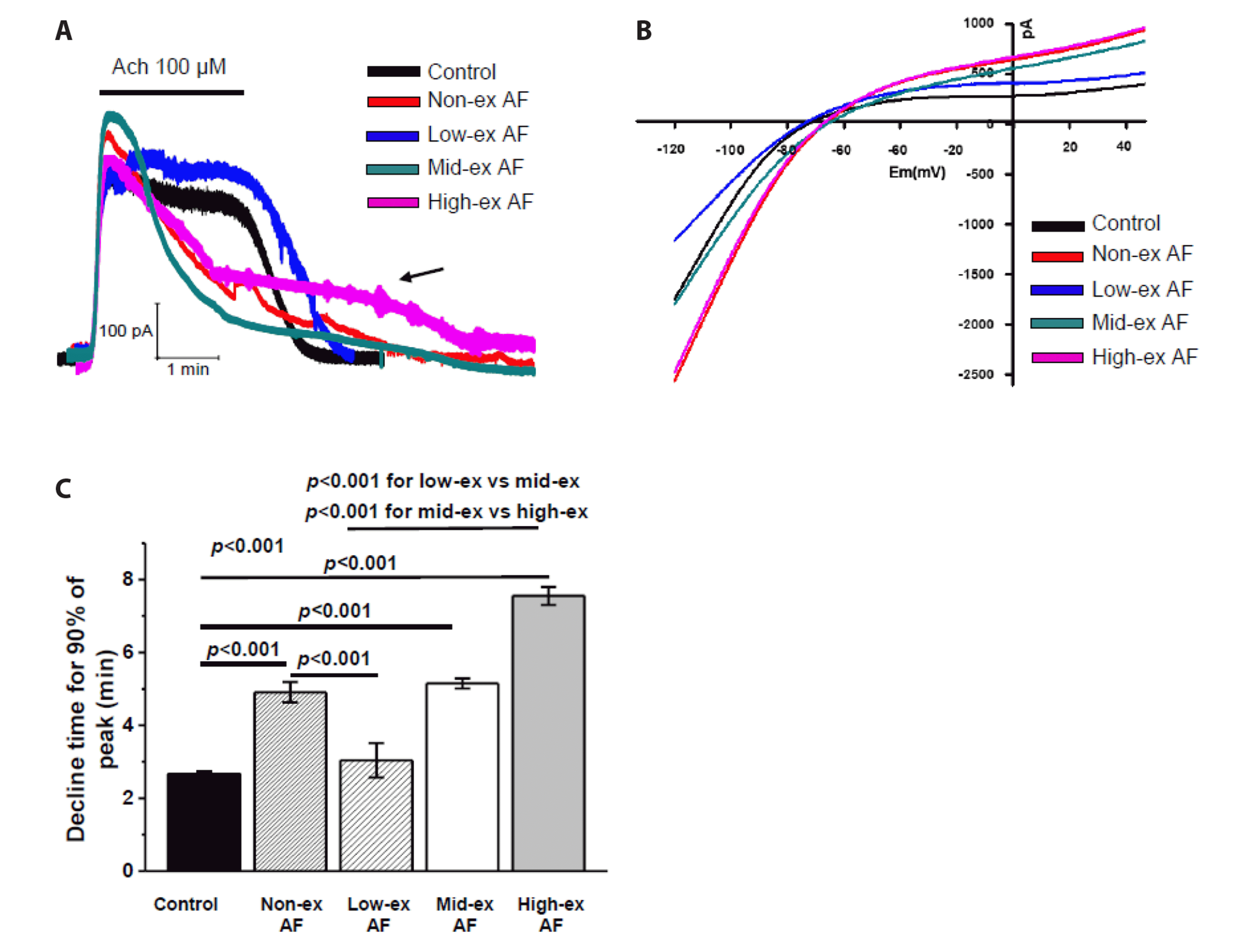
GIRK currents play a pivotal role in inducing atrial repolarization and correlate highly with AF [25], and we observed changes in GIRK currents in each group as well. GIRK currents were evoked by applying ACh (100 µM, for 2 min) at –40 mV holding potential (Fig. 4A) and then washing out the ACh. We then examined the I–V relationships of the GIRK currents with representative ramp pulses from –120 mV to 50 mV (Fig. 2B) and found that the currents rapidly deactivated after the ACh was removed in the control and low-ex AF groups. However, in high-ex and mid-ex AF, GIRK currents deactivation was markedly delayed, and we observed significant amounts of persistent currents after the ACh removal (Fig. 4A, indicated by arrow).
The I–V relationships in Fig. 4B were measured at the peaks of the ACh-induced GIRK currents at –40 mV, and peak currents in non-ex, mid-ex, and high-ex AF were higher than in control and low-ex AF. We compared the persistent currents between groups by measuring the times when the GIRK currents declined to 10% of their peaks for each group (T90; Fig. 4C). We observed that the GIRK currents decrease was significantly delayed after ACh stimulation dependent on exercise intensity (p < 0.001 for all). As shown in Fig. 4C, T90 of the control and low-ex AF was 2.66 ± 0.08 min and 3.04 ± 0.47 min, respectively (not significantly different to each other). However, T90 of high-ex AF was significantly delayed (7.55 ± 0.25 min) compared to those of other groups (Fig. 4C). T90 for the non-ex AF and mid-ex AF was 4.91 ± 0.28 min and 5.15 ± 0.14 min, respectively.
In these results, the potential target ion channels in AF induction after high-intensity exercise appeared to involve the GIRK channels, and low-intensity exercise had the greatest AF protective effect by preventing the AF-related channel remodeling.
L-type Ca2+ currents
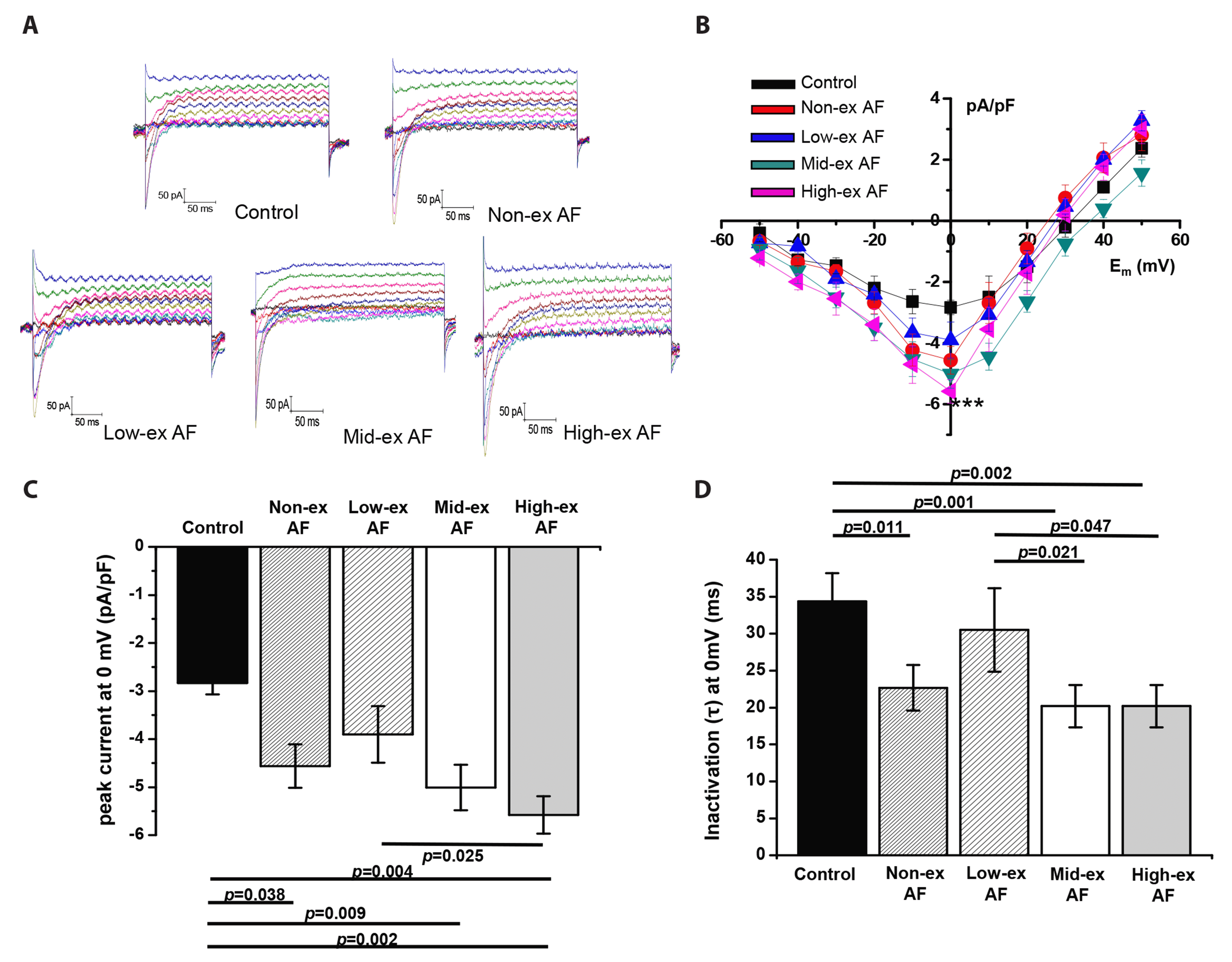
We measured whole-cell ICaL currents to determine whether Ca2+ channels following AF induction were affected by exercise intensity, and Fig. 5 show that the Ca2+ currents increased with exercise intensity. Specifically, in mid-ex and high-ex AF, the ICaL amplitudes at 0 mV were significantly higher than those in the control group (Fig. 5B, C; p < 0.05). In contrast, the inactivation time constants (tau) of Ca2+ channels at 0 mV were shorter in non-ex, mid-ex, and high-ex AF (Fig. 5D; p < 0.05) but returned to normal in low-ex AF (Fig. 5D); there was no significant difference between control and low-ex AF.
In these results, the accelerated inactivation of Ca2+ currents contributed to inducing AF in the non-ex, mid-ex, and high-ex groups. In addition, the absence of statistical differences in the magnitude of Ca2+ currents and inactivation time course between control and low-ex AF suggests that low-intensity exercise contributes to preventing AF-related remodeling of ICaL.
Action potential
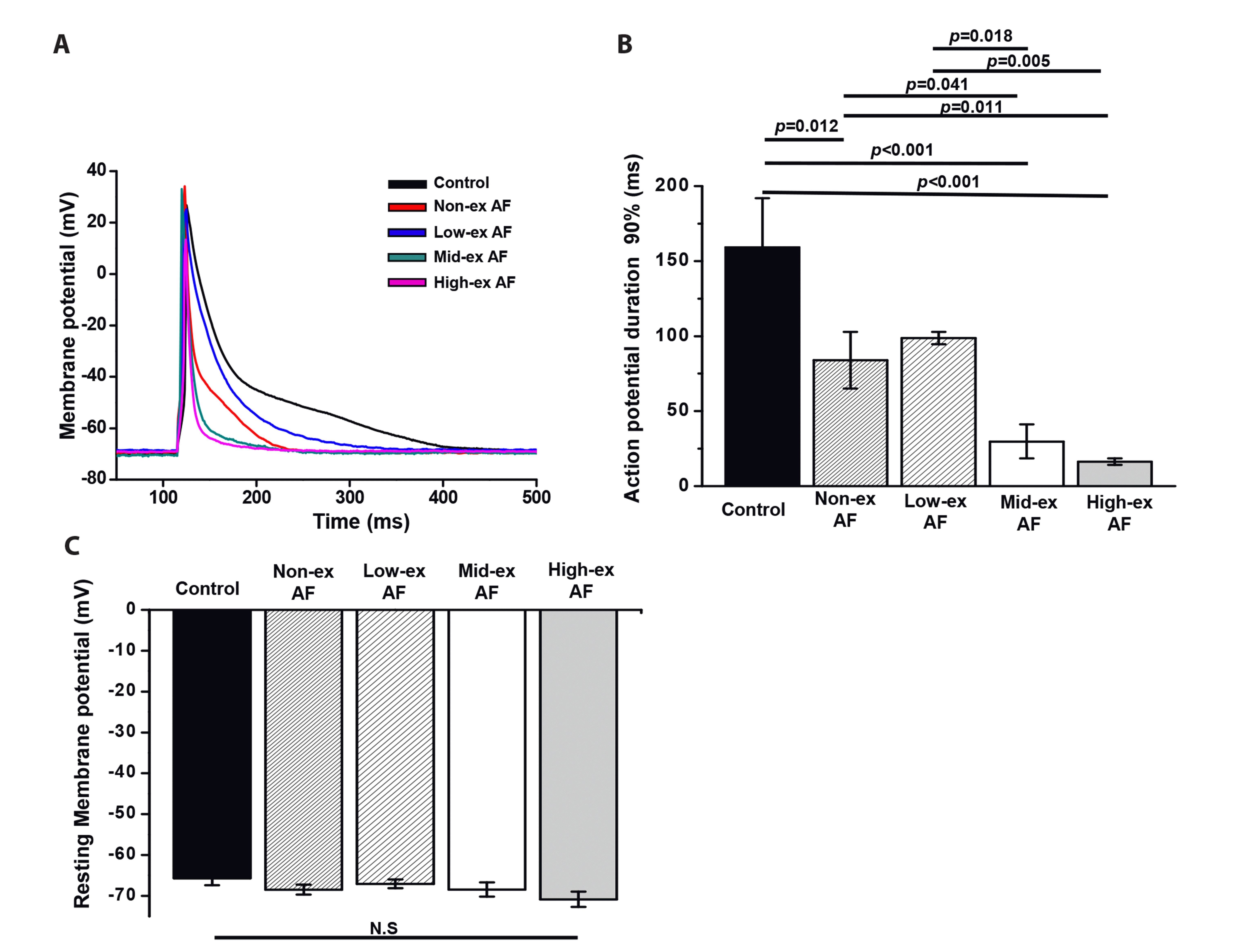
Finally, we observed the changes in AP after AF induction by exercise intensity, and Fig. 6A shows shorter AP durations in the AF groups depending on the exercise intensity. AP duration 90% (APD90) was got shorter depending on exercise intensity (Fig. 6B; p < 0.05). Especially, APD90 of high-ex AF was 16.29 ± 2.17 msec, which was much shorter than 98.78 ± 4.10 msec of low-ex AF. However, there were no differences in resting membrane potential between groups (Fig. 6C). These results indicate that APD90 shortens in an exercise intensity-dependent manner, leading to an increase in heart rate.
DISCUSSION
In this study, we investigated the effect of exercise intensity on the AF, which was induced by ACh + CaCl2 injection for 7 days after the 4-weeks of treadmill exercise. We used ECG, immunohistochemistry, and patch clamp to measure degree of AF and found that low-intensity treadmill exercise had a protective effect against the (ACh + CaCl2)-induced AF, whereas high-intensity exercise exacerbated the AF and its associated atrial remodeling.
Although there are established benefits to the cardiovascular system of regular exercise, exercise can be associated with cardiac remodeling [26]. High-intensity exercise caused arrhythmogenic cardiomyopathy-like manifestations [27] and was associated with ventricular arrhythmias with right ventricular fibrosis and dilation in a rat model [28]. Interestingly, in some reports, AF risk was particularly high for cross-country skiers [3], elite cyclists [29], and marathon runners [30], but the risk is limited to high-intensity exercise; there was a dose-response relationship in healthy non-athlete men younger than 50 years and joggers [31]. These results suggest high-intensity exercise as a potential AF risk factor even in healthy subjects.
Our results clearly showed that ACh + CaCl2 evoked AF-like characteristics such as increased heart rate and irregular RR interval in ECG accompanied with atrial fibrosis; ion channel remodeling such as persistent GIRK currents and rapid inactivation of ICaL currents. All of this remodeling was lessened by low-intensity exercise and exacerbated by high-intensity exercise, indicating a preventive effect for the former and increased risk with the latter.
It is not yet clear how exercise intensity affects AF generation. Aschar-Sobbi et al. [32] reported TNF-alpha as a key factor. High-intensity endurance exercise promotes ion channel remodeling and atrial fibrosis through vagal tone [33]. In this study, because we induced AF with parasympathomimetic stimulus (ACh + CaCl2), it makes sense that GIRK channels, which is activated by parasympathetic stimulation, contributed to AF generation as well as its exaggeration by high-intensity exercise.
As we mentioned above, GIRK channel activity is a very strong candidate for explaining AF generation: Even though we found no strong correlation between the peak GIRK currents amplitudes and the AF induction, markedly persistent GIRK currents remained after the ACh removal in the high-ex AF group but not in low-ex or control. The results may indicate that the GIRK-activating signaling is enhanced or facilitated in the ACh-CaCl2-induced AF groups and reflecting upregulation of parasympathetic tone. In support of this, in patients with chronic AF, the GIRK currents were continuously activated through remodeling the GIRK pathway in the isolated myocytes of right atrium, and this remodeling contributed significantly to ongoing AF [34]. Zou et al. [24] also reported significantly higher (ACh + CaCl2)-induced AF in rats and K+ channel activation through ACh in the AF model than in control. Other researchers showed that regular exercise induces AF by increasing vagal tone in a time-dependent manner [35]. Interestingly, in GIRK channel knock-out mice, AF was not induced by increasing vagal tone [36]. Additionally, high-intensity exercise-induced increase in vagal tone [37] induces atrial arrhythmias by inducing GIRK channel overactivation [38]. Therefore, our findings suggest that enhanced activity of GIRK currents plays a pivotal role in the generation of AF, and this enhanced GIRK activity is still persistent even in the isolated cardiac myocytes. This indicates that remodeling of GIRK channels contributed to the AF generation at the cardiomyocytes level (i.e., increase of constitutive GIRK) without systemically increased parasympathetic stimulation. In support this, spontaneous openings of the GIRK single-channels were observed only in the chronic AF group, which were inhibited with the GIRK channel inhibitor tertiapin [34].
Although we found higher ICaL currents in both non-ex and high-ex AF, previous researchers reported lower ICaL currents in AF with the suppressed α1 subunit [39,40] and ICaL density [16,41]. Because ICaL is involved in the AP plateau phase [42], decreasing ICaL should accelerate AP repolarization to make it short. Therefore, the increased ICaL observed in this study could compensate to prevent pathologic AP shortening. However, the accelerated inactivation time course of ICaL, together with the increased GIRK activity, could have contributed to the AP shortening despite the increased ICaL peak amplitude. Interestingly, even the increased ICaL was suggested to contribute to AF generation [41]. The precise mechanism of the ICaL contribution in AF generation or maintenance warrants future study.
Dzeshka et al. [43] found that the representative morphological change in AF patients is cardiac fibrosis, and we here measured fibrosis degree and myocyte size to examine histological changes after AF induction. Our results clearly showed more fibrosis in mid-ex and high-ex AF than in low-ex AF, which was consistent with the results by Aschar-Sobbi et al. [32] Atrial fibrosis, together with atrial hypertrophy, can induce AF through abnormal electrical conduction [44]. Because there were no significant changes in myocyte size in the AF groups in this study, it is likely that the induced AF and various arrhythmias were attributable to cardiac fibrosis rather than hypertrophy.
In this study, APD90 was significantly lower in the non-ex, mid-ex, and high-ex AF groups, but low-intensity exercise normalized these abnormal changes. As predicted by Rosenberg et al. [45], as AF progresses, AP duration and the heart refractory period decrease, which contribute to maintaining AF through incomplete excitation-contraction coupling. Here, low-intensity exercise normalized the electrophysiological variables such as GIRK and L-type Ca2+ channels. We interpret that these composite effects from low-intensity exercise contributed to consequently normalizing heart rate and AP duration
In conclusion, in this study, by injecting ACh-CaCl2 for 7 days, we successfully generated AF in a mouse model and showed atrial tachycardia/fibrillation with irregular RR interval in ECG. AF featured electrical remodeling such as enhanced GIRK channel activity and shortened action potential and morphological remodeling like atrial fibrosis. In addition, low-intensity exercise clearly lessened all these AF-related changes whereas high- intensity exercise exacerbated them. Future researchers should establish a clearer scientific basis by observing the association between various exercise-intensity-dependent variables and ion channels.
This study has the following limitations: The AF model we applied did not explain the overall cause of AF, we did not study sport specificity or changes in AF-time course, and we did not address AF induced by atrial remodeling with increasing age. In addition, we observed no atrial changes by exercise period. Therefore, it is necessary to study AF induction mechanisms in various disease models and under various exercise conditions


 XML Download