

 PDF
PDF Citation
 Print
Print


Dear Editor,
ETV6–RUNX1 (previously TEL–AML1) is the most common fusion gene associated with childhood ALL, which occurs in approximately 25% of pediatric ALL patients receiving diagnoses between the ages of 2–10 years and is associated with favorable prognoses [1, 2]. ETV6 rearrangement contributes to leukemogenesis in lymphoid neoplasms but is not well described in myeloid leukemia, and very few cases have been reported [3-6]. We report the first case of AML with the ETV6–RUNX1 fusion gene. The Institutional Review Board of Chonnam National University Hwasun Hospital, Hwasun, Korea, approved this study (CNUHH-2022-013) and waived the need for informed consent.
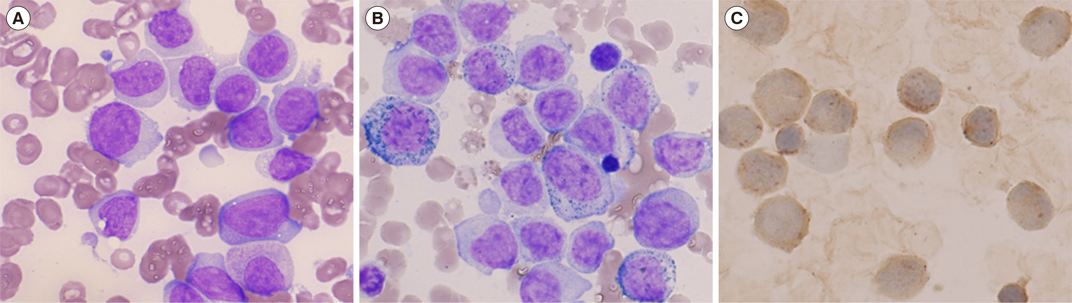
In January 2021, a 66-year-old man presented with newly developed thrombocytopenia and underlying dyslipidemia. Complete blood count showed leukocytosis (13.3×10⁹/L) and thrombocytopenia (82×109/L). Peripheral blood smear comprised ~78% abnormal-appearing monocytes and nucleated RBCs. The bone marrow (BM) aspirate showed 63%, 7%, and 7% monoblasts, promonocytes, and monocytes, respectively (Fig. 1A). Myeloperoxidase (MPO) and non-specific esterase were positive in cytochemical staining (Fig. 1B, C). Flow cytometry analysis (Navios, Beckman Coulter, Miami, FL, USA) with BM cells identified two cell populations: the blast population showed a positive reaction in anti-CD33, anti-CD64, anti-CD117, and anti-HLA-DR fractions, whereas the monocyte population was positive for anti-CD13, anti-CD14, anti-CD33, anti-CD64, anti-HLA-DR, and anti-MPO. Both populations were negative for the lymphoid markers CD2, CD5, CD7, CD10, CD19, CD20, and CD22; only cytoplasmic CD3 showed aberrant dim expression in the monocyte population (~11.3%).
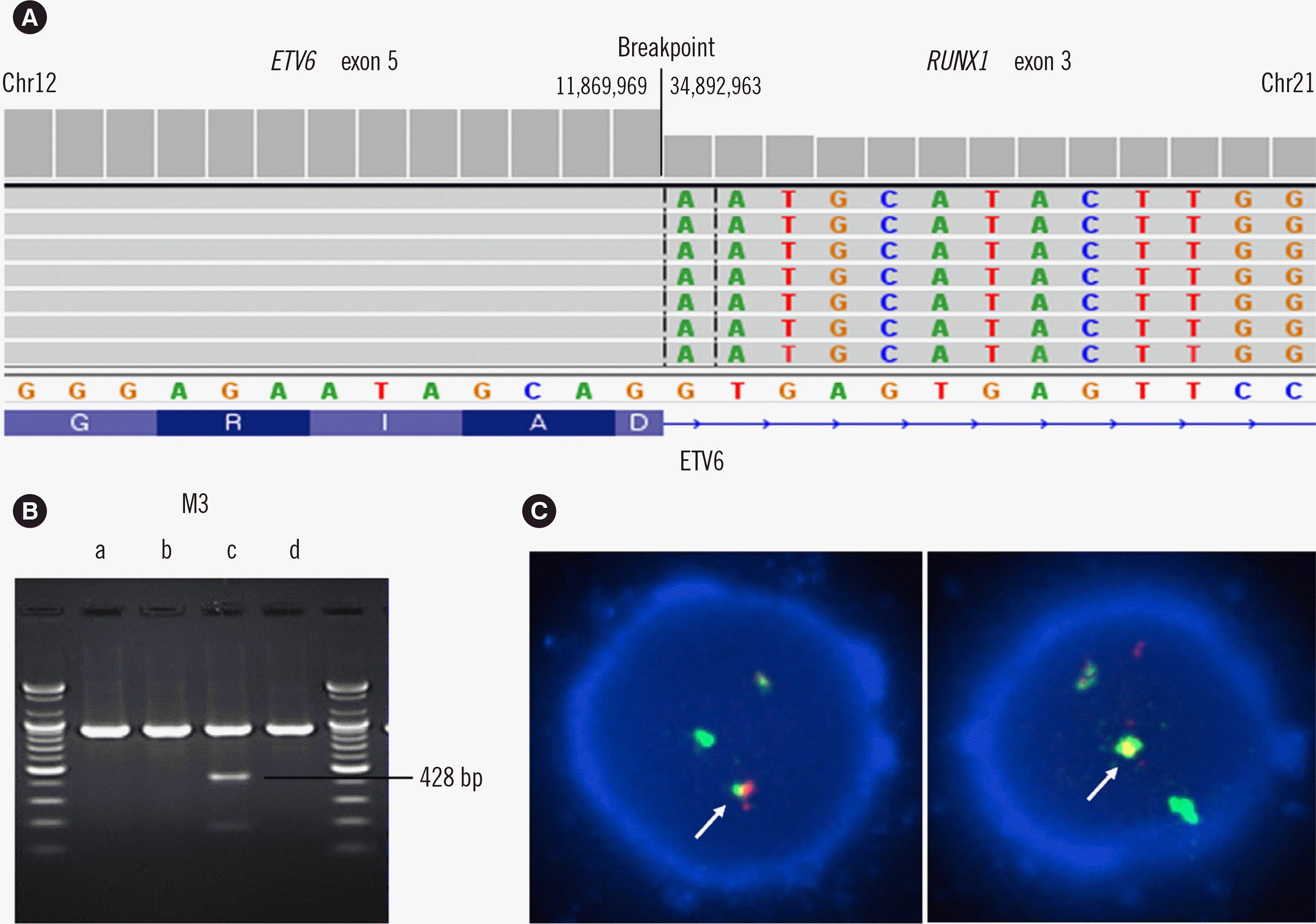
Conventional cytogenetic analysis demonstrated a normal karyotype. Based on morphologic and immunophenotypic results, a diagnosis of AML-M5a was made. The ETV6–RUNX1 fusion gene was detected using next-generation sequencing (NGS) RNA panel (KBB-RNAseq NGS-Leukemia-PHB; KBlueBio Inc., Hwasun, Korea) and confirmed with multiplex reverse transcription (RT)-PCR (HemaVision Kit; DNA Technology, Aarhus, Denmark). Other genetic variants, including SETBP1, JAK2, PHF6, and STAT3, were detected with an NGS DNA sequencing panel (HEMEaccu Test DNA; Ngenebio, Seoul, Korea); these variants are known to be related to myeloid rather than lymphoid neoplasms (Fig. 2A, B) [7].
Twenty-six days after induction chemotherapy, follow-up BM biopsy showed 49% monoblasts and 19% promonocytes. The samples were positive (10/200) for the reciprocal translocation t(12;21)(p13;q22), as demonstrated by FISH with a t(12;21) probe (Vysis, Abbott Molecular, Des Plaines, IL, USA) (Fig. 2C). Repeated BM biopsy 47 days after reinduction chemotherapy showed no monoblasts or promonocytes, and the sample was negative for t(12;21)(p13;q22) in FISH analysis. Consolidation chemotherapy was continued for five days. Despite long-term additional treatment because of neutropenic fever, neurologic symptoms became worse, accompanied by a confused mental state without clear reason. The patient was discharged without curative hope on day 226 after the diagnosis.
RUNX1 encodes a transcription factor (AML1) that controls the expression of its target genes involved in hematopoietic differentiation, and its functional disruption is a major causative factor of leukemia development [8, 9]. The RUNX1–RUNX1T1 fusion product in AML contains only the N-terminal portion of AML1, whereas the ETV6–RUNX1 fusion product includes almost the entire AML1 sequence, containing both the Runt homology domain and C-terminal transactivating and repressor domains [4, 9]. ETV6–RUNX1 translocation results in the production of a fusion protein that likely acts in a dominant-negative fashion to interfere with the normal function of RUNX1 and is considered an early lesion in B-cell leukemogenesis [2, 9]. A two-hit model of ETV6–RUNX1 leukemogenesis is assumed: ETV6–RUNX1 expression and a second genetic or environmental hit must occur at the hematopoietic progenitor stage for leukemic transformation [10]. ETV6–RUNX1 fusion is considered to be absent in leukemias of myeloid lineage because the fusion protein severely interferes with myeloid development and enforced expression of ETV6–RUNX1 in myeloid cells leads to apoptosis, possibly by interfering with autoregulatory loops essential for the maintenance of myeloid identity [3]. Presence of the ETV6–RUNX1 fusion gene in AML patients is very rare, and we found no report describing this association. The ETV6–RUNX1 fusion is considered to occur in adult patients, resulting in leukemogenesis to myeloid leukemia caused by uncommon second hits, representing a distinct mechanism from pediatric ALL.
In this case, the ETV6–RUNX1 fusion gene was detected at diagnosis and follow-up status but disappeared at morphological remission status. We tentatively conclude that the ETV6–RUNX1 fusion played an important role in the pathogenesis of AML in this case; however, the mechanism is unclear, which is the study’s limitation. Although further studies are needed to determine the leukemogenesis of the ETV6–RUNX1 fusion gene in AML, our case demonstrates the possibility of this fusion gene in adult de novo AML.
 XML Download