Abstract
Extraction of premolars for orthodontic purposes may prove challenging when the tooth is blocked or lingualised. The standard buccal approach may prove difficult in such cases. A novel technique was used for 16 patients with healthy linguoverted mandibular premolars using maxillary extraction forceps. The ease of extraction increased and resulted in uneventful postoperative healing in all patients. The authors suggest this as a preferred technique for extracting mandibular premolars in linguoversion.
Go to : 
Intra-alveolar extraction has always been a challenge when the tooth is not in an ideal anatomical position. Difficulty of extraction increases based on the anatomic and pathologic conditions of the teeth and jaw1. Orthodontic extraction performed to gain space in the arch for correction of crowding or proclination is one of the most common reasons for removal of healthy teeth2. Removal of premolars is a trend in orthodontic treatment planning. Extraction of teeth is a challenge when the tooth is either blocked or in a lingualised position due to limited access to the area and inability to properly use forceps to grasp the tooth. This article explains in detail the technique of extracting lingualised/linguoverted premolars in the arch.
Go to : 
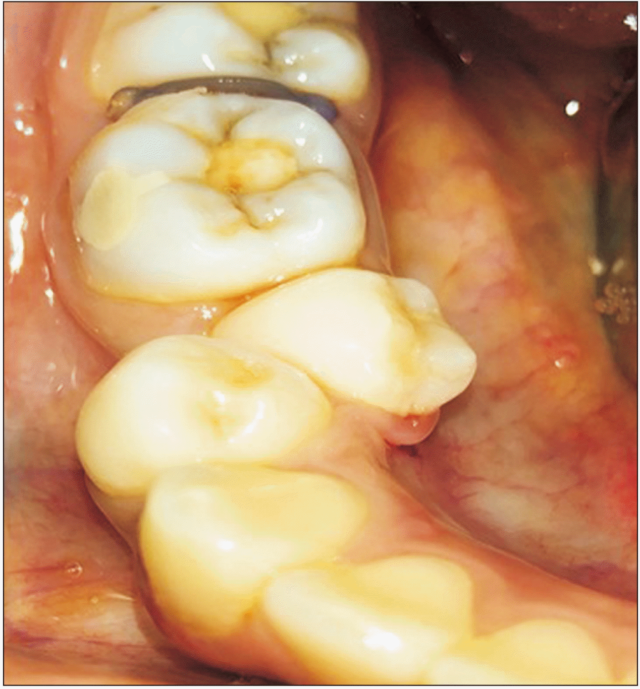
This technique was used for lower premolar teeth in a lingualised position where access to the teeth through the standard buccal approach was difficult or not feasible.(Fig. 1)
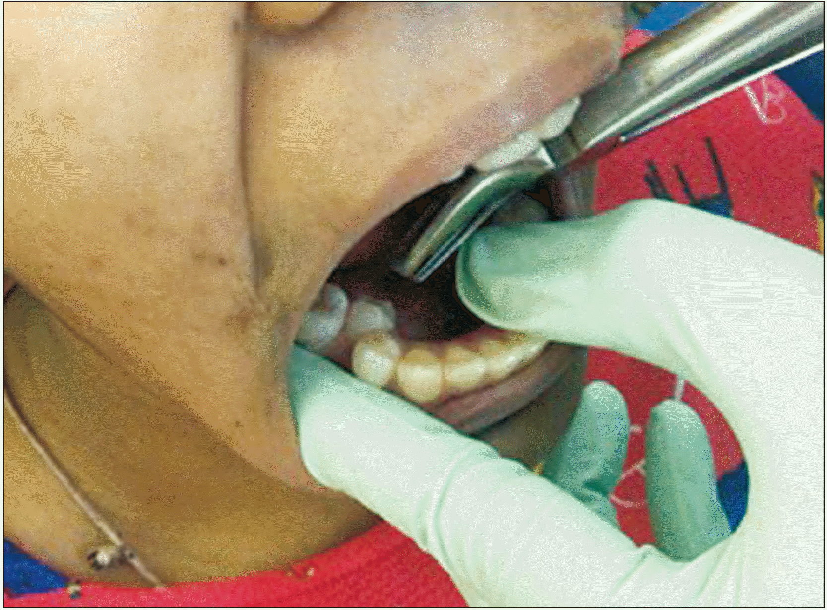
A total of 16 patients met the inclusion criteria for this study, which involved healthy first and second mandibular linguoverted premolars requiring extraction for orthodontic purposes. Since the teeth were healthy with no caries, upper premolar forceps were used to extract the lower premolars. Following local anaesthesia, only the lingual periodontal ligament (PDL) fibres were severed to gain access to the teeth. The chair was positioned two inches lower than the ideal position for extracting lower teeth. The approach to the teeth was lingual and from the opposite direction (for example, approaching from the right side for a left mandibular premolar).(Fig. 2)
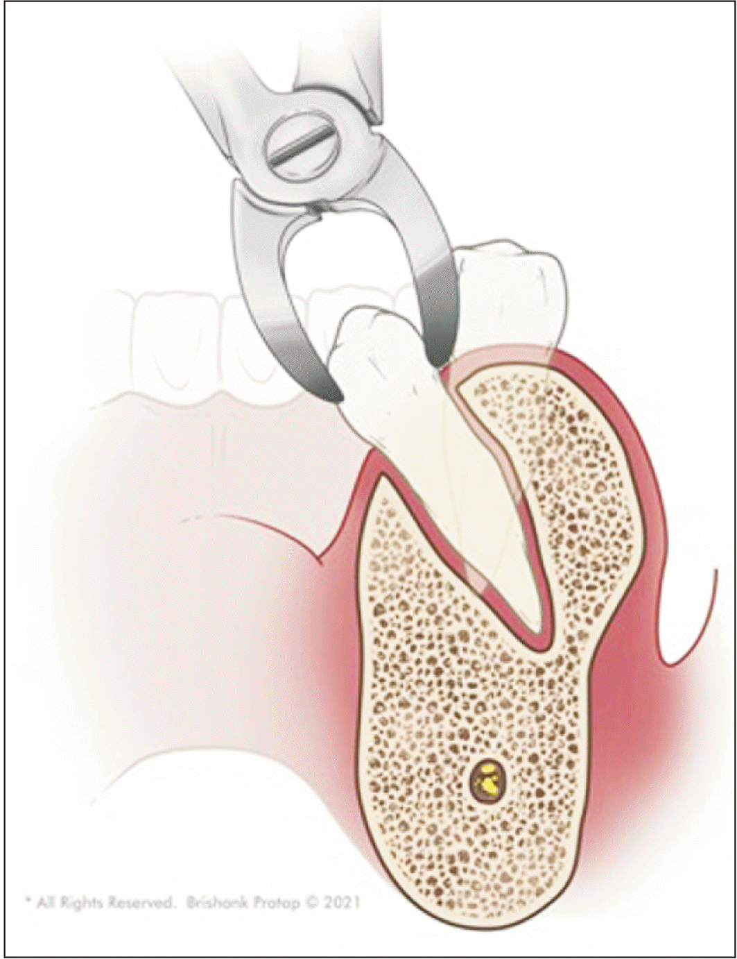
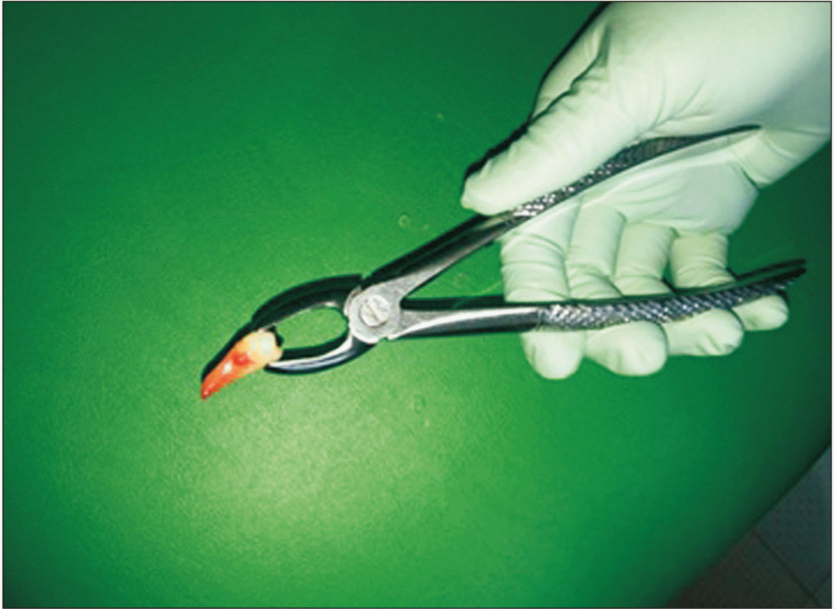
To extract lower right premolars, the operator will stand on the left side at the 4 o’clock position. Upper premolar forceps provide an ideal grip to hold the tooth versus two-point contacts of the crown with forceps, as seen with the buccal approach.(Fig. 3) Initially, apical pressure was applied to the tooth, followed by rotational and tractional forces to remove the tooth from the socket. The rationale behind this technique was consideration of lower premolars as similar to upper second premolars extracting them in the reverse plane. The surgeon should apply pressure only from the shoulder and arm and not the wrist. The single-rooted mandibular premolar was extracted with ease.(Fig. 4)
• Drawback: The technique is difficult in cases of gross destruction of the crown and tooth anomalies such as multiple roots or dilacerations. Gripping the crown and delivering the tooth in such situations may not be possible using maxillary premolar forceps.
• Results: The ease of extraction using this novel technique was high, resulting in uneventful postoperative healing.
Go to : 
Several studies have been performed to determine whether to extract teeth for orthodontic purposes. Extraction in orthodontics was reintroduced scientifically in the 1930s and reached its peak in the 1960s with the advent of Begg’s technique3. Simple extraction of the tooth requires separation of the PDL, expansion of the bony socket, and use of coronal forceps to deliver the tooth. The ideal forceps for removal of mandibular premolars are English/Asch forceps or No. 151 (mandibular universal) forceps4.
Even a skilful and experienced surgeon may face difficulties when extracting teeth. Sound knowledge of the surrounding anatomy along with basic principles can result in atraumatic extraction in some cases. Premolars in the mandibular arch pose a greater challenge than their counterparts in the maxillary arch due to the greater bone density of the mandible. The difficulty index increases with crowding, positioning of the tooth in the arch, root pattern, and health of the tooth. Fractured cortical plates can lead to narrowed-out ridges that may interfere with complete closure of the extraction space2.
In this study, extracting linguoverted teeth is a difficult task through the normal buccal approach. The rationale behind the use of maxillary forceps is to obtain a better grip; since bicuspids are single rooted, apical pressure followed by rotation and tractional movements can be carried out to deliver the tooth. Minimal severing of the PDL fibres is necessary. The major drawbacks of this technique are that it is difficult in cases of gross decay of the crown or of anomalies such as multiple roots or dilaceration. In addition, case selection in this study was very specific, with a small sample size and possible selection bias. A larger sample size is necessary to obtain better results. There were no complications noted, and all cases healed uneventfully.
In conclusion, adherence to the ideal principles of extraction while using maxillary forceps should be used to deliver linguoverted premolars in the mandibular arch.
Go to : 
Notes
Authors’ Contributions
O.A.S. participated in data collection and wrote the manuscript. O.A.S., P.N.M., and R.V.K. participated in the study design and performed the statistical analysis. T.B.F., V.D., and F.A. participated in the study design and coordination and helped to draft the manuscript. All authors read and approved the final manuscript.
Go to : 
References
1. Coleman F. 1922; Types of difficult extraction and their treatment. Proc R Soc Med. 15(Odontol Sect):76–82. DOI: 10.1177/003591572201501014. PMID: 19982444. PMCID: PMC2102172.

2. Rai AK, Yadav BK. 2016; Facilitating orthodontic teeth extraction-a technique suggestion. Saudi J Dent Res. 7:96–100. https://doi.org/10.1016/j.sjdr.2015.10.001. DOI: 10.1016/j.sjdr.2015.10.001.

3. Al-Ani MH, Mageet AO. 2018; Extraction planning in orthodontics. J Contemp Dent Pract. 19:619–23. DOI: 10.5005/jp-journals-10024-2307. PMID: 29807975.

4. Dym H, Weiss A. 2012; Exodontia: tips and techniques for better outcomes. Dent Clin North Am. 56:245–66. xhttps://doi.org/10.1016/j.cden.2011.07.002. DOI: 10.1016/j.cden.2011.07.002. PMID: 22117954.

Go to : 


 PDF
PDF Citation
 Print
Print


 XML Download