

 PDF
PDF Citation
 Print
Print


I. Introduction
A ranula is a pseudocyst (not lined by epithelium) that originates from extravasation and accumulation of mucus from the sublingual gland following a trauma or obstruction1-3. It can be classified as a simple or plunging ranula based on its extent2. A simple ranula is confined to the sublingual space2, whereas a plunging ranula develops when mucus herniates through or behind the mylohyoid muscle along the facial planes into the cervical, submandibular, or submental space, with or without intraoral collection4. Although several methods to treat ranulas have been reported, theoretically, removal of the sublingual gland may be most beneficial because it plays a key role in the formation of a ranula1,4. A ranula typically presents as a painless, gradually enlarging swelling on the floor of the mouth or neck; however, few cases of acute, rapidly developing ranulas causing respiratory distress have been reported2,5.
Holoprosencephaly (HPE) is a developmental defect that results from incomplete cleavage of the embryonic forebrain structures during early embryogenesis6. Depending on the severity of HPE, a variety of clinical findings (including craniofacial malformations, motor and developmental dysfunctions, and oromotor dysfunctions) can be seen in children with HPE7. Mild craniofacial malformations can be seen in all types of HPE, and these malformations include median cleft lip and palate (premaxillary agenesis), midface hypoplasia and hypotelorism, congenital nasal pyriform aperture stenosis, and a single maxillary central incisor7. Therefore, children with HPE can experience upper airway obstruction due to these malformations and dysfunctions7.
In this report, we present a case of HPE with a plunging ranula that manifested as an acute and progressive course and was characterized by respiratory distress with difficult perioperative management.
II. Case Report
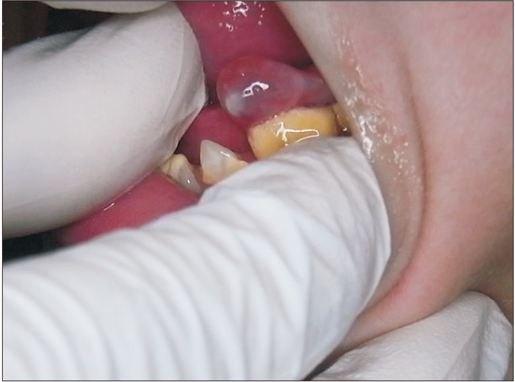
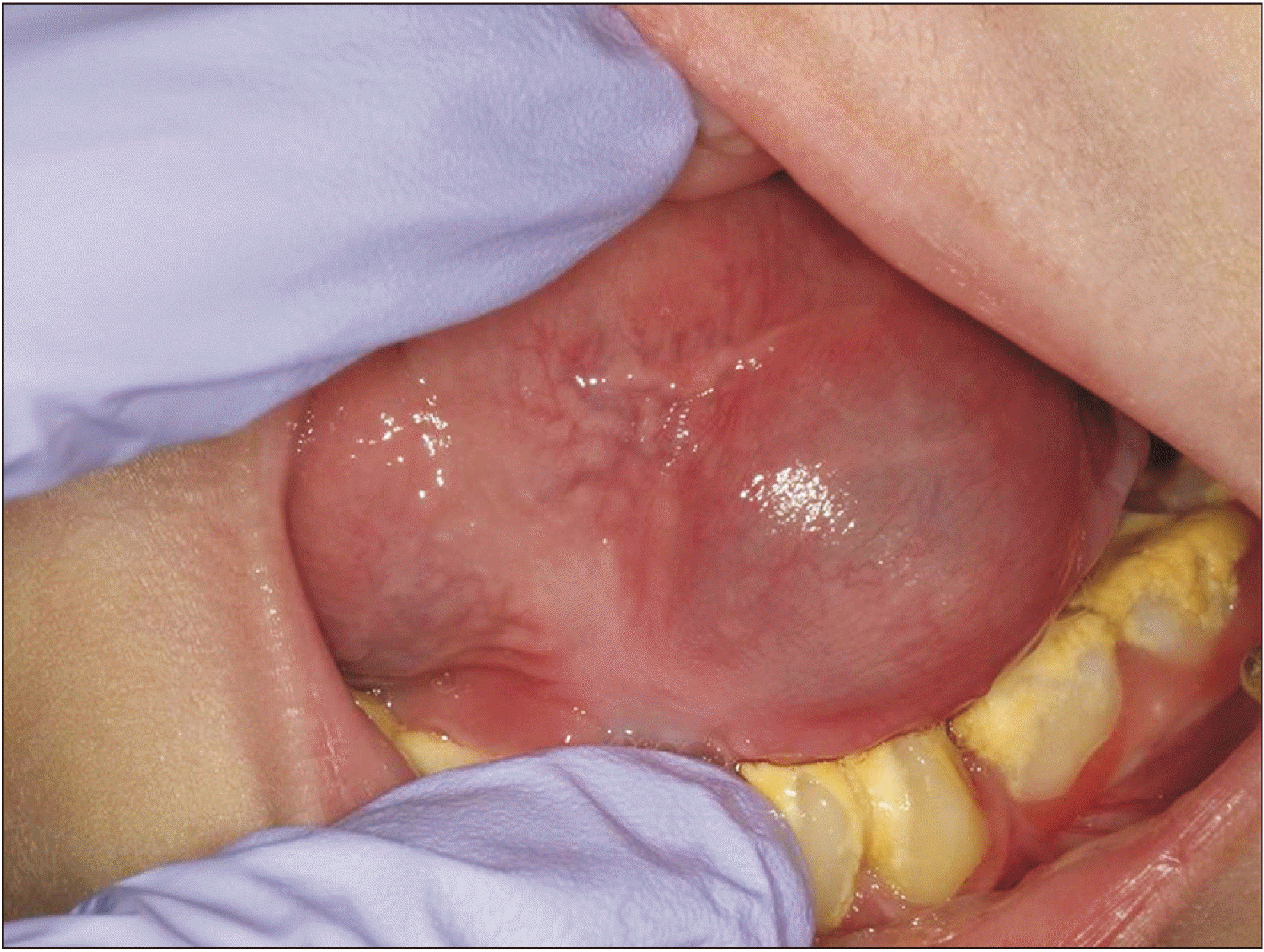
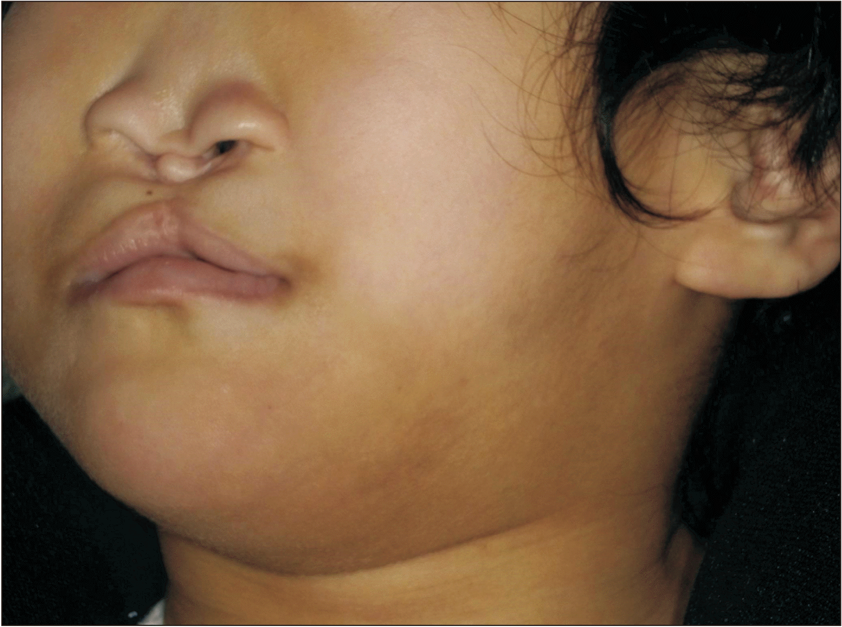
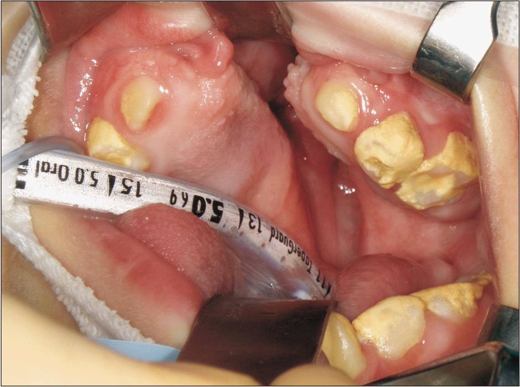
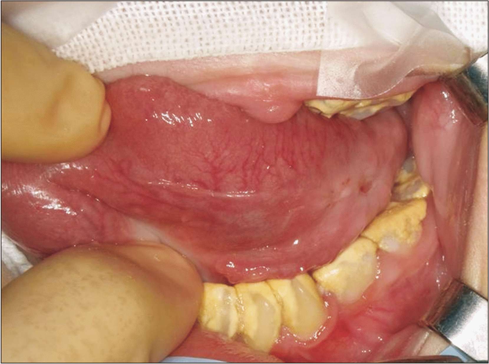
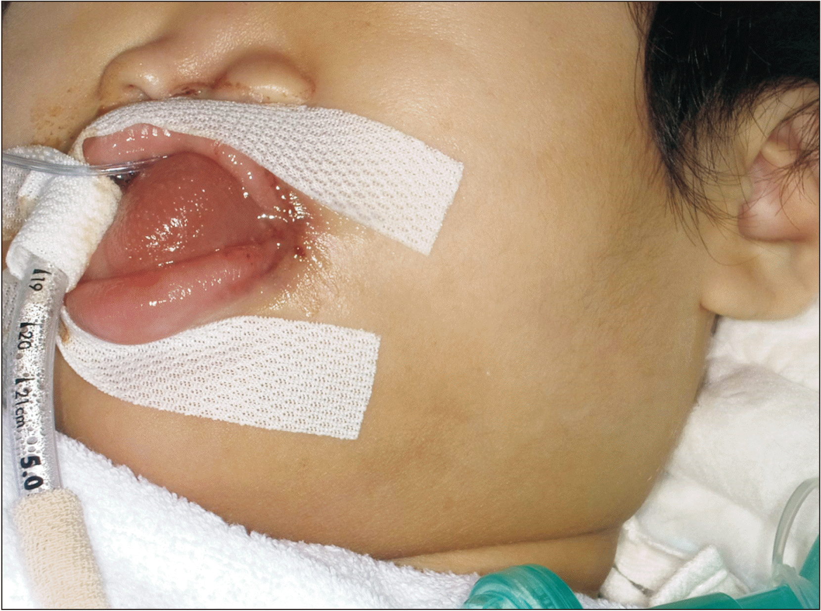
In 2016, an eight-year-old female patient presented to our department with a mass on the floor of the mouth. Her medical history included HPE with intellectual disability, cleft lip and palate, and pyriform aperture stenosis. She required gastrostomy for oromotor dysfunctions related to feeding and swallowing and difficulty in managing oral secretions. Clinical examination revealed a soft, fluctuant, and translucent mass with a diameter of 7 mm on the left side of the floor of the mouth.(Fig. 1) The mass, which was clinically diagnosed as a mucocele, grew and then subsequently shrank and disappeared spontaneously within a few months. In 2020, she was referred to our department for intra- and extra-oral swellings. Upon clinical examination, we noted a swelling located in the left upper neck region that was covered with normal overlying skin. Intraorally, on the left side of the floor of the mouth, there was a fluctuant, translucent, dome-shaped swelling with a diameter of approximately 30 mm.(Fig. 2) The swelling pushed the tongue up toward the cleft palate, making it difficult to visualize the oropharynx. The patient experienced unsteady breathing and respiratory distress, and blood gas analysis showed hypercapnia. Her oxygen saturation improved to 90%-99% with 2 L/min of oxygen inhalation. A clinical diagnosis of plunging ranula was made. With the cooperation of a pediatrician, incisional drainage was performed intraorally with the patient under mild sedation with midazolam owing to non-compliance due to her intellectual disability. The swelling shrank markedly, and the respiratory distress resolved. The ranula contained light yellow mucus that revealed no evidence of malignancy on cytology. Amylase content could not be examined because of the high viscosity of the fluid. Subsequently, she presented with frequent recurrences of the swelling that were accompanied by respiratory distress (Fig. 3); as a result, incisional drainage was performed four times in total. Based on close consultations with a pediatrician and an anesthetist regarding the patient’s clinical course, a consensus was reached that the optimal treatment choice was a radical surgical approach under general anesthesia. Approximately two months after the initial incisional drainage, removal of the sublingual gland and ranula was performed under general anesthesia, during which the anesthetist had difficulty intubating the patient orotracheally.(Fig. 4) Given that the surgical field was insufficient due to narrowing of the tongue space by lingual inclination of the mandibular teeth, only partial excision of the left sublingual gland was performed.(Fig. 5) The light yellow fluid that extravasated from the pseudocyst was assessed, and there were no signs of active infection. After the surgical procedure, the patient underwent trial extubation; however, she required urgent reintubation for upper airway obstruction due to persistent swelling on the floor of the mouth. Subsequently, she was closely monitored in the intensive care unit. As visible facial swelling was present on the first day after surgery (Fig. 6), the patient underwent extubation on the second postoperative day when the facial swelling decreased. She was transferred to the pediatric ward, and her swelling gradually resolved. Eventually, she was discharged with steady breathing on the ninth postoperative day. Histopathological analysis of the specimen revealed features of a pseudocyst consistent with those of a ranula and sublingual gland. The ranula did not recur during one year of follow-up.
III. Discussion
The present case provided two important clinical suggestions. First, the patient presented with a plunging ranula that arose from the sublingual gland following a mucocele that originated from the minor salivary glands. Both of these entities can occur due to trauma to the floor of the mouth in association with lingually inclined mandibular teeth, a type of dentoalveolar compensation frequently seen in patients with maxillary hypoplasia associated with HPE. Second, careful perioperative airway management is essential in a patient with a massive ranula and oromotor dysfunction because the ranula could exacerbate airway obstruction during induction of general anesthesia and also because postoperative swelling can cause potentially lethal upper airway obstruction after extubation as the tongue space is limited by lingual inclination of the mandibular teeth.
The main causes of a ranula are congenital anatomical variations, eating, dental procedures, and external blunt trauma2,3,8. The cause of a plunging ranula may be correlated with congenital partial dehiscence or fragility of the mylohyoid muscle4. In the present case, the ranula probably developed due to trauma to the floor of the mouth owing to lingual inclination of the mandibular teeth that is typically associated with maxillary hypoplasia in HPE.
Management of pediatric ranula may include observation for five to six months to monitor for spontaneous resolution1,4. If the lesion does not resolve spontaneously or recurs repeatedly, surgical treatment may be recommended1,4. Many surgical techniques to manage ranulas, including placement of a silk suture into the dome of the pseudocyst, cryosurgery, marsupialization, excision of the ranula alone, and excision of the ranula and the ipsilateral sublingual and/or submandibular gland, have been described in the literature1,4. Than et al.8 reported that intraoral removal of the sublingual gland was the most effective treatment for both simple and plunging ranulas. Complete removal of the ipsilateral sublingual gland via an intraoral approach is expected to cure both simple and plunging ranulas since the gland is the lone source of the ranula3,4. Alternatively, a previously published review suggested that conservative excision of only the involved part of the sublingual gland from which extravasation occurs could be a successful approach for management of a ranula9. Moreover, it is conceivable that total removal of the ranula itself is unnecessary because it is a pseudocyst and consists of mucus contained by inflamed granulation tissue and will eventually resolve3,4. In our case, incisional drainage of the ranula was required repeatedly for relieving acute respiratory distress; the best time to schedule a radical approach could not be decided immediately due to the coronavirus disease 2019 (COVID-19) pandemic. Eventually, the planned surgery was performed because spontaneous resolution did not occur. A preparatory decompression procedure that is carried out by aspiration and reduction of the tension and size of a ranula before the planned surgery can ensure that the involved discrete unit of the sublingual gland is excised completely3. Although the planned surgery was performed after the ranula had shrunk following incisional drainage, partial excision of the left sublingual gland was eventually performed because the surgical field was insufficient. No recurrence has been observed since.
Various malformations have been observed in patients with HPE10. Midfacial defects, including agnathia or micrognathia, midline or lateral cleft lip and/or palate, flat nose, and hypoplasia of the pyriform aperture are frequently seen craniofacial malformations10. Intellectual disability and abnormal movements have been described as common neurological signs10. Dysphagia and dysphonia are also frequently encountered as oromotor dysfunctions10. Common symptoms of oropharyngeal dysphagia include choking, coughing, or gagging while feeding or increased respiratory symptoms following feeding, such as wheezing, coughing, and increased secretions7. Therefore, children with oromotor dysfunctions have difficulty in managing their oral secretions and are at risk of aspiration and recurrent respiratory problems7. In the present case, the ranula itself and oromotor dysfunctions led to respiratory distress, while postoperative swelling on the floor of the mouth and oromotor dysfunctions complicated upper airway management.
Plunging ranulas in children with craniofacial malformations (such as HPE) and oromotor dysfunctions require prompt treatment since there is a risk of respiratory distress. A pediatrician assisted during the sedation and airway management procedures of the patient in the hospital ward, while an anesthetist provided perioperative and postoperative airway management in the intensive care unit. We successfully managed this patient with a plunging ranula and HPE with close cooperation of these specialists in our hospital.
 XML Download