Journal List > J Korean Assoc Oral Maxillofac Surg > v.48(4) > 1516079927

Kim, Choi, Youn, Ko, Oh, Lee, Park, and Choi: Treatment outcomes and prognostic factors in oral tongue cancer: a 20-year retrospective study at the National Cancer Center, South Korea

Abstract

Objectives

This study aimed to analyze the treatment outcomes and to evaluate the clinicopathological prognostic factors of oral tongue cancer.
Patients and Methods We retrospectively analyzed treatment results and prognostic factors in 205 patients with oral tongue squamous cell carcinoma who were admitted to the National Cancer Center, South Korea, between January 2001 and December 2020. The patients were treated with surgery and postoperative, definitive radiotherapy (RT) or chemoradiotherapy (CRT).

Results

Eighteen patients (8.8%) were treated with curative RT or CRT, while the rest (91.2%) were treated with surgery with or without postoperative RT or CRT. The median follow-up period was 30 months (range, 0-234 months). The 5-year overall survival (OS) and 5-year disease-free survival (DFS) were 72% and 63%, respectively. Multivariate analysis revealed that a positive neck nodal status (N1, N2-3) was significantly associated with poorer 5-year OS and DFS, while perineural invasion was associated with poorer 5-year DFS.

Conclusion

Cervical metastasis and perineural invasion are significant prognostic predictors, and combination treatments are necessary for improving OS and DFS in patients with these factors.

I. Introduction

Oral cavity cancer is common, with an estimated 350,000 new cases and 170,000 deaths annually worldwide1. The most common histological type of oral cavity cancer is squamous cell carcinoma (SCC), with the tongue being the most frequent site of occurrence2,3. The standard treatment for oral cavity cancer is surgery. However, in locally or locoregionally advanced disease, combined-modality therapy with surgery followed by adjuvant radiotherapy (RT) or chemoradiotherapy (CRT) is indicated4. Despite a stable annual incidence of oral cavity cancer in South Korea, the incidence of tongue cancer has increased significantly in both men and women, particularly in younger patients (≤40 years)5. Furthermore, there have been no improvements in 5-year overall survival (OS) and disease-free survival (DFS), which remain in the range of 50%-60%6,7. Several factors that negatively affect the clinicopathological prognosis of oral tongue squamous cell carcinoma (OTSCC) have been identified, including pathological T-stage, N-stage perineural invasion (PNI), and extranodal extension (ENE)8,9. Moreover, OTSCC is characterized by a high frequency of lymphatic metastasis, a high risk of recurrence, and the possibility of developing drug resistance to chemotherapy during treatment10. Since the incidence of OTSCC is on the rise and treatment outcomes in OTSCC are reportedly poorer compared to those of carcinomas arising in other oral cavity regions11,12, it is important to identify the clinicopathological factors associated with carcinomas in this site. Therefore, this retrospective study aimed to assess the potential clinicopathological factors that could affect treatment outcomes in OTSCC with the goal of making recommendations for the management of tongue cancers.

II. Patients and Methods

1. Patient selection

This study was a retrospective, monocentric, independent analysis of 205 patients with primary OTSCC who were treated and followed-up at the Oral Oncology Clinic of the National Cancer Center (Goyang, Korea) from January 2001 to December 2020. This study was reviewed and approved by the Institutional Review Board of the National Cancer Center, South Korea (NCC2022-0095). Demographic data, risk factors, histopathological features, and disease status were obtained from the medical records. The inclusion criteria involved patients with primary OTSCC, without second primaries other than mobile tongue cancer, and who had undergone primary surgical resection of the primary tumor and regional lymph nodes or definitive RT or CRT. The exclusion criteria included neoadjuvant chemotherapy administered prior to primary surgical resection, surgical resection performed with palliative or debulking intent, recurrent OTSCC, and a history of treatment including surgery or RT/CRT at another center. Although the seventh edition of the American Joint Committee on Cancer’s (AJCC) staging manual was used for staging and management, the eighth edition was used for analyzing the prognostic factors in this study. Patients treated with definitive RT or CRT were staged clinically, while those treated with primary surgery were staged pathologically. Adjuvant RT was administered within 4-6 weeks of surgery depending on the high-risk features found on the histopathology of the resected specimen. As per institutional policy, pathological T3 or T4 stages, node positivity, and critical resection margins within 5.0 mm were considered indications for adjuvant RT based on the decision of the treating physician. Adjuvant CRT was administered to patients with positive resection margins or ENE.

2. Statistical analysis

Statistical analyses were performed using SAS 9.4 (SAS Institute Inc., Cary, NC, USA) and Stata 17.0 (Stata Corp., College Station, TX, USA). The endpoints were OS and DFS, which were calculated through the Kaplan–Meier method. Univariate analysis was performed using demographic and clinicopathological factors, while multivariate Cox proportional hazard models were used to identify factors independently associated with OS and DFS. A P-value <0.05 was considered significant.

III. Results

1. Demographic characteristics

The median age of the patients was 57 years (range, 19-92 years), and the median follow-up period was 30 months (range, 0-234 months). Table 1 presents an overview of the demographic and clinicopathological data of the cohort. A total of 187 patients (91.2%) underwent primary surgical resection with or without adjuvant therapy (RT/CRT), while 18 patients (8.8%) were treated with definitive RT or CRT. Overall, disease control failed in 60 patients (29.3%) as observed during follow-up. The median duration of disease control failure was 6 months in this group. The most frequent type of recurrence was locoregional (25 patients, 41.7%). The most common site of distant metastasis was the lung.

2. Survival outcomes and prognostic factors associated with OS

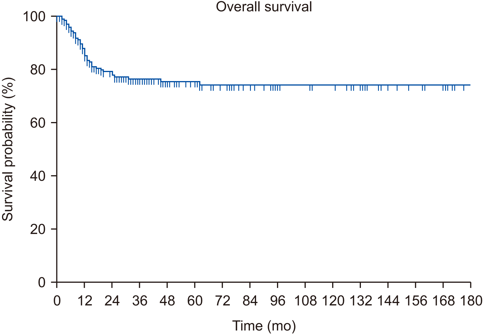
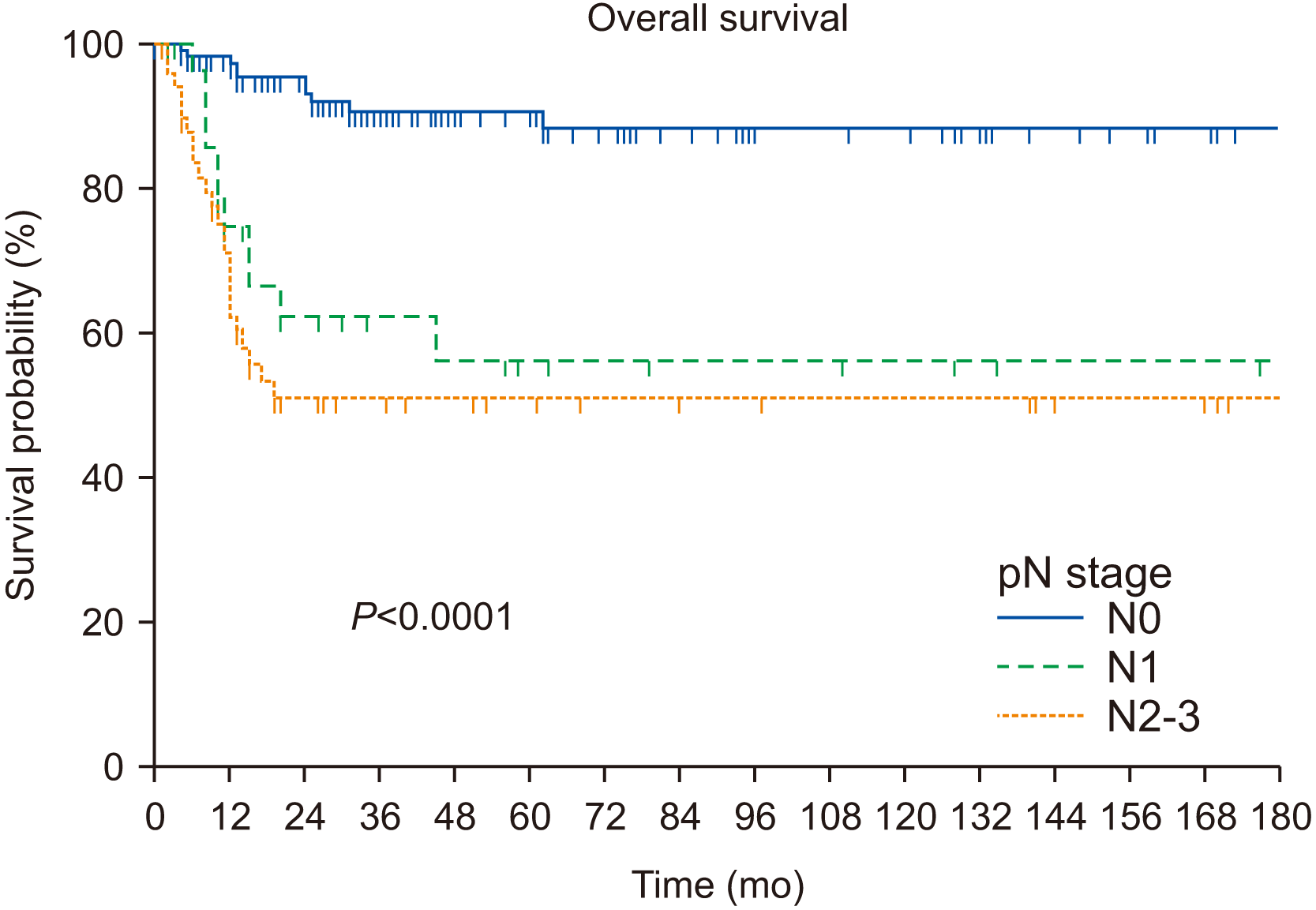
The median duration of OS was 30 months. The 5-year OS was 72%.(Fig. 1) The univariate analysis revealed that the following factors were significantly associated with poor prognosis in terms of OS: increasingly positive pN stages (N1, N2-3); advanced-stage disease (III-IV); treatment modality (adjuvant RT or CRT, definitive RT/CRT); moderately (G2) to poorly differentiated (G3) tumors; depth of invasion (DOI) >10 mm; advanced T stage (T3-4); PNI, lymphovascular invasion (LVI), and ENE. However, the multivariate analysis revealed that pN1 (hazard ratio [HR], 4.684; 95% confidence interval [CI], 1.975-11.111; P<0.001) and pN2-3 (HR, 7.640; 95% CI, 3.588-16.266; P<0.001) were significantly associated with poorer OS.(Table 2, Fig. 2) Unlike other studies, age, sex, and addictions (smoking and drinking) were not significant predictors of OS.

3. Survival outcomes and prognostic factors associated with DFS

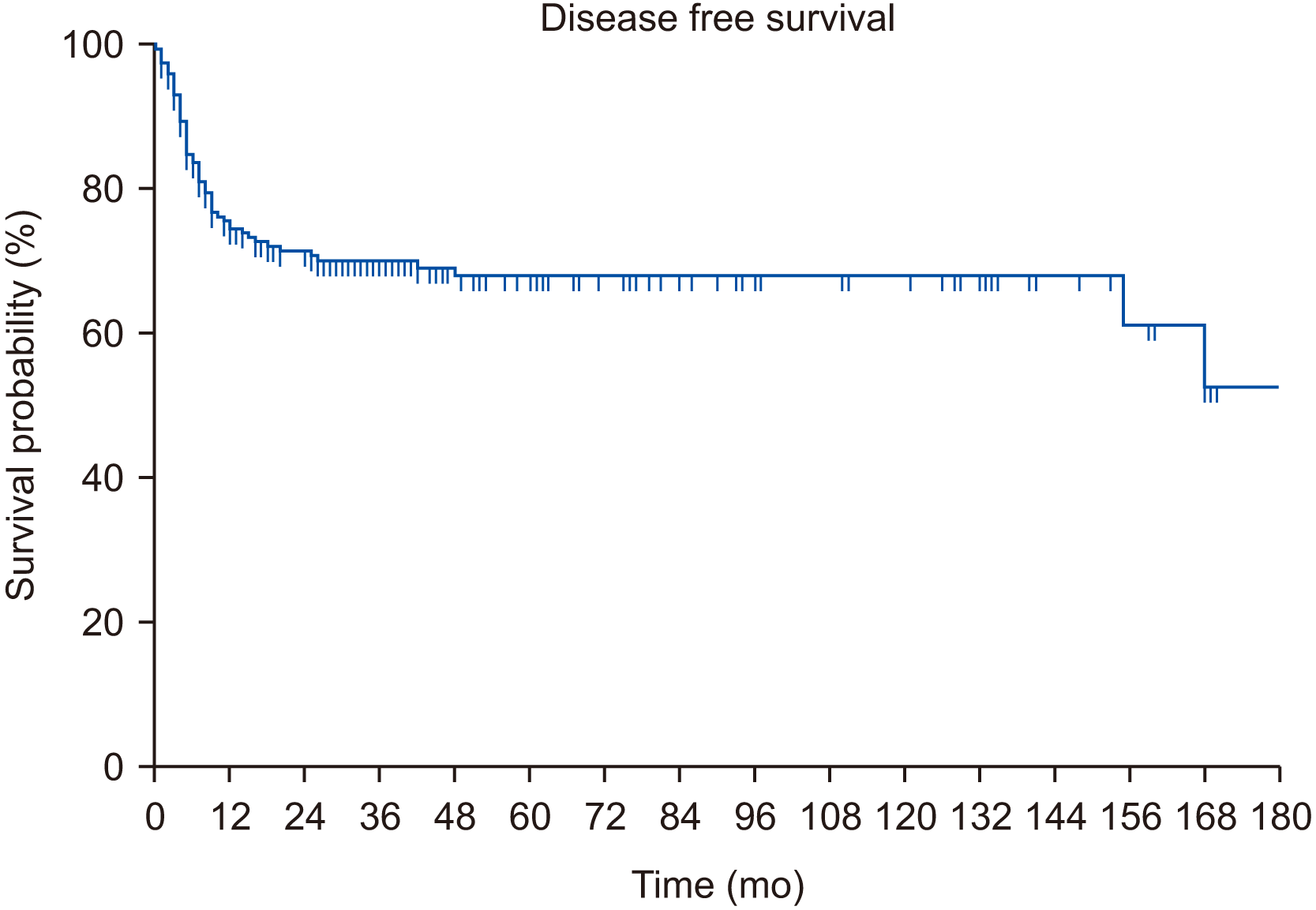
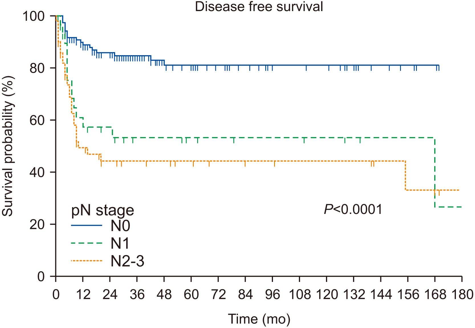
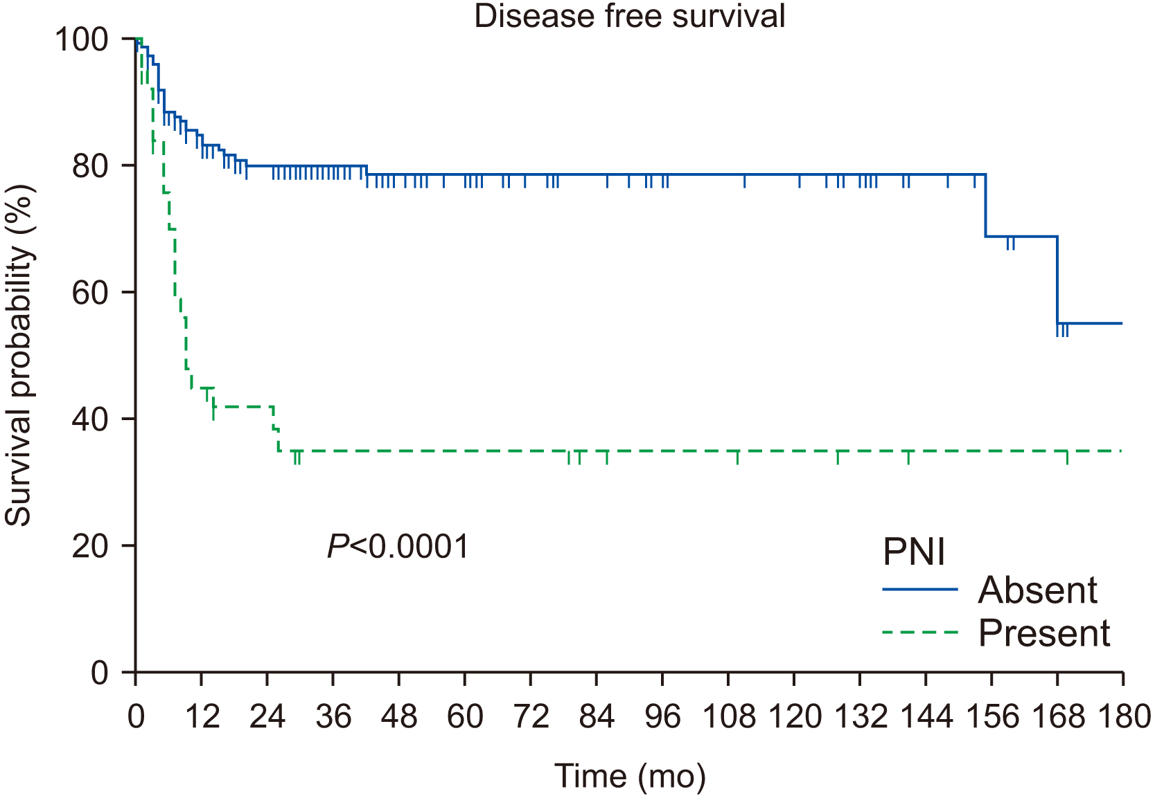
The median duration of DFS was 25 months. The 5-year DFS was 63%.(Fig. 3) The univariate analyses revealed that the following factors were significantly associated with poor prognosis in terms of DFS: increasing pN stages (N1, N2-3), treatment modality (adjuvant RT or CRT, definitive RT/CRT), PNI, advanced disease stage (III-IV), DOI >10 mm, G3 tumors, ENE, advanced T stage (T3-4), and marginal status (close or involved). However, multivariate analysis revealed that pN1 (HR, 2.499; 95% CI, 1.148-5.442; P=0.021), pN2-3 (HR, 4.022; 95% CI, 2.124-7.614; P<0.001), and PNI (HR, 2.145; 95% CI, 1.192-3.860; P=0.0109) exhibited significant negative effects on DFS.(Table 3, Fig. 4, 5) Similar to OS, age, sex, and addictions (smoking and drinking) did not significantly affect DFS.

IV. Discussion

In this study, we aimed to ascertain OS and DFS rates in tongue cancer and to identify the clinicopathological factors predicting treatment outcomes in this disease. Our study of survival outcomes revealed 5-year OS and DFS of 72% and 63%, respectively. These results are encouraging given that the corresponding results in previous reports are in the range of 5%-60%6-8,11. These results could be attributed to the postoperative treatment strategies, including adjuvant therapy (RT/CRT), implemented by a multidisciplinary team, as well as to surgical treatment with concomitant microvascular reconstruction, which enabled complete (R0) resection and improved patient quality of life. Moreover, it is believed that the relatively high frequency at which elective neck dissection was performed in this cohort also contributed to this favorable outcome.
Previous reports have discussed several clinicopathological parameters in relation to survival and disease progression in oral squamous cell carcinoma (OSCC), but only a few focused on tongue cancer. For patients with high-risk factors, multimodality treatments are mandatory for better prognoses6,13-15. In the univariate analysis, factors significantly affecting prognosis included pN status (N1, N2-3), advanced disease stage (III-IV), treatment modality (adjuvant RT or CRT, definitive RT/CRT), moderately (G2) to poorly differentiated (G3) tumors, DOI >10 mm, advanced T stage (T3-4), PNI, ENE, and close marginal status. Multivariate analysis also revealed that the pN status (N1, N2-3) was the most important predictor of both OS and DFS. Moreover, PNI had a significant negative effect on DFS.
Cervical metastasis is reportedly a significant predictor of poor survival in OTSCC. Both univariate and multivariate analyses revealed that nodal metastasis with increasing pN stages significantly affected OS and DFS. In locoregionally advanced disease, the survival rate decreased by <50%11. Our study showed that nodal metastasis decreased 5-year OS and DFS from 93.7% to 50.7% and 85.1% to 47%, respectively, confirming these findings. Considering that approximately 60% of the instances of disease control failure occurred locoregionally and regionally in this study and considering the high rate of occult metastasis at this site8,16-18, active management of regional lymphatics with elective neck dissection should be considered. In the present study, occult nodal metastasis was observed in 17.3% of patients, including patients with T1-2 tumors. The 5-year DFS was better in patients who underwent neck dissections compared to those who did not undergo the procedure, even with early-stage disease (I, II).
PNI and LVI have been reported to adversely affect treatment outcomes in OSCC. However, a high level of heterogeneity exists in the detection criteria among studies. PNI is classified as tumor cells invading nerve sheaths or surrounding at least one-third of the circumference of the nerve, while LVI is defined as the presence of tumor cells within or adjacent to the vessels. Both of these factors can involve subjectivity in the diagnosis19. Several studies have confirmed that PNI and LVI possess a high prognostic value for decreased survival in OTSCC8,20,21. The results of the univariate analyses in our study were consistent with such findings. However, the results of the multivariate analysis, apart from those related to PNI in DFS, were not significant in terms of OS and DFS. The identification of PNI as an independent prognosticator in our study supports the finding that the perineural space acts as a low-resistance route for progression of SCC within connective tissue-innervating peripheral nerves22.
Regarding the effect of tumor grade on OS and DFS, it is widely accepted that well-differentiated tumors (G1) have a better prognosis in OSCC compared with moderately to poorly differentiated tumors (G2-3)18. However, most studies do not consider the histological grade to be a significant predictor of treatment outcomes9,14,20,23. Our study showed that OS and DFS decreased with increasing tumor grade (from G1 to G3). This finding was significant only in the univariate analysis. Further randomized multi-institutional prospective studies are necessary to ascertain the effect of tumor grade on prognosis.
Age and sex have been suggested to be possible risk factors affecting treatment outcomes. Some studies have reported that the prognosis in younger patients (≤40 years of age) is poorer compared to the elderly, justifying more aggressive treatment in this subgroup23-25. However, other studies have highlighted the role of aging in poor prognosis and the negative effect of comorbidities in the elderly26,27. Likewise, regarding sex as a prognostic factor, various studies have produced varying results, ranging from those indicating that female sex is associated with better survival28,29 to those exhibiting no significant difference between the sexes30. The current study revealed no significant effects of age and sex on OS and DFS, in line with the findings of the latter studies. Therefore, further well-defined studies are warranted to elucidate the prognostic effects of age and sex on OTSCC.
Many studies have shown that smoking and alcohol consumption are prognostic predictors in head and neck cancers16,31,32. However, many other studies have presented conflicting results, revealing no significant association between survival time and habitual factors such as smoking and alcohol consumption33,34. Upon examining OTSCC cases, no association was observed between these habits and an increased risk of reduced survival time20,21. Similar to the findings in the latter studies, these habits were not identified as prognostic factors in our study. However, the high number of cases in which the association with these habits was falsely classified as negative has to be considered.
In OTSCC, the T and N stages were independent indicators of prognosis even though they are interrelated. A higher T stage leads to an increase in the rate of occult metastases20,21,35, while a higher N-stage is associated with the development of distant metastases, particularly with multiple organ involvement or ENE36. Our study found that the prognostic value of a higher T stage is high in predicting decreased OS and DFS in cases when the T stage increases from early (T1-2) to advanced (T3-4) stages. This result is consistent with those of other studies20,21,37. However, this factor was not a significant independent predictor in multivariate analysis.
Since DOI was a significant prognostic predictor and was included in the AJCC tumor-node-metastasis system (eighth edition), the staging system was redefined based on the DOI cut off values of 5 and 10 mm. A DOI greater than 10 mm was significantly associated with decreased 5-year survival and increased risk of occult metastasis17. In this cohort, increasing DOI was significantly associated with poor prognosis in terms of decreased 5-year OS and DFS in the univariate analysis. However, in multivariate analysis, this finding was not significant. This result could be attributed to the inconsistency and subjectivity in histopathological reporting by different pathologists.
ENE is associated with poor prognosis in terms of locoregional failure and distant metastasis in OSCC, indications for concurrent CRT19,38. This was observed in several retrospective studies on OTSCC6,8,9,11, including our cohort. Most patients with ENE in this cohort, along with involved resection margins, underwent CRT. Although the univariate analysis revealed a strong association between ENE and poor outcomes in terms of both OS and DFS in our cohort, the same association was found only in relation to OS in the multivariate analysis.
Surgery with or without adjuvant RT or CRT is the standard treatment for resectable OTSCC, with a high survival rate of up to 70% in the early stages38. The importance of clear surgical margins has been the main focus of surgical oncology, considering that positive and close margins have a strong negative effect on survival. Several studies have shown the benefit of clear margins in OTSCC8. The results of our univariate analysis indicated that clear margins exhibited a significant effect on OS and DFS compared to close (5 mm) margins. The multivariate analysis did not demonstrate a significant effect of surgical margins on OS and DFS.
This study had several limitations. A potential bias could have resulted from its retrospective study design. Although our clinic adopted standardized treatment guidelines, differences may have arisen depending on the individual experiences of the treating oncologists. Since the histopathological characteristics of the analyzed samples were recorded by different pathologists over an extended period of time, standardization of pathologic assessments should be a subject for future studies.

V. Conclusion

In conclusion, the univariate analyses in this study revealed that survival outcomes in OTSCC were affected by increasing T stage, pN status, ENE, moderately to poorly differentiated tumors, LVI, PNI, and marginal status. These factors were reported in previous studies30,31,38. However, our multivariate analysis revealed that pN status (N1, N2-3) is significantly associated with poor prognosis in terms of OS and DFS and with PNI in terms of DFS. The rate of primary cervical metastases in this cohort was greater than 39%, while that of occult metastases was 17%. These results advocate the importance of regional control along with the need for elective neck dissection and postoperative adjuvant therapy in patients with neck node positivity.

Notes

Authors’ Contributions

M.G.K. and Y.S.C. participated in data collection and wrote the manuscript. S.M.Y., J.H.K., and H.J.O. participated in the design of the study. J.H.L., J.Y.P., and S.W.C. participated in the study design, coordination, and helped to draft the manuscript. All authors read and approved the final manuscript.

Ethics Approval and Consent to Participate

This study was conducted after obtaining approval from the Institutional Review Board of the National Cancer Center, South Korea (NCC2022-0095), and informed consent was waived.

Conflict of Interest

No potential conflict of interest relevant to this article was reported.

References

1. Bray F, Ferlay J, Soerjomataram I, Siegel RL, Torre LA, Jemal A. 2018; Global cancer statistics 2018: GLOBOCAN estimates of incidence and mortality worldwide for 36 cancers in 185 countries. CA Cancer J Clin. 68:394–424. https://doi.org/10.3322/caac.21492. DOI: 10.3322/caac.21492. PMID: 30207593.
crossref
2. Shah JP, Gil Z. 2009; Current concepts in management of oral cancer--surgery. Oral Oncol. 45:394–401. https://doi.org/10.1016/j.oraloncology.2008.05.017. DOI: 10.1016/j.oraloncology.2008.05.017. PMID: 18674952. PMCID: PMC4130348.
crossref
3. Pulte D, Brenner H. 2010; Changes in survival in head and neck cancers in the late 20th and early 21st century: a period analysis. Oncologist. 15:994–1001. https://doi.org/10.1634/theoncologist.2009-0289. DOI: 10.1634/theoncologist.2009-0289. PMID: 20798198. PMCID: PMC3228039.
crossref
4. Zanoni DK, Montero PH, Migliacci JC, Shah JP, Wong RJ, Ganly I, et al. 2019; Survival outcomes after treatment of cancer of the oral cavity (1985-2015). Oral Oncol. 90:115–21. https://doi.org/10.1016/j.oraloncology.2019.02.001. DOI: 10.1016/j.oraloncology.2019.02.001. PMID: 30846169. PMCID: PMC6417804.
crossref
5. Choi SW, Moon EK, Park JY, Jung KW, Oh CM, Kong HJ, et al. 2014; Trends in the incidence of and survival rates for oral cavity cancer in the Korean population. Oral Dis. 20:773–9. https://doi.org/10.1111/odi.12251. DOI: 10.1111/odi.12251. PMID: 24735459.
crossref
6. Bello IO, Soini Y, Salo T. 2010; Prognostic evaluation of oral tongue cancer: means, markers and perspectives (II). Oral Oncol. 46:636–43. https://doi.org/10.1016/j.oraloncology.2010.06.008. DOI: 10.1016/j.oraloncology.2010.06.008. PMID: 20637679.
crossref
7. van Dijk BA, Brands MT, Geurts SM, Merkx MA, Roodenburg JL. 2016; Trends in oral cavity cancer incidence, mortality, survival and treatment in the Netherlands. Int J Cancer. 139:574–83. https://doi.org/10.1002/ijc.30107. DOI: 10.1002/ijc.30107. PMID: 27038013.
crossref
8. Faisal M, Dhanani R, Ullah S, Bakar MA, Irfan N, Malik KI, et al. 2021; Prognostic outcomes of treatment naïve oral tongue squamous cell carcinoma (OTSCC): a comprehensive analysis of 14 years. Eur Arch Otorhinolaryngol. 278:3045–53. https://doi.org/10.1007/s00405-020-06482-x. DOI: 10.1007/s00405-020-06482-x. PMID: 33236214.
crossref
9. Marra A, Violati M, Broggio F, Codecà C, Blasi M, Luciani A, et al. 2019; Long-term disease-free survival in surgically-resected oral tongue cancer: a 10-year retrospective study. Acta Otorhinolaryngol Ital. 39:84–91. https://doi.org/10.14639/0392-100X-2336. DOI: 10.14639/0392-100X-2336. PMID: 31097825. PMCID: PMC6522864.
crossref
10. Han G, Xu C, Yu D. 2018; Mechanisms correlated with chemotherapy resistance in tongue cancers. J Cancer Res Ther. 14:1–5. https://doi.org/10.4103/jcrt.JCRT_763_17. DOI: 10.4103/jcrt.JCRT_763_17. PMID: 29516950.
crossref
11. Rusthoven K, Ballonoff A, Raben D, Chen C. 2008; Poor prognosis in patients with stage I and II oral tongue squamous cell carcinoma. Cancer. 112:345–51. https://doi.org/10.1002/cncr.23183. DOI: 10.1002/cncr.23183. PMID: 18041071.
crossref
12. Cheng YJ, Tsai MH, Chiang CJ, Tsai ST, Liu TW, Lou PJ, et al. 2018; Adjuvant radiotherapy after curative surgery for oral cavity squamous cell carcinoma and treatment effect of timing and duration on outcome - a Taiwan Cancer Registry national database analysis. Cancer Med. 7:3073–83. https://doi.org/10.1002/cam4.1611. DOI: 10.1002/cam4.1611. PMID: 29905028. PMCID: PMC6051157.
crossref
13. Asakage T, Yokose T, Mukai K, Tsugane S, Tsubono Y, Asai M, et al. 1998; Tumor thickness predicts cervical metastasis in patients with stage I/II carcinoma of the tongue. Cancer. 82:1443–8. https://doi.org/10.1002/(sici)1097-0142(19980415)82:8<1443::aid-cncr2>3.0.co;2-a. DOI: 10.1002/(SICI)1097-0142(19980415)82:8<1443::AID-CNCR2>3.0.CO;2-A. PMID: 9554518.
crossref
14. Po Wing Yuen A, Lam KY, Lam LK, Ho CM, Wong A, Chow TL, et al. 2002; Prognostic factors of clinically stage I and II oral tongue carcinoma - a comparative study of stage, thickness, shape, growth pattern, invasive front malignancy grading, Martinez-Gimeno score, and pathologic features. Head Neck. 24:513–20. https://doi.org/10.1002/hed.10094. DOI: 10.1002/hed.10094. PMID: 12112547.
crossref
15. Huang SH, Hwang D, Lockwood G, Goldstein DP. OʼSullivan B. 2009; Predictive value of tumor thickness for cervical lymph-node involvement in squamous cell carcinoma of the oral cavity: a meta-analysis of reported studies. Cancer. 115:1489–97. https://doi.org/10.1002/cncr.24161. DOI: 10.1002/cncr.24161. PMID: 19197973.
crossref
16. Inoue M, Nagata C, Tsuji I, Sugawara Y, Wakai K, Tamakoshi A, et al. 2012; ; Research Group for the Development and Evaluation of Cancer Prevention Strategies in Japan. Impact of alcohol intake on total mortality and mortality from major causes in Japan: a pooled analysis of six large-scale cohort studies. J Epidemiol Community Health. 66:448–56. https://doi.org/10.1136/jech.2010.121830. DOI: 10.1136/jech.2010.121830. PMID: 21148820.
crossref
17. Faisal M, Abu Bakar M, Sarwar A, Adeel M, Batool F, Malik KI, et al. 2018; Depth of invasion (DOI) as a predictor of cervical nodal metastasis and local recurrence in early stage squamous cell carcinoma of oral tongue (ESSCOT). PLoS One. 13:e0202632. https://doi.org/10.1371/journal.pone.0202632. DOI: 10.1371/journal.pone.0202632. PMID: 30133515. PMCID: PMC6105019.
crossref
18. Montoro JRMC, Hicz HA, de Souza L, Livingstone D, Melo DH, Tiveron RC, et al. 2008; Prognostic factors in squamous cell carcinoma of the oral cavity. Braz J Otorhinolaryngol. 74:861–6. https://doi.org/10.1016/S1808-8694(15)30146-4. DOI: 10.1016/S1808-8694(15)30146-4. PMID: 19582342.
crossref
19. Dolens EDS, Dourado MR, Almangush A, Salo TA, Gurgel Rocha CA, da Silva SD, et al. 2021; The impact of histopathological features on the prognosis of oral squamous cell carcinoma: a comprehensive review and meta-analysis. Front Oncol. 11:784924. https://doi.org/10.3389/fonc.2021.784924. DOI: 10.3389/fonc.2021.784924. PMID: 34858861. PMCID: PMC8631280.
crossref
20. Sharma K, Ahlawat P, Gairola M, Tandon S, Sachdeva N, Sharief MI. 2019; Prognostic factors, failure patterns and survival analysis in patients with resectable oral squamous cell carcinoma of the tongue. Radiat Oncol J. 37:73–81. https://doi.org/10.3857/roj.2018.00577. DOI: 10.3857/roj.2018.00577. PMID: 31266288. PMCID: PMC6610009.
crossref
21. Thiagarajan S, Nair S, Nair D, Chaturvedi P, Kane SV, Agarwal JP, et al. 2014; Predictors of prognosis for squamous cell carcinoma of oral tongue. J Surg Oncol. 109:639–44. https://doi.org/10.1002/jso.23583. DOI: 10.1002/jso.23583. PMID: 24619660.
crossref
22. Fagan JJ, Collins B, Barnes L, DʼAmico F, Myers EN, Johnson JT. 1998; Perineural invasion in squamous cell carcinoma of the head and neck. Arch Otolaryngol Head Neck Surg. 124:637–40. https://doi.org/10.1001/archotol.124.6.637. DOI: 10.1001/archotol.124.6.637. PMID: 9639472.
crossref
23. Hilly O, Shkedy Y, Hod R, Soudry E, Mizrachi A, Hamzany Y, et al. 2013; Carcinoma of the oral tongue in patients younger than 30 years: comparison with patients older than 60 years. Oral Oncol. 49:987–90. https://doi.org/10.1016/j.oraloncology.2013.07.005. DOI: 10.1016/j.oraloncology.2013.07.005. PMID: 23927849.
crossref
24. Zhang YY, Wang DC, Su JZ, Jia LF, Peng X, Yu GY. 2017; Clinicopathological characteristics and outcomes of squamous cell carcinoma of the tongue in different age groups. Head Neck. 39:2276–82. https://doi.org/10.1002/hed.24898. DOI: 10.1002/hed.24898. PMID: 28842932.
crossref
25. Jeon JH, Kim MG, Park JY, Lee JH, Kim MJ, Myoung H, et al. 2017; Analysis of the outcome of young age tongue squamous cell carcinoma. Maxillofac Plast Reconstr Surg. 39:41. https://doi.org/10.1186/s40902-017-0139-8. DOI: 10.1186/s40902-017-0139-8. PMID: 29302590. PMCID: PMC5742317.
crossref
26. Cadoni G, Giraldi L, Petrelli L, Pandolfini M, Giuliani M, Paludetti G, et al. 2017; Prognostic factors in head and neck cancer: a 10-year retrospective analysis in a single-institution in Italy. Acta Otorhinolaryngol Ital. 37:458–66. https://doi.org/10.14639/0392-100X-1246. DOI: 10.14639/0392-100X-1246. PMID: 28663597. PMCID: PMC5782422.
crossref
27. Molteni G, Valerini S, Alicandri-Ciufelli M, Sprio AE, Crosetti E, Berta GN, et al. 2018; Unravelling the risk factors that underlie oral and oropharyngeal surgery in elderly. Acta Otorhinolaryngol Ital. 38:409–16. https://doi.org/10.14639/0392-100X-1507. DOI: 10.14639/0392-100X-1507. PMID: 29393927. PMCID: PMC6265672.
crossref
28. Listl S, Jansen L, Stenzinger A, Freier K, Emrich K, Holleczek B, et al. 2013; ; GEKID Cancer Survival Working Group. Survival of patients with oral cavity cancer in Germany. PLoS One. 8:e53415. https://doi.org/10.1371/journal.pone.0053415. DOI: 10.1371/journal.pone.0053415. PMID: 23349710. PMCID: PMC3548847.
crossref
29. Funk GF, Karnell LH, Robinson RA, Zhen WK, Trask DK, Hoffman HT. 2002; Presentation, treatment, and outcome of oral cavity cancer: a National Cancer Data Base report. Head Neck. 24:165–80. https://doi.org/10.1002/hed.10004. DOI: 10.1002/hed.10004. PMID: 11891947.
crossref
30. Garavello W, Spreafico R, Somigliana E, Gaini L, Pignataro L, Gaini RM. 2008; Prognostic influence of gender in patients with oral tongue cancer. Otolaryngol Head Neck Surg. 138:768–71. https://doi.org/10.1016/j.otohns.2008.02.026. DOI: 10.1016/j.otohns.2008.02.026. PMID: 18503852.
crossref
31. Leoncini E, Vukovic V, Cadoni G, Pastorino R, Arzani D, Bosetti C, et al. 2015; Clinical features and prognostic factors in patients with head and neck cancer: results from a multicentric study. Cancer Epidemiol. 39:367–74. https://doi.org/10.1016/j.canep.2015.02.004. DOI: 10.1016/j.canep.2015.02.004. PMID: 25770642.
crossref
32. Li Y, Mao Y, Zhang Y, Cai S, Chen G, Ding Y, et al. 2014; Alcohol drinking and upper aerodigestive tract cancer mortality: a systematic review and meta-analysis. Oral Oncol. 50:269–75. https://doi.org/10.1016/j.oraloncology.2013.12.015. DOI: 10.1016/j.oraloncology.2013.12.015. PMID: 24405883.
crossref
33. Koo K, Barrowman R, McCullough M, Iseli T, Wiesenfeld D. 2013; Non-smoking non-drinking elderly females: a clinically distinct subgroup of oral squamous cell carcinoma patients. Int J Oral Maxillofac Surg. 42:929–33. https://doi.org/10.1016/j.ijom.2013.04.010. DOI: 10.1016/j.ijom.2013.04.010. PMID: 23702369.
crossref
34. Loeffelbein D, Ritschl LM, Güll FD, Roth M, Wolff KD, Mücke T. 2017; Influence of possible predictor variables on the outcome of primary oral squamous cell carcinoma: a retrospective study of 392 consecutive cases at a single centre. Int J Oral Maxillofac Surg. 46:413–21. https://doi.org/10.1016/j.ijom.2016.11.014. DOI: 10.1016/j.ijom.2016.11.014. PMID: 28007325.
crossref
35. Sparano A, Weinstein G, Chalian A, Yodul M, Weber R. 2004; Multivariate predictors of occult neck metastasis in early oral tongue cancer. Otolaryngol Head Neck Surg. 131:472–6. https://doi.org/10.1016/j.otohns.2004.04.008. DOI: 10.1016/j.otohns.2004.04.008. PMID: 15467620.
crossref
36. Jones HB, Sykes A, Bayman N, Sloan P, Swindell R, Patel M, et al. 2009; The impact of lymphovascular invasion on survival in oral carcinoma. Oral Oncol. 45:10–5. https://doi.org/10.1016/j.oraloncology.2008.03.009. DOI: 10.1016/j.oraloncology.2008.03.009. PMID: 18620889.
crossref
37. Shim SJ, Cha J, Koom WS, Kim GE, Lee CG, Choi EC, et al. 2010; Clinical outcomes for T1-2N0-1 oral tongue cancer patients underwent surgery with and without postoperative radiotherapy. Radiat Oncol. 5:43. https://doi.org/10.1186/1748-717X-5-43. DOI: 10.1186/1748-717X-5-43. PMID: 20504371. PMCID: PMC2887888.
crossref
38. Grégoire V, Lefebvre JL, Licitra L. Felip E; EHNS-ESMO-ESTRO Guidelines Working Group. 2010; Squamous cell carcinoma of the head and neck: EHNS-ESMO-ESTRO clinical practice guidelines for diagnosis, treatment and follow-up. Ann Oncol. 21 Suppl 5:v184–6. https://doi.org/10.1093/annonc/mdq185. DOI: 10.1093/annonc/mdq185. PMID: 20555077.
crossref

Fig. 1
Kaplan–Meier curve of overall survival.
jkaoms-48-4-192-f1.tif
Fig. 2
Kaplan–Meier curve of overall survival by neck status.
jkaoms-48-4-192-f2.tif
Fig. 3
Kaplan–Meier curve of overall disease-free survival.
jkaoms-48-4-192-f3.tif
Fig. 4
Kaplan–Meier curve of disease-free survival by neck status.
jkaoms-48-4-192-f4.tif
Fig. 5
Kaplan–Meier curve of disease-free survival by presence of perineural invasion (PNI).
jkaoms-48-4-192-f5.tif
Table 1
Demographic and clinicopathological data of the cohort
Variable Value
Sex
Female 89 (43.4)
Male 116 (56.6)
Age
≤40 yr 33 (16.1)
>40 yr 172 (83.9)
Smoking
No 106 (51.7)
Yes 99 (48.3)
Drinking
No 103 (50.2)
Yes 102 (49.8)
T stage
T1 or T2 128 (62.4)
T3 or T4 77 (37.6)
N stage
N0 124 (60.5)
N1 31 (15.1)
N2 or N3 50 (24.4)
pTNM stage
Stage I or II (early) 100 (48.8)
Stage III or IV (advanced) 105 (51.2)
Treatment
Surgery only 110 (53.7)
Surgery+RT/CT 77 (37.6)
RT/CT 18 (8.8)
Extranodal extension
Absent 165 (80.5)
Present 40 (19.5)
Differentiation grade
Well 97 (47.3)
Moderate 76 (37.1)
Poor 24 (11.7)
N/A 8 (3.9)
Depth of invasion
≤5 mm 65 (31.7)
6-10 mm 37 (18.0)
>10 mm 71 (34.6)
N/A 32 (15.6)
Perineural invasion
Absent 149 (72.7)
Present 38 (18.5)
N/A 18 (8.8)
Lymphovascular invasion
Absent 144 (70.2)
Present 43 (21.0)
N/A 18 (8.8)
Resection status
Close (<5 mm) 63 (30.7)
Safe (≥5 mm) 124 (60.5)
N/A 18 (8.8)
Recurrence
No 145 (70.7)
Yes 60 (29.3)

(RT: radiotherapy, CT: chemotherapy, N/A: not available)

Values are presented as number (%).

Table 2
Cox regression for overall survival
Variable No. of patients No. of events Univariable Cox regression Multivariable Cox regression (backward selection)


HR (95% CI) P-value HR (95% CI) P-value
Sex
Female 89 22 1 (ref.)
Male 116 25 0.962 (0.543-1.707) 0.8959
Age
≤40 yr 33 11 1 (ref.)
>40 yr 172 36 0.685 (0.349-1.347) 0.2727
Addictions
Absent 83 16 1 (ref.)
Present 122 28 1.313 (0.720-2.394) 0.3739
T stage
T1 or T2 128 17 1 (ref.)
T3 or T4 77 30 3.302 (1.820-5.993) <0.0001
N stage
N0 124 10 1 (ref.) 1 (ref.)
N1 31 11 5.316 (2.256-12.528) 0.0001 4.684 (1.975-11.111) 0.0005
N2 or N3 50 26 8.616 (4.148-17.896) <0.0001 7.640 (3.588-16.266) <0.0001
TNM stage
Stage I or II (early) 100 6 1 (ref.)
Stage III or IV (advanced) 105 41 7.925 (3.361-18.686) <0.0001
Treatment
Surgery only 110 10 1 (ref.)
Surgery+RT/CT 77 32 5.433 (2.668-11.063) <0.0001
RT/CT 18 5 5.219 (1.778-15.319) 0.0026
Differentiation (missing 8 cases)
Well 97 14 1 (ref.) 1 (ref.)
Moderate 76 21 2.233 (1.133-4.399) 0.0203 1.932 (0.972-3.840) 0.0604
Poor 24 12 4.220 (1.946-9.149) 0.0003 2.181 (0.981-4.852) 0.0558
Depth of invasion (missing 32 cases)
≤5 mm 65 6 1 (ref.)
6-10 mm 37 7 2.005 (0.674-5.968) 0.2113
>10 mm 71 26 4.653 (1.913-11.315) 0.0007
Lymphovascular invasion (missing 18 cases)
Absent 144 27 1 (ref.)
Present 43 15 1.980 (1.053-3.724) 0.0339
Perineural invasion (missing 18 cases)
Absent 149 23 1 (ref.)
Present 38 19 3.951 (2.146-7.276) <0.0001
Extranodal extension
Absent 165 22 1 (ref.)
Present 40 25 5.701 (3.209-10.126) <0.0001
Resection status (missing 18 cases)
Close (<5 mm) 63 21 1 (ref.)
Safe (≥5 mm) 124 21 0.508 (0.278-0.931) 0.0283

(HR: hazard ratio, CI: confidence interval, ref.: reference, RT: radiotherapy, CT: chemotherapy)

Table 3
Cox regression for disease-free survival
Variable No. of patients No. of events Univariable Cox regression Multivariable Cox regression (backward selection)


HR (95% CI) P-value HR (95% CI) P-value
Sex
Female 89 34 1 (ref.)
Male 116 34 0.762 (0.474-1.227) 0.2638
Age
≤40 yr 33 13 1 (ref.)
>40 yr 172 55 0.867 (0.472-1.592) 0.6456
Addictions
Absent 83 21 1 (ref.)
Present 122 39 1.337 (0.796-2.246) 0.2718
T stage
T1 or T2 128 32 1 (ref.)
T3 or T4 77 36 2.072 (1.285-3.341) 0.0028
N stage
N0 124 23 1 (ref.) 1 (ref.)
N1 31 14 2.956 (1.519-5.753) 0.0014 2.499 (1.148-5.442) 0.0210
N2 or N3 50 31 4.553 (2.645-7.836) <0.0001 4.022 (2.124-7.614) <0.0001
TNM stage
Stage I or II (early) 100 18 1 (ref.)
Stage III or IV (advanced) 105 50 3.150 (1.834-5.412) <0.0001
Treatment
Surgery only 110 19 1 (ref.)
Surgery+RT/CT 77 39 3.357 (1.932-5.833) <0.0001
RT/CT 18 10 4.899 (2.271-10.566) <0.0001
Differentiation (missing 8 cases)
Well 97 25 1 (ref.)
Moderate 76 27 1.626 (0.942-2.808) 0.0812
Poor 24 13 2.486 (1.270-4.864) 0.0078
Depth of invasion (missing 32 cases)
≤5 mm 65 12 1 (ref.)
6-10 mm 37 11 1.625 (0.716-3.693) 0.2459
>10 mm 71 32 2.894 (1.488-5.628) 0.0017
Lymphovascular invasion (missing 18 cases)
Absent 144 40 1 (ref.)
Present 43 18 1.612 (0.924-2.814) 0.0926
Perineural invasion (missing 18 cases)
Absent 149 35 1 (ref.) 1 (ref.)
Present 38 23 3.375 (1.987-5.732) <0.0001 2.145 (1.192-3.860) 0.0109
Extranodal extension
Absent 165 41 1 (ref.)
Present 40 27 3.836 (2.345-6.274) <0.0001
Resection status (missing 18 cases)
Close (<5 mm) 63 27 1 (ref.)
Safe (≥5 mm) 124 31 0.547 (0.326-0.918) 0.0224

(HR: hazard ratio, CI: confidence interval, ref.: reference, RT: radiotherapy, CT: chemotherapy)

TOOLS
Similar articles