

 PDF
PDF Citation
 Print
Print


INTRODUCTION
Type V choledochal cysts are rare. They account for less than 1% of all choledochal cysts [1]. Here, we report a case of a diverticular variant of type V choledochal cyst arising from both hepatic ducts at the hilar region. This appeared to be a biliary cystadenoma on the preoperative imaging. We have discussed the preoperative imaging features, intraoperative cholangiogram, and the management of this cystic lesion.
Go to : 
CASE
A 28-year-old lady with no comorbidities presented with right upper quadrant abdominal pain. The abdominal examination was normal. Ultrasound of the abdomen done elsewhere revealed an echogenic cystic lesion with internal separations in segment IV, V of the liver. A presumptive diagnosis of hydatid cyst was made, and she had received a course of albendazole prior to the referral. Further evaluation at our center revealed a normal liver function test and complete blood counts. Hydatid serology was negative.
Contrast enhanced computed tomography (CT) of the abdomen revealed a multiloculated cystic lesion in segment IV, V of the liver, with enhancing walls and septa (Fig. 1). The rest of the liver parenchyma was normal. The portal vein at the confluence was stretched by the lesion. The right hepatic artery was abutting the cyst wall over a length of 1.5 cm. Intra-hepatic biliary radicles, common bile duct, common hepatic duct, and the cystic duct along with the gallbladder were also normal. Magnetic resonance imaging revealed a T1 hypo-intense and T2 hyper-intense multilocular lesion with no mural nodules (Fig. 2). The liver parenchyma, biliary radicles, and hepatic veins were normal. A diagnosis of biliary cystadenoma was made.
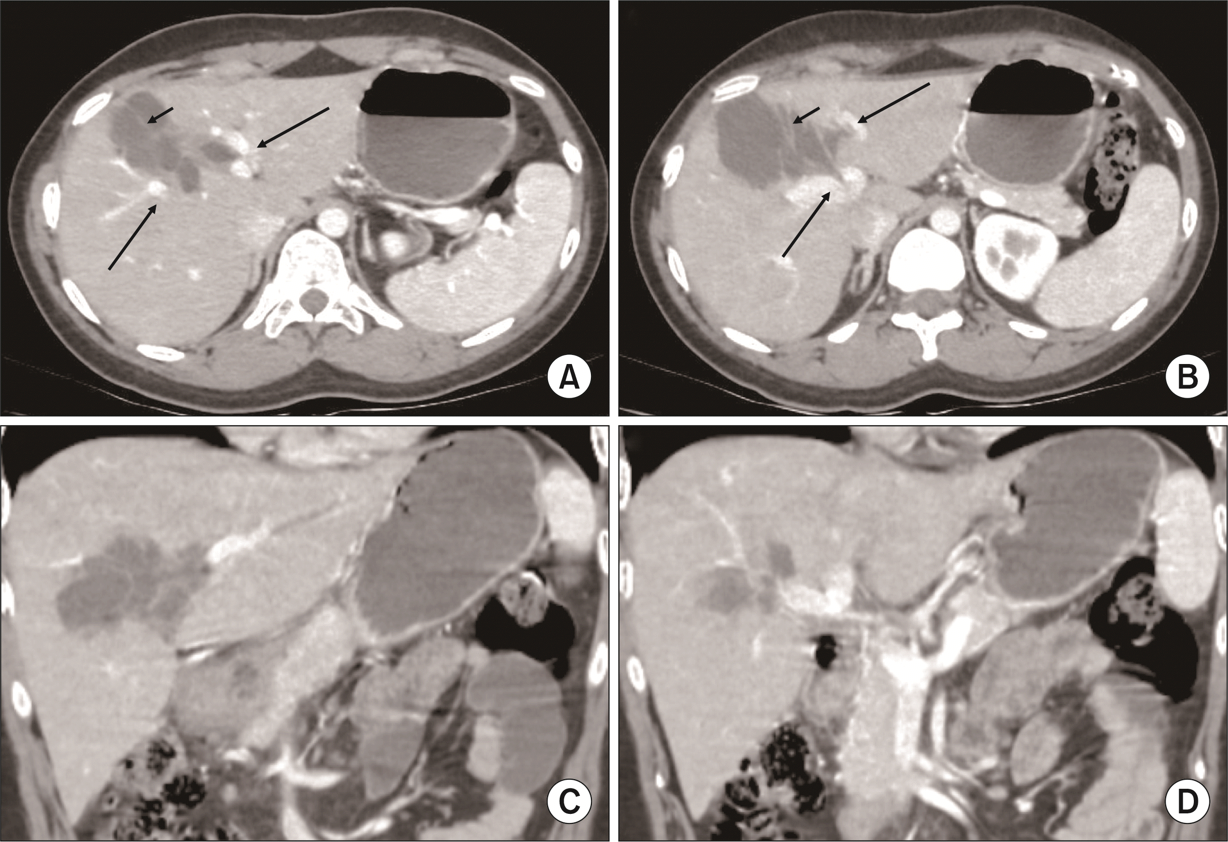 | Fig. 1Contrast enhanced computed tomography images. (A) Multiloculated cyst involving segment IVa and V of the liver with enhancing internal septa (short arrow), the cyst is in close proximity to the tributaries of the portal vein just above the confluence (long arrows). (B) At the level of the portal confluence (long arrows), cyst is seen extending into the segment IVb of the liver with multiple internal septations (short arrow). There are no other cystic lesions in the rest of the liver and other solid organs such as the kidney, pancreas and spleen. (C) Coronal image of the cyst anterior to the hilum in relation to the left portal pedicle. (D) Coronal image of the cyst near the hilum.
|
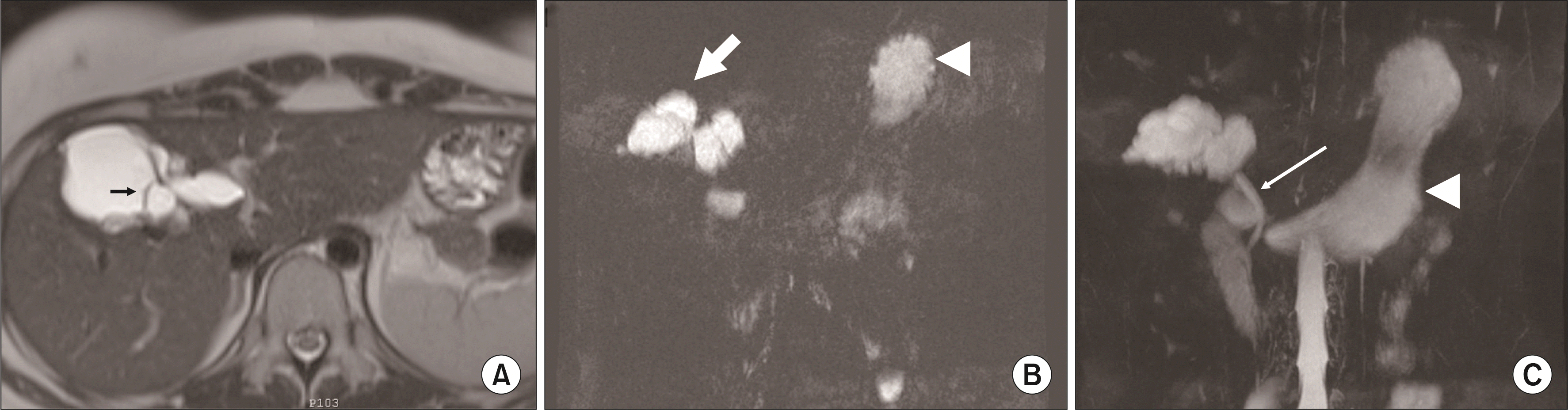 | Fig. 2Magnetic resonance images. (A) T2-weighted image showing a hyper-intense multilocular cyst with internal septa (short arrow). There is no biliary dilatation in the rest of the liver parenchyma. (B) Single-shot fast spin-echo sequence showing the bilobed butterfly like cystic structure (thick arrow), also seen is the fundic shadow (arrowhead). (C) Undilated distal common bile duct (long arrow), gastric shadow (arrowhead).
|
The patient was taken up for surgical excision after a written informed consent. On laparoscopy, there was a cyst in segments IV and V of the liver. The gallbladder was distended, and the rest of the liver parenchyma was normal (Fig. 3). There was a cleft in the liver from the gallbladder bed till the cyst. A trial dissection in an enucleation plane was performed. However, in view of dense adhesions of the cyst with the liver parenchyma, an oozy plane and slow progress of dissection, a decision was made to convert to an open procedure. After mobilization of the cystic structure, an intraoperative cholangiogram was done through the cystic duct (Fig. 3). Real-time visualization under fluoroscopy revealed a butterfly-like bilobed cystic structure filling up with contrast simultaneously from the right and left hepatic ducts. The peripheral ducts were not visualized.
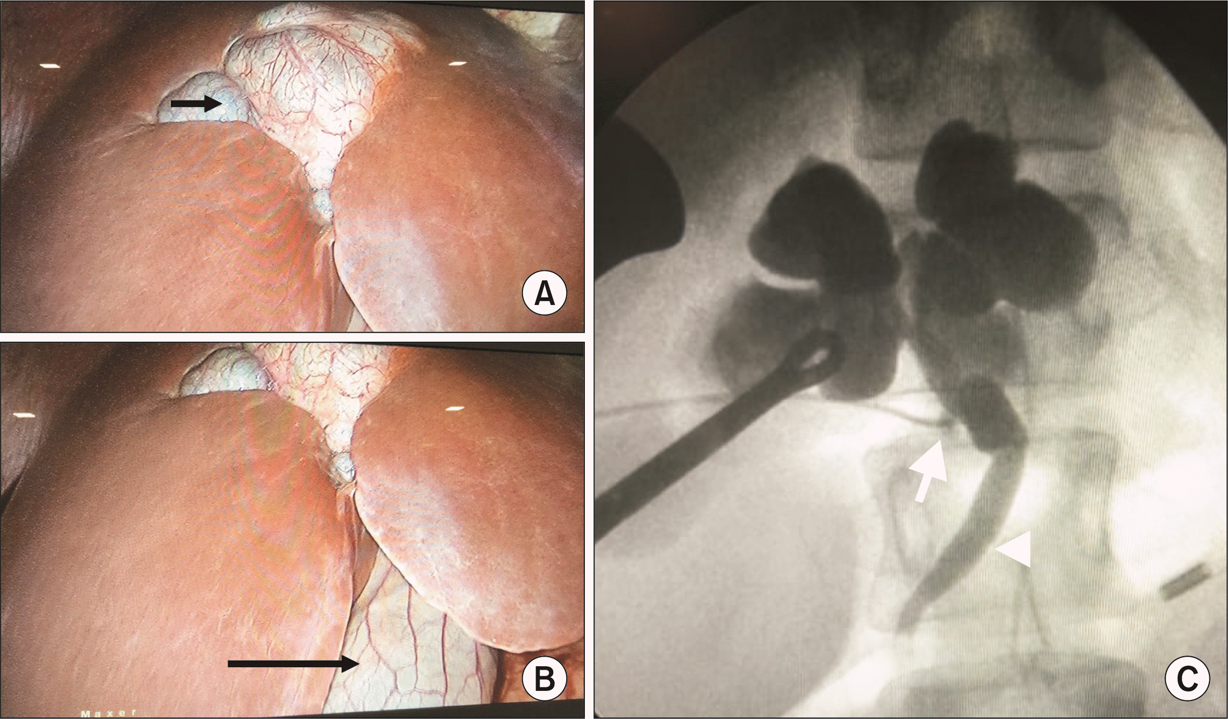 | Fig. 3(A) Laparoscopic image of the cyst (short arrow). (B) Gallbladder (long arrow) in relation to the cyst. Note a cleft in the liver at the region of the cyst. (C) Intraoperative cholangiogram done through the cystic duct (arrow) after cholecystectomy showing a bilobed structure in communication with the biliary system (arrowhead). The ducts distal to the cysts were not visualized.
|
The location of biliary communication of the cyst could not be discerned with certainty. Dense pericystic inflammation, particularly near the hilar plate, made it difficult to dissect around to get to the communication with the biliary system. Any attempt at dissection to achieve complete excision was fraught with risk of injury to the right hepatic artery and the biliary bifurcation. This could have subsequently warranted two separate anastomosis to the undilated right and the left hepatic ducts. Therefore, a decision was made to leave a 4-mm rim of the cyst wall at the base of the cyst to enable drainage into a Roux limb of jejunum. The lack of any mural nodules or solid component on preoperative CT imaging enabled the decision to drain than risk a biliary injury during resection. On opening the bilobed cystic structure, clear bile was seen with no mucin. There were multiple small pigmented stones of 2–3 mm with sludge. The cyst was trimmed down, leaving a 4-mm rim near the suspected biliary communication. An isolated Roux limb of the jejunum was anastomosed to this rim of the cyst. The patient made an uneventful recovery. She was started on an oral diet on the second postoperative day and was discharged by seventh postoperative day.
The pathology of the cyst wall showed a lining of cuboidal biliary-type epithelium with fibrous stroma. There were no ovarian stroma, dysplasia, or malignant cells. Thus, a diagnosis of a diverticular variant of a choledochal cyst was made. At one-year follow-up, the patient is asymptomatic with normal liver function tests and follow-up CT and magnetic resonance cholangiopancreatography evaluation were essentially normal (Fig. 4).
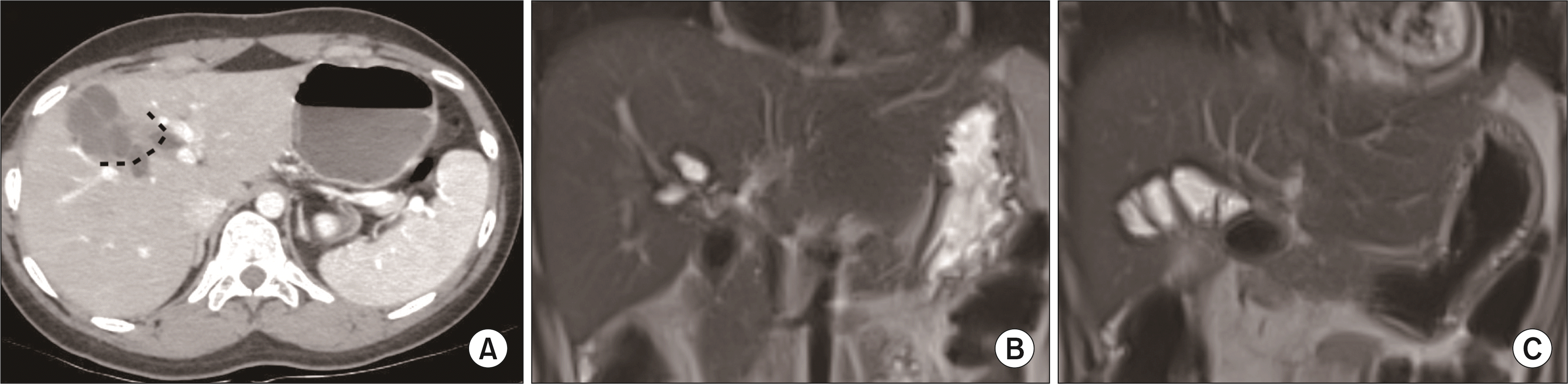 | Fig. 4(A) Representation of the extent of resection of the cyst on the computed tomography image (dashed lines). (B) Coronal image of the postoperative magnetic resonance imaging (MRI) shows the residual cystic structure near the hilum. (C) Coronal image of the postoperative MRI shows the cystojejunostomy.
|
Go to :
DISCUSSION
Cystic lesions of the liver are commonly encountered in routine clinical practice with a reported prevalence of 15%–18% [2]. They may range from a benign simple developmental cyst to a malignancy. Therefore, an accurate diagnosis is essential for adequate management. The varied pathologies may make the diagnosis difficult. An adequate clinical history may provide a clue to the diagnosis in cases of infectious, inflammatory, and traumatic cysts. Evaluation with ultrasound of the abdomen and radiological studies such as CT and magnetic resonance imaging to look for the number, location, loculations, contents, solid nodules, status of the biliary radicles, and presence of concomitant cysts in other solid organs such as the kidney and spleen clinches the diagnosis in most of the cases.
Cystic tumors of the liver are classified based on the content (mucin containing or not), presence of ovarian stroma, and biliary communication. Biliary cystadenoma are a group of hepatobiliary neoplasia which by definition must be multilocular, lined by a columnar epithelium, and have a densely cellular ovarian stroma [3]. This definition was later revised to include unilocular tumors with cuboidal epithelial lining not containing ovarian stroma [4]. The current understanding of cystic tumors is based on the biliary communication and ovarian stroma. Those with ovarian stroma and lack biliary communication are classified as mucinous cystic neoplasms, whereas those communicating with the biliary system without ovarian stroma are classified as intraductal papillary mucinous neoplasms (IPMN-B). IPMN-B may present with tubular or fusiform dilatations of the involved ducts or as cystic structures known as the cyst-forming IPMN-B. Dilatation of the proximal and the distal biliary system is a hallmark of IPMN-B.
In this case, the multiloculated cyst with enhancing wall and the septa in the absence of biliary dilatation made us consider mucinous cystic neoplasm as the preoperative diagnosis. The surgical plan with this diagnosis was enucleation of the cyst. The mobilization revealed a bilobed butterfly-like cystic lesion. Intraoperative cholangiogram showed a communication with the biliary system without upstream and downstream biliary dilatation. Given the location of the lesion just anterior to the biliary bifurcation along with dense peri-cystic adhesions near the hilar plate and the absence of biliary dilatation, a decision was made to open the cyst. With bile and no mucin draining from the cyst, the diagnosis was narrowed to a diverticular variant of type V choledochal cyst communicating with both hepatic ducts near the hilum. Pathology of the cyst wall confirmed the above.
The management of type V choledochal cyst depends on the distribution of the cysts [5]. Cysts which are localized to one segment or hemi-liver could be managed with liver resections. However, the management of bilobar, diffuse cysts is problematic. Here, we encountered a cyst in segment IV, V abutting the hilar plate with risk of injury to the critical hilar structures such as the portal vein branches, hepatic artery, and the biliary radicles. Cyst excision with drainage by a Roux-en-Y cystojejunostomy provided the safer option for completion of the procedure as compared to a total cyst excision and a Roux-en-Y hepaticojejunostomy in undilated ductal systems. Prudence dictates a close long-term follow-up with liver function tests and imaging in such patients.
In conclusion, type V choledochal cysts are rare. We report a rare variant of type V choledochal cyst arising from both hepatic ducts. Cysts at the hilar region in proximity to the biliary bifurcation are better managed with a near total cyst excision and bilio-enteric anastomosis.
Go to :
 XML Download