

 PDF
PDF Citation
 Print
Print


INTRODUCTION
Todani type II choledochal cysts are bile duct diverticula, diagnosed in 60% of children under 10 years of age [1]. The etiology of these cysts is still debated, with the most widely recognized one argued by Babbitt [2]. According to his theory, choledochal cysts are caused by an anomalous pancreaticobiliary ductal junction, found in 50% to 80% of cases of choledochal cysts [2,3]. Other hypotheses, according to which the abnormality is due to the occurrence of pancreatic fluid reflux into the common bile duct (CBD), include the obstruction and the congenital presence of a few ganglion cells in distal CBD, as well as the dysfunction of the sphincter of Oddi [4-6]. In the Western population, the reported incidence of choledochal cysts is 1 case per 100,000 to 150,000 live births [7] and is variable around the world. For example, it is higher in the United States (1 in 13,500) than in Australia (1 in 15,000) [7], while the country with the highest number of registered cases is Japan, with an incidence of 1 per 1,000, which alone accounts for about two-thirds of all the described cases [8]. This malformation has been reported particularly in males compared with females, on the order of 4 : 1 to 3 : 1, except for the Asian population, where it has a higher prevalence among females [1,9]. Most studies describing biliary malformations are dated, but because of the introduction of current imaging methods, such as magnetic resonance cholangiopancreatography (MRCP), the diagnosis of these malformations is on the rise, so we can expect to see an increase in their prevalence. Herein, we present the case of a 53-year-old female with a Todani type II choledochal cyst, associated with calculous cholecystitis, who was successfully treated by embedding the laparoscopic resection.
Go to : 
CASE
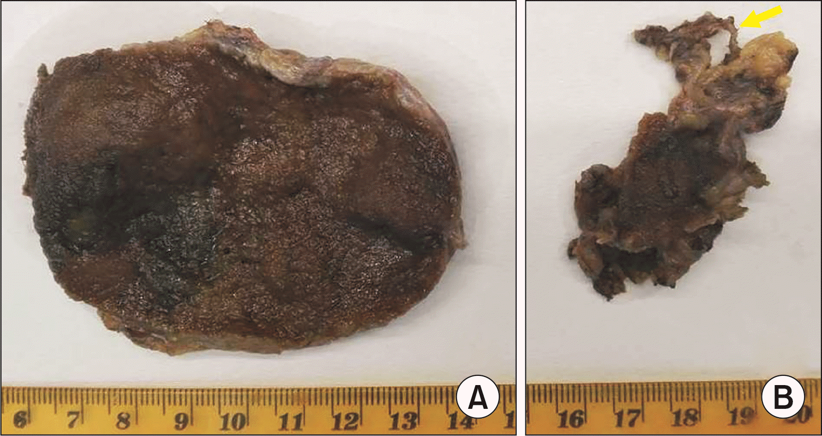
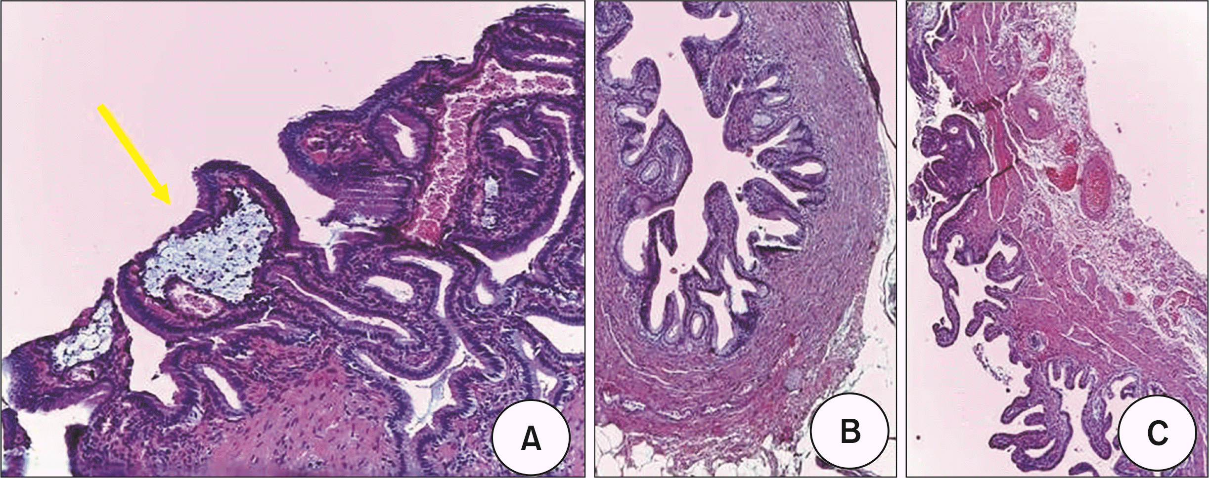
A 53-year-old female was admitted to the emergency room (ER) for pain localized in the right hypochondrium. On admission, her temperature was 37.8°C, blood pressure 125/85 mmHg, pulse rate 75/minute, and respiratory rate 15/minute. Increased white blood cell count (15.94/mm3; neutrophils 90.9%), hepatocellular necrosis (alanine aminotransferase, 412 U/L and aspartate aminotransferases, 401 U/L), and cholestasis (serum bilirubin, 3.2 mg/dL) were present. Physical examination showed pain on palpation in the right hypochondrium. The patient reported that she had been suffering from biliary colic for about 2 years, with ever-closer episodes. An ultrasound performed in the ER revealed multiple microcalculations in the gallbladder lumen, associated with thickened walls (Fig. 1). Given the persistence of the symptoms, and to better investigate them, an MRCP was performed. This examination confirmed the ultrasound finding and showed the presence of a diverticulum of about 3 cm of the CBD (Fig. 1). A diagnosis of a Todani type II choledochal cyst associated with a lithiasic cholecystitis was made. A surgical approach was suggested and was accepted by the patient. After giving informed consent for the surgical procedure, the patient underwent laparoscopic cholecystectomy and simultaneous excision of the diverticular formation of the choledochus and its entire pedicle. The patient also underwent excision of a lozenge of the CBD with a transversal reconstruction of the biliary breach using an absorbable monofilament (Fig. 2). Since the extemporaneous histological examination of the diverticulum collar and the biliary lozenge was negative for the presence of atypical or neoplastic cells, biliodigestive drainage was not performed. The final anatomopathological and histological results showed a Todani type II malformation with normal biliary epithelium and chronic cholecystitis with cholesterol deposits (Fig. 3, 4). The postoperative course was uneventful, and the patient was discharged 7 days later in good condition. After more than 2 years of follow-up with annual imaging and blood tests, she continues to be asymptomatic.
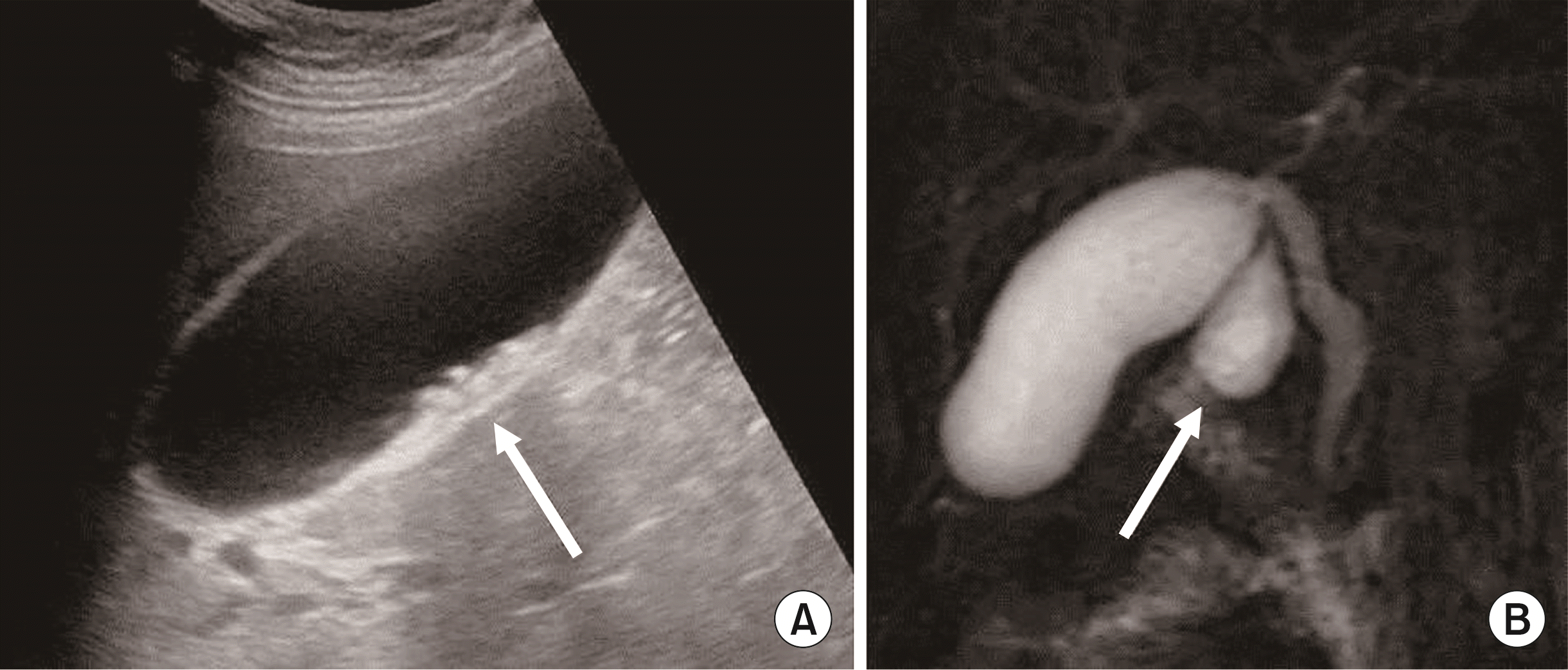 | Fig. 1Preoperative images. Microcalculations in the gallbladder lumen (arrow), associated with biliary sludge on ultrasound (A), and a Todani type II (arrow) with a 2-cm-wide peduncle, with relative insertion below the cystic duct on resonance (B).
|
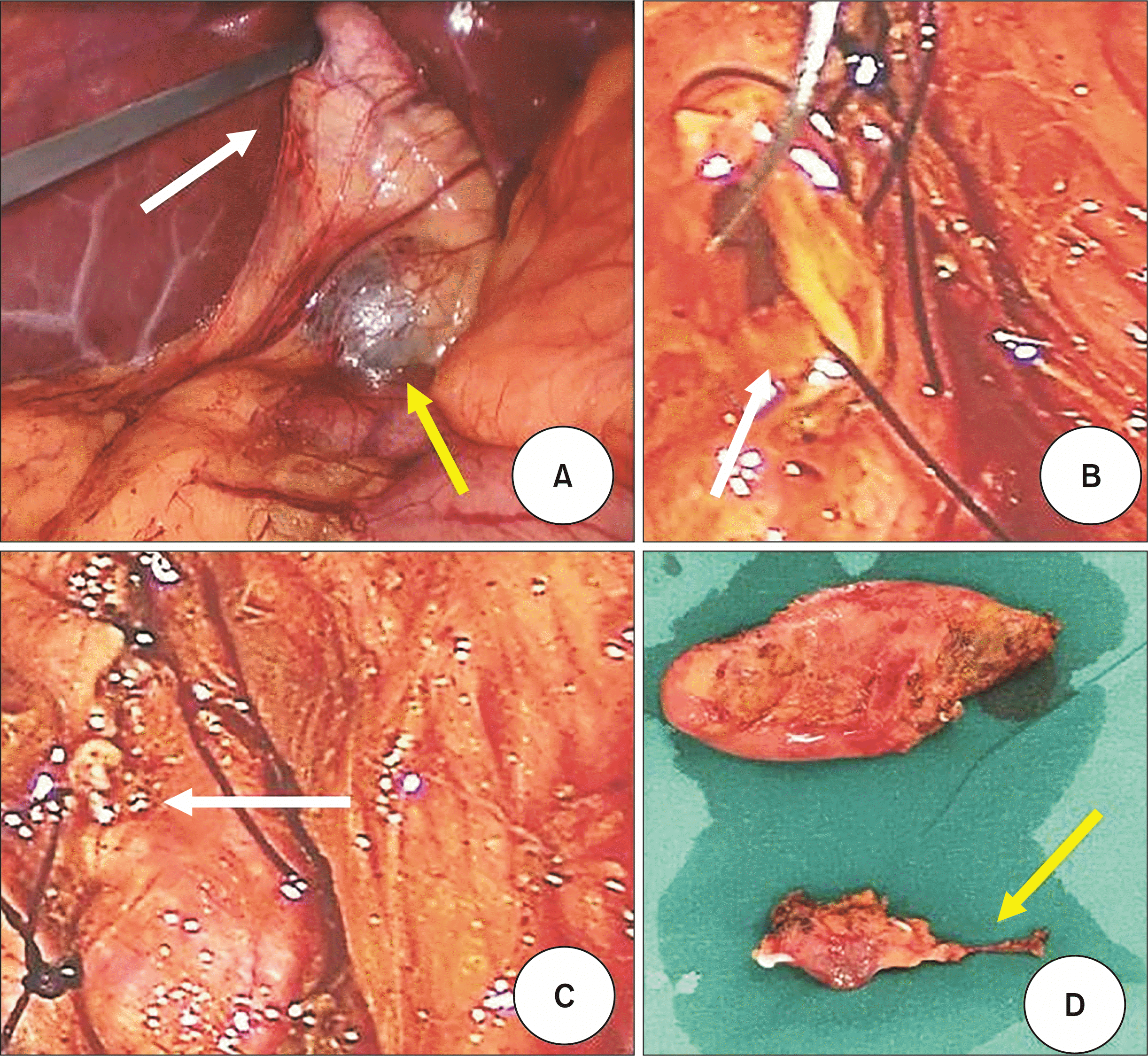 | Fig. 2Intraoperative images. Gallbladder (white arrow) and Todani type II cyst (yellow arrow) on laparoscopic view (A); closure of the lozenge of the main bile channel with absorbable monofilament (white arrows) (B, C); anatomical structures just excised, gallbladder at the top and cyst at the bottom (D), where the diverticulum’s peduncle (yellow arrow) ending with the biliary lozenge can be seen.
|
Go to :
DISCUSSION
The first classification of biliary congenital anomalies, by Alonso-Lej et al. [10], dates back to 1959. In 1977, Todani et al. [11] modified this categorization, making it currently the most used and recognized categorization. Choledochal diverticula (named Todani type II) are extrahepatic supraduodenal outpouchings of the CBD lined with biliary epithelium [1]. These represent a very rare finding, with an incidence of less than 1/1,000,000 and account for 2% of all the types of choledochal malformation [1,12]. Oftentimes, the diagnosis is difficult because this kind of diverticula histologically is similar to gallbladder duplications [1,12]. As evidenced by a systematic review including all the articles on this topic from 1990 to 2017, this type of malformation was confused with multiple gallbladders, and the correct diagnosis of Todani type II was made only intraoperatively [13]. As in our case, the correct definition of the biliary malformation (Todani type II) was made intraoperatively due to the absence of an anomalous right hepatic artery or abnormal right posterior sectoral hepatic duct [14].
The clinical presentation is consistent with the existing literature showing that the main symptoms in adults, whether presenting singly or in association, are pain, nausea, vomiting, fever, and jaundice [15]. The main complaint of our patient was abdominal pain located in the right hypochondrium.
There is an increased risk of cancer among people who have biliary cysts, particularly for cholangiocarcinoma but also for pancreatic and gallbladder neoplasms [16]. Patients with type I and type IV cysts, according to the Todani classification, are more likely to develop cancer [11,16]. Whenever these types of cysts are found, the recommended treatment is complete excision [17]. The laparoscopic surgical approach for the treatment of a Todani type II choledochal malformation is quite recent. Generally, it is characterized by a shorter hospitalization time than the open intervention, in which it can always be reconverted in case of intraoperative complications. To the best of our knowledge, Liu et al. [18] described the first case report regarding this technique in 2000. However, it is necessary to underline that the disadvantages of laparoscopy may include incomplete cyst excision and potential abdominal dissemination in case there is neoplastic degeneration [19]. Choledochal diverticula (type II) are excised prophylactically to prevent sequelae of mass effect on adjoining structures despite their low malignant potential [20]. As a precaution, in our case, we removed the diverticulum in its entirety, including its peduncle and part of the main biliary tract insertion site, without finding either intra- or postoperative cellular atypia or neoplasms on histological examination. Given the low prevalence of this disease, there is no appropriate follow-up for operated patients. Reading the literature, a longer follow-up of 24 months after laparoscopic resection of a type II cyst, with no alteration of hepatobiliary function, is described [21]. In our case, the patient is followed up every year by complete serum liver tests and by a noninvasive imaging study, specifically for MRCP associated with abdominal magnetic resonance imaging with intravenous contrast.
In conclusion, Todani type II cyst is a rare disease that is difficult to diagnose before surgery, despite availability of imaging studies. Although the likelihood of associated neoplastic transformation is low, complete removal of the diverticulum is imperative. The most recent laparoscopic approach offers both a full field of view during the surgery and a shorter recovery period.
Go to :
 XML Download