

 PDF
PDF Citation
 Print
Print


INTRODUCTION
Small bowel obstruction is a common presentation of internal hernias, and accounts for 4.1% of all intestinal obstructions. Transomental hernia (TOH) is rare, constituting 1-4% of all internal hernia.1-3 TOH often occurs due to trauma, surgery, or peritoneal inflammation. however, it can occur spontaneously due to senile atrophy of the omentum. The postoperative mortality rate of TOH is high. Therefore, early diagnosis and treatment are important.4,5 Herein, we report a case of a spontaneous TOH presenting with small bowel obstruction in a patient without history of any surgery, trauma, or peritoneal inflammation.
CASE REPORT
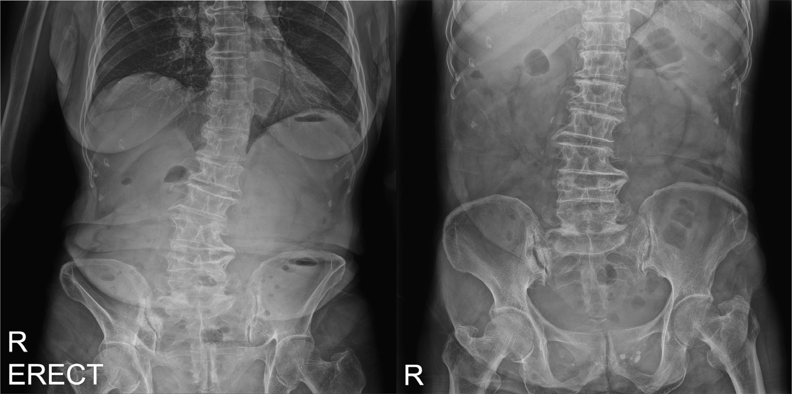
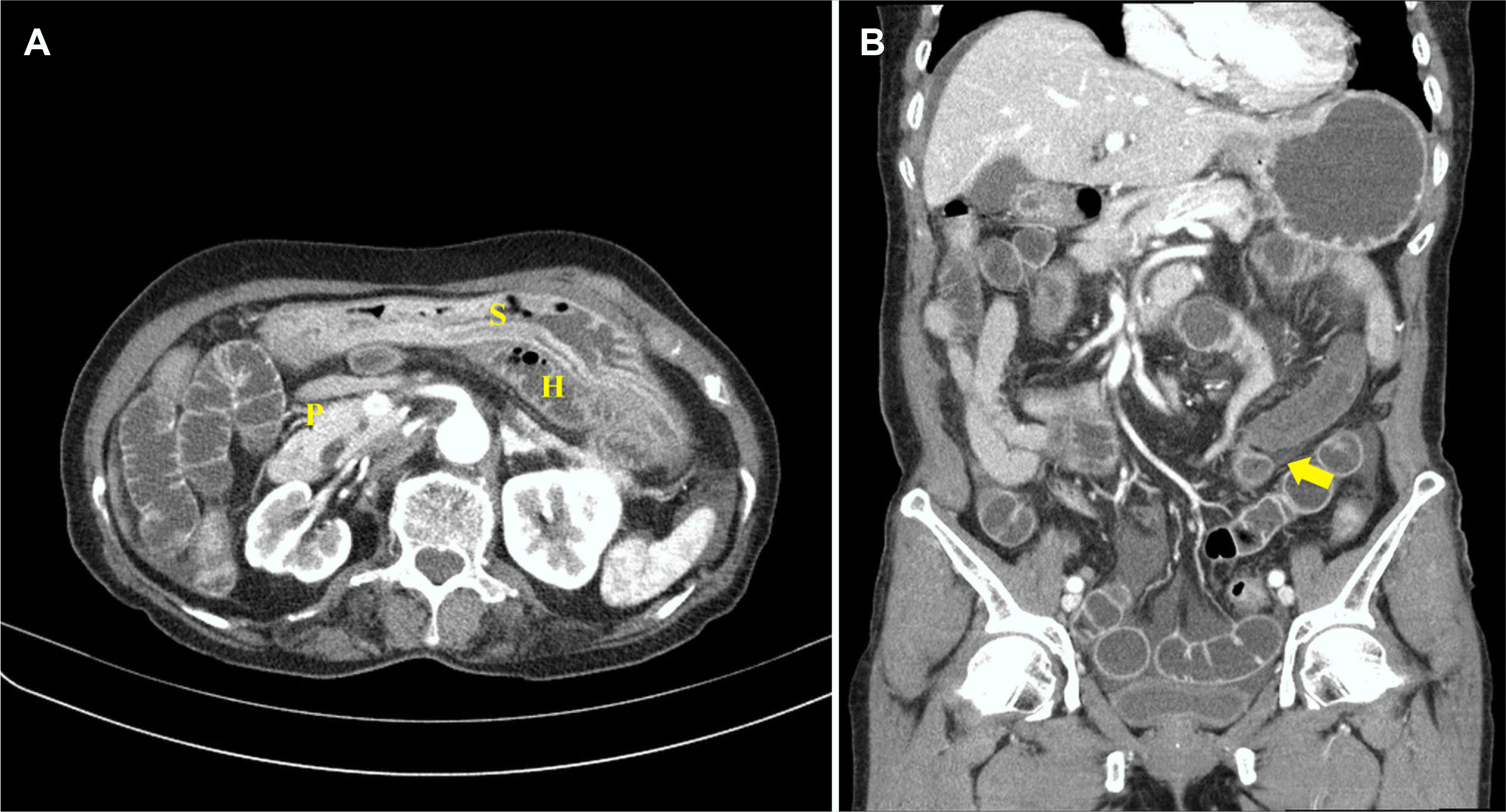
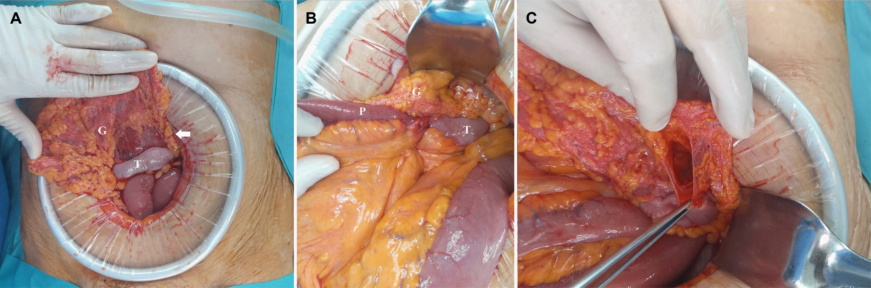
This study was approved by the Institutional Review Board (IRB) of Wonkwang University Hospital (IRB No. WKUH 2022-04-026) and informed consent was waived. A 77-year-old female visited emergency department of Wonkwang University Hospital with hematochezia for the previous 1 day. She also complained of a mild, dull and intermittent abdominal pain. The vital signs were stable. She had a history of hypertension, diabetes mellitus, and thyroid surgery, but no history of abdominal surgery, peritoneal inflammation, or trauma. Abdominal physical examination demonstrated direct tenderness in left upper and lower quadrant, but no rebound tenderness. Since abdominal pain and hematochezia appeared as first symptoms, additional tests were performed under suspicion of bleeding due to ischemic colitis or diverticulitis initially. The laboratory test results showed: hemoglobin, 15.7 g/dL; white blood cell count, 11.61×10³/mm3; and CRP, 23.24 mg/L, with other parameters within normal limits. Abdominal X-ray showed no abnormal findings (Fig. 1). Abdomen-pelvis CT revealed dilated small bowel loops located within the lesser sac, and a beak sign at the suspected hernia orifice located in the left lower quadrant. The herniated small bowel loop showed ischemic changes, and some fluid was also present in the peritoneal cavity (Fig. 2). On the CT findings, small bowel obstruction caused by internal hernia and small bowel ischemia were suspected, so open exploration was performed immediately. During the operation, approximately 70 cm of the small bowel loop was found herniated into the lesser sac through a 2 cm defect in the greater omentum (GO), 50 cm distal to the ileocolic valve, and partial necrosis of the small bowel was also observed (Fig. 3). TOH was finally diagnosed base on the surgical findings. We performed a gentle reduction of the herniated small bowel and resection of the necrosed portion. A side-to-side anastomosis was performed using staplers. The patient had a delayed recovery due to postoperative ileus, but was discharged without other complications.
DISCUSSION
An internal hernia is a protrusion of visceral contents through a defect in the mesentery or peritoneum, within the abdominal cavity. The most common type of internal hernia is the paraduodenal hernia (53%), followed by the pericecal hernia (13%) and foramen of Winslow hernia (8%). TOH is exceptionally rare, accounting for approximately 1-4% of all internal hernias.1,6
TOH is usually reported in patients above 50 years of age. In adults, it is commonly iatrogenic, mostly due to a Roux-en-Y anastomosis after gastric surgery. Less often, it results from a blunt trauma or peritoneal inflammation. However, it can occur spontaneously and probably due to senile atrophy of the omentum.3,4,7 In children, TOH can occur in congenital defects of the omentum and is seen more commonly in males. In TOH, incarceration may depend on the defect size and length of the herniated bowel loop.4,5,8 Since our patient was elderly and without history of any surgery, trauma, or peritoneal inflammation, the TOH was most probably spontaneous, due to senile atrophy of the GO.
Yamaguchi9 classified TOH into type A, B, and C according to the path of the herniated intestine. Type A is an internal hernia through a defect in the fused part of the GO. Type B refers to a case in which the bowel prolapses through a defect in the GO, via the omental bursa, and toward the opposite defect site in the GO. Regarding type C, it is divided into three subtypes: C0 is the intestine herniating into the omental bursa through a defect in the GO, C1 is when the herniated bowel of C0 prolapses into the peritoneal cavity via the Winslow’s pouch, and C2 is via the lesser omentum instead of the Winslow’s pouch in C1. In our case, the hernia corresponds to type C0 because the small bowel herniated into the lesser sac through the defect in the GO.
According to a study that analyzed 188 patients with TOH by Tsuchida et al.10 in Japan, type A occurred more in elderly patients than type C did (60.0 vs. 42.1 years) and was slightly more common than type C (107 vs. 70). Only 6.9% of cases were diagnosed by preoperative examination, and 38.8% of cases (73) underwent bowel resection. They also reported that patients who underwent bowel resection for TOH had a tendency to be elderly, have a narrow hernia orifice, and a long-herniated intestine. Although statistical significance was not shown, it is meaningful in that an earlier diagnosis and treatment can reduce the possibility of intestinal resection when TOH is suspected in elderly patients.
Preoperative diagnosis of TOH is difficult because of symptoms similar to that of acute small bowel obstruction due to other causes, including abdominal pain, abdominal distention, nausea, vomiting, and constipation. TOH does not have a hernia sac, and the orifice size is small; therefore, it tends to be strangulated more often than other types of internal hernias, and the postoperative mortality rate is as high as 30%. Therefore, early diagnosis and management are important.7,11,12
CT plays an important role in the diagnosis of TOH. CT scan may show a dilated bowel loop with a “beak sign”, which is a triangle shaped transition zone between the dilated proximal loop and herniated loop, or between the herniated, dilated loop and collapsed distal loop. In addition, it may reveal the “whirl sign” of engorged mesenteric vessels, accompanied by bowel wall thickening. TOH might also be suspected when dilated bowel loops are located within the lesser sac. However, in most cases, the diagnosis is made definitively during laparotomy.7,13,14
Surgical treatment is similar to that for other types of internal hernias. The herniated bowel loops are reduced; and if irreversible ischemia, necrosis, or perforation of the herniated bowel is observed, resection of the bowel is performed. The hernial orifice must be closed to prevent recurrence. Laparoscopic surgery has recently been used to treat small bowel obstructions. Therefore, if an appropriate view of the surgical field is secured, it can be helpful in diagnosing and treating TOH early with minimal invasiveness.4,6,12,15
In conclusion, spontaneous TOH is a rare disease that is difficult to diagnose before surgery because of its non-specific symptoms. Therefore, when the specific findings are suspected in radiologic examinations, diagnosis and treatment using a rapid surgical approach can reduce the incidence of complications.
 XML Download