

 PDF
PDF Citation
 Print
Print


INTRODUCTION
Skeletal Class III malocclusions can present with various combinations of skeletal and dental discrepancies.1 Non-growing patients with a skeletal Class III malocclusion can either be treated with a combination of orthodontic and orthognathic surgery or receive orthodontic camouflage treatment, depending on the severity of the malocclusion. For mild-to-moderate skeletal Class III malocclusions, various camouflage treatment protocols have been proposed, including using fixed appliances with Class III intermaxillary elastics,2,3 incorporating the multiloop edgewise archwire technique,4 and using temporary anchorage devices.5,6 However, when the discrepancy is beyond the limit of dental compensation, orthognathic surgery may be the only option for achieving normal occlusion.
Stellzig-Eisenhauer et al.7 suggested that the vertical dimension is an important factor when determining whether Class III malocclusion should be corrected by camouflage treatment or surgery. A reduced lower anterior facial height, deep overbite, or passive lip seal can predict a better prognosis in the treatment of a Class III malocclusion, because clockwise rotation of the mandible can assist in camouflaging the anteroposterior discrepancy.8 Thus, the range of camouflage treatment could be expanded in hypodivergent Class III patients. In this regard, Hisano et al.9 reported two cases of adult patients with skeletal Class III malocclusions and deep bites with predisposing upper incisor problems, including root resorption and severe mobility, who were treated using a monoblock appliance in combination with vertical elastics that reduced the overbite and avoided excessive force on the incisor by controlling the vertical relationship.
The risk–benefit factor should also be considered when selecting between surgery or camouflage treatment. Treatments, including invasive procedures, should be carefully planned or modified for specific medical problems such as bleeding and immune disorders. Aplastic anemia is a disease in which the body fails to produce sufficient numbers of red blood cells, white blood cells, and platelets. A low platelet count is associated with an increased risk of bleeding, while a low white blood cell count can increase the risk of infection.
This case report presents orthodontic camouflage treatment for patient with a severe skeletal Class III malocclusion and aplastic anemia. Backward rotation of the mandible by orthodontic extrusion of the posterior teeth improved the maxillomandibular anteroposterior relationship and facial esthetics. The dentoskeletal and soft-tissue effects of the camouflage treatment and its long-term stability are discussed.
DIAGNOSIS AND ETIOLOGY
A 19.5-year-old man visited the Department of Orthodontics, Gangneung-Wonju National University Dental Hospital with a complaint of an anterior crossbite; he was willing to undergo surgery if necessary. The patient had a medical history of aplastic anemia and had received annual blood tests for six years; however, he did not take any medication. His family history revealed the presence of a sister with mandibular prognathism.
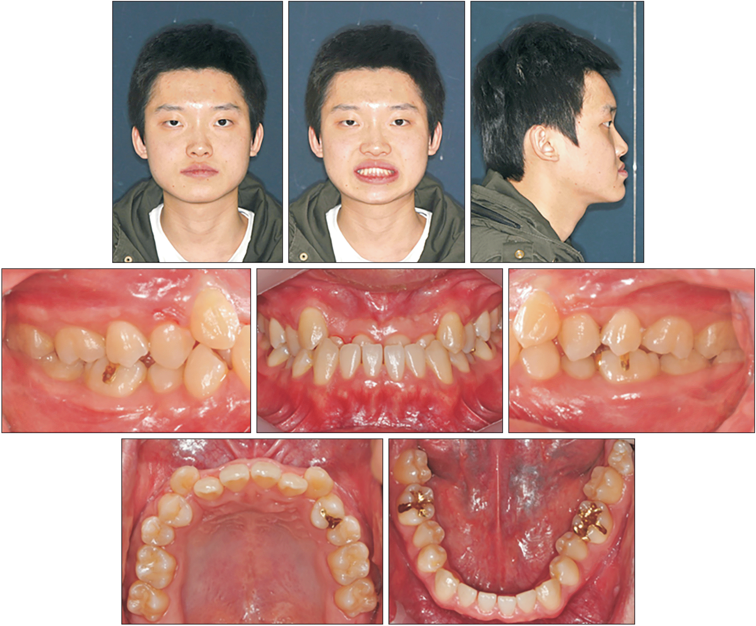
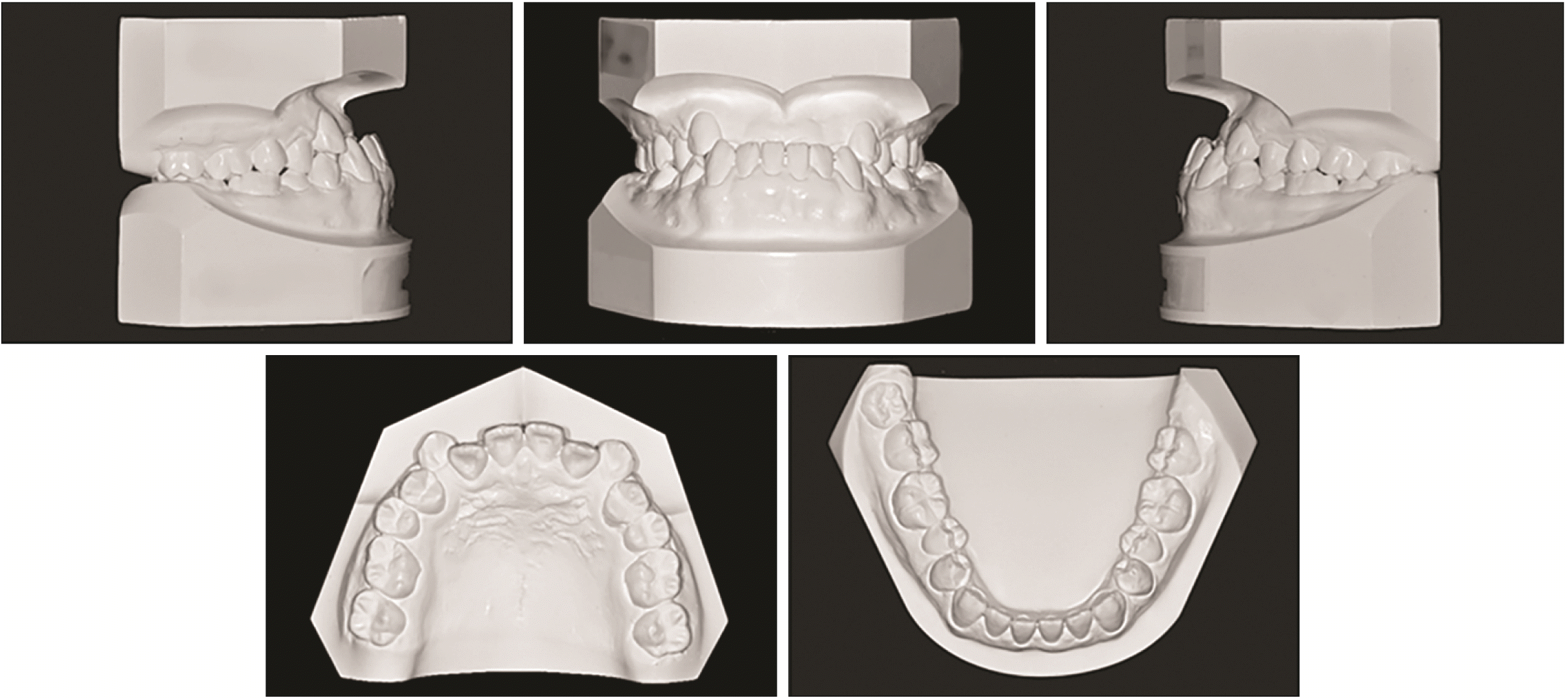
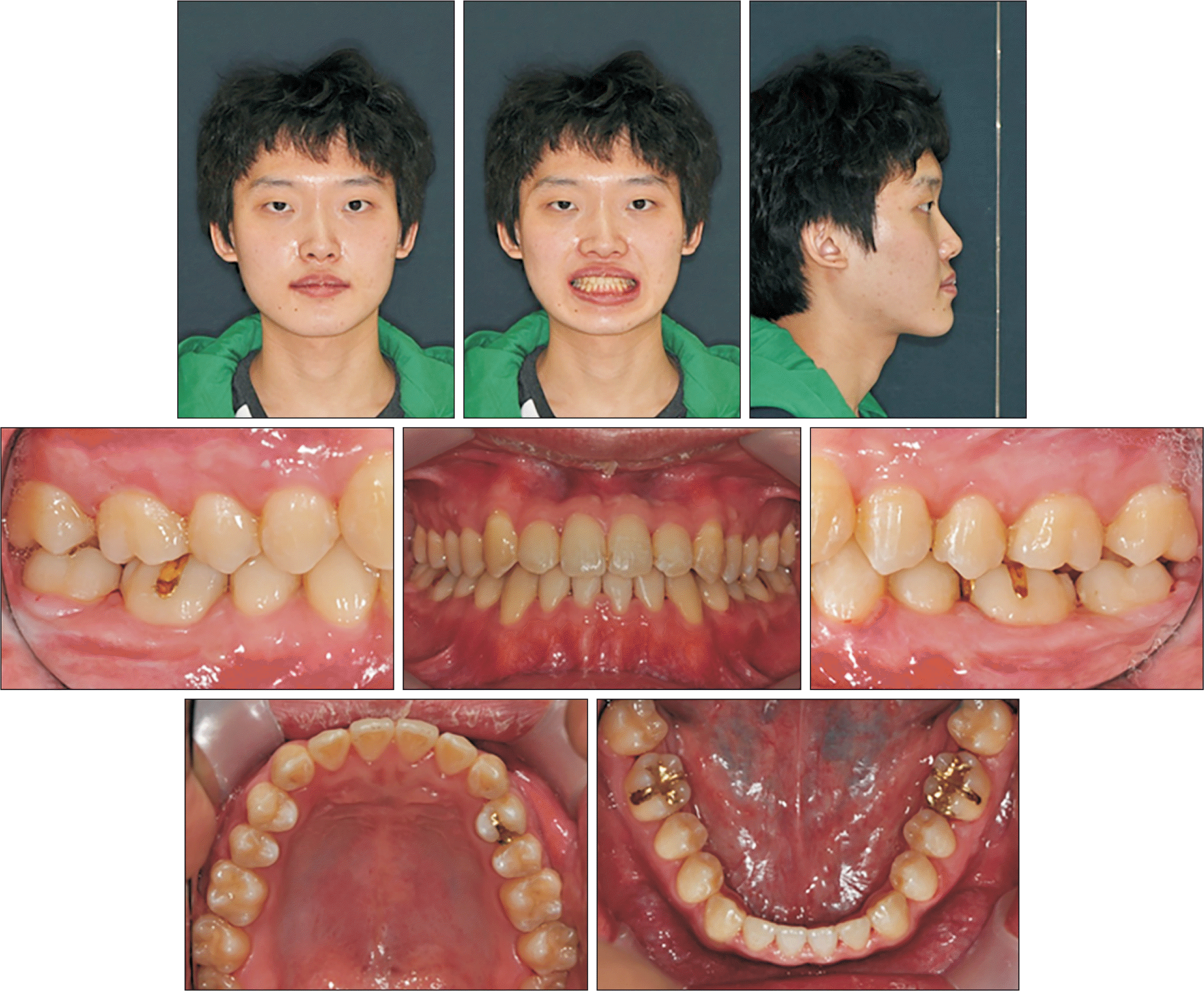
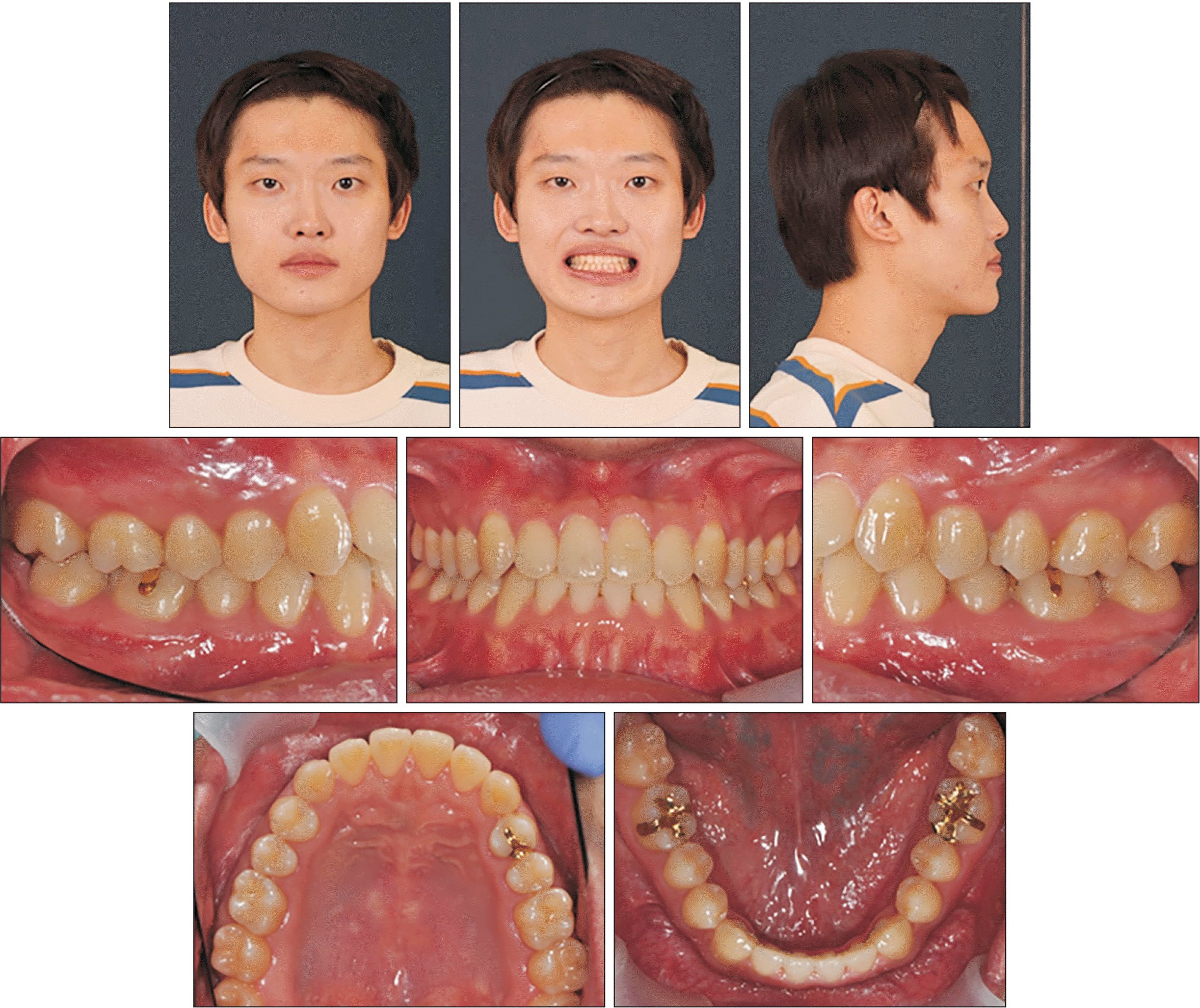
The patient presented with a severe concave facial profile with a prognathic mandible and brachyfacial type. He exhibited slight mandibular asymmetry, and his anterior crossbite caused an unsatisfactory smile. An acute nasolabial angle and lip redundancy were observed because of overclosure of the mandible (Figure 1). Intraoral examination revealed a Class III molar relationship, an anterior crossbite (overjet, –5.2 mm), and a deep bite (overbite, 9.8 mm). The maxillary arch exhibited moderate crowding and supraversion of the maxillary incisors. The maxillary dental midline was coincident with the facial midline, but the mandibular dental midline deviated to the left by 1 mm (Figures 1 and 2).
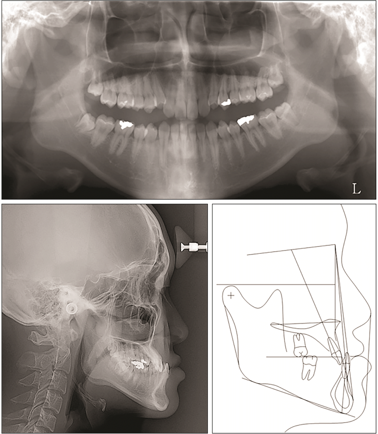
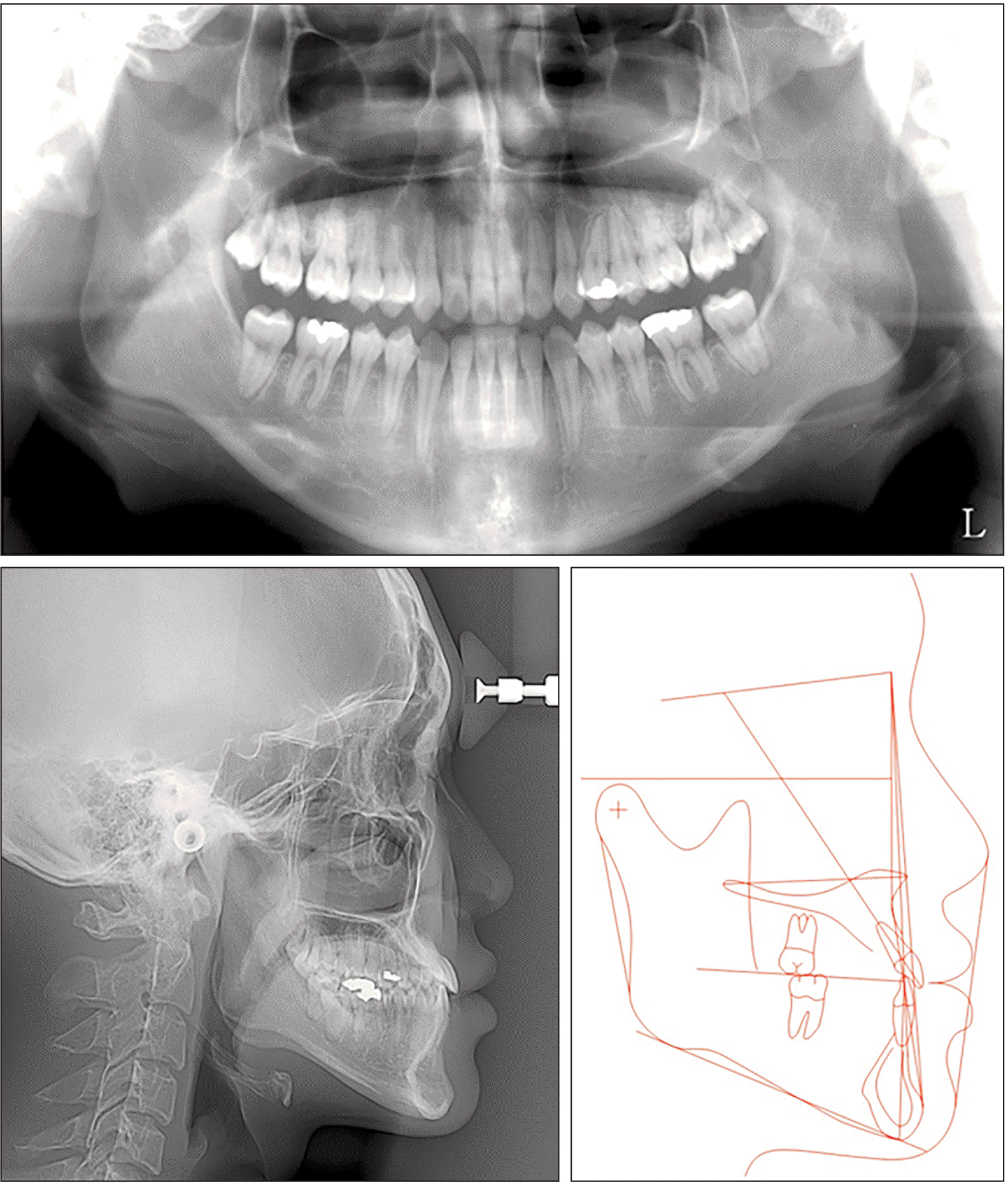
A panoramic radiograph showed four developing third molars. Lateral cephalometric analysis indicated severe maxillomandibular discrepancy (A point-nasion-B point [ANB], –5.0°) and a hypodivergent growth pattern with a mandibular plane angle (gonion-menton [Go-Me] to Frankfort horizontal plane [FH plane]) of 19.1°. The mandibular incisors showed linguoversion, whereas the maxillary incisors showed normal labiolingual inclination (Figure 3 and Table 1). There was no discrepancy between the centric occlusion (CO) and the maximal intercuspal position (MI).
Informed consent was obtained form the patient for release of the patient’s records. This case report was approved by the ethics committee of Gangneung-Wonju National University Dental Hospital (GWNUDH-IRB2020-A002).
TREATMENT OBJECTIVES
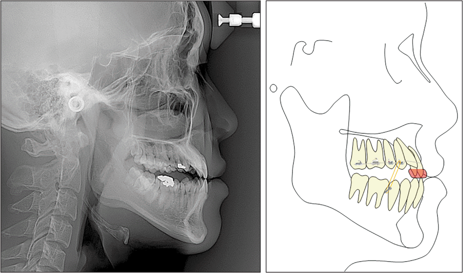
The treatment goal was to establish normal occlusion and improve the facial profile. A camouflage treatment plan was established in consideration of the patient’s medical problems. An additional lateral cephalometric radiograph was performed in an edge-to-edge incisal position to simulate the effects that backward and downward rotation of the mandible would have for reducing the anteroposterior discrepancy and improving the patient’s facial profile. It was confirmed that clockwise rotation of the mandible might improve the anteroposterior skeletal relationship (ANB, from –5.0° to –0.6°) and lip profile (nasolabial angle, from 81.2° to 105.9°) (Figure 4).
The treatment plan included orthodontic extrusion of the maxillary posterior teeth using vertical intermaxillary elastics and a removable anterior bite plate to maintain an intentionally increased vertical dimension (Figure 4). The preadjusted edgewise appliances with 0.022 × 0.028-inch slots would be bonded at only the maxillary posterior teeth. The leveling and alignment of both dental arches would be delayed until after the extrusion of the maxillary posterior teeth. Flaring of the maxillary incisors, which would occur during alignment of the upper anterior teeth, would contribute to correcting the anterior crossbite. Distalization of the mandibular teeth with extraction of the mandibular third molars would be required to correct the Class III molar relationship.
TREATMENT ALTERNATIVES
Orthognathic surgery, including maxillary inferior repositioning and mandibular setback, could also be considered for the patient’s severe skeletal discrepancy and mandibular prognathism. In this case, extraction of the maxillary bilateral first premolars was planned to relieve crowding and decompensate the maxillary dental arch. However, the anesthetist for orthognathic surgery did not recommend surgery under general anesthesia in consideration of risk factors associated with the patient’s aplastic anemia, including bleeding and infections. The patient selected orthodontic camouflage treatment after considering the risk-benefit factors.
There is another alternative camouflage treatment plan that could be considered. The maxillary incisors could be protruded by activating an expansion screw in the anteroposterior direction mounted in the removable appliance. The fixed appliances could then be used to protrude the maxillary incisors more and extrude the posterior teeth.
TREATMENT PROGRESS
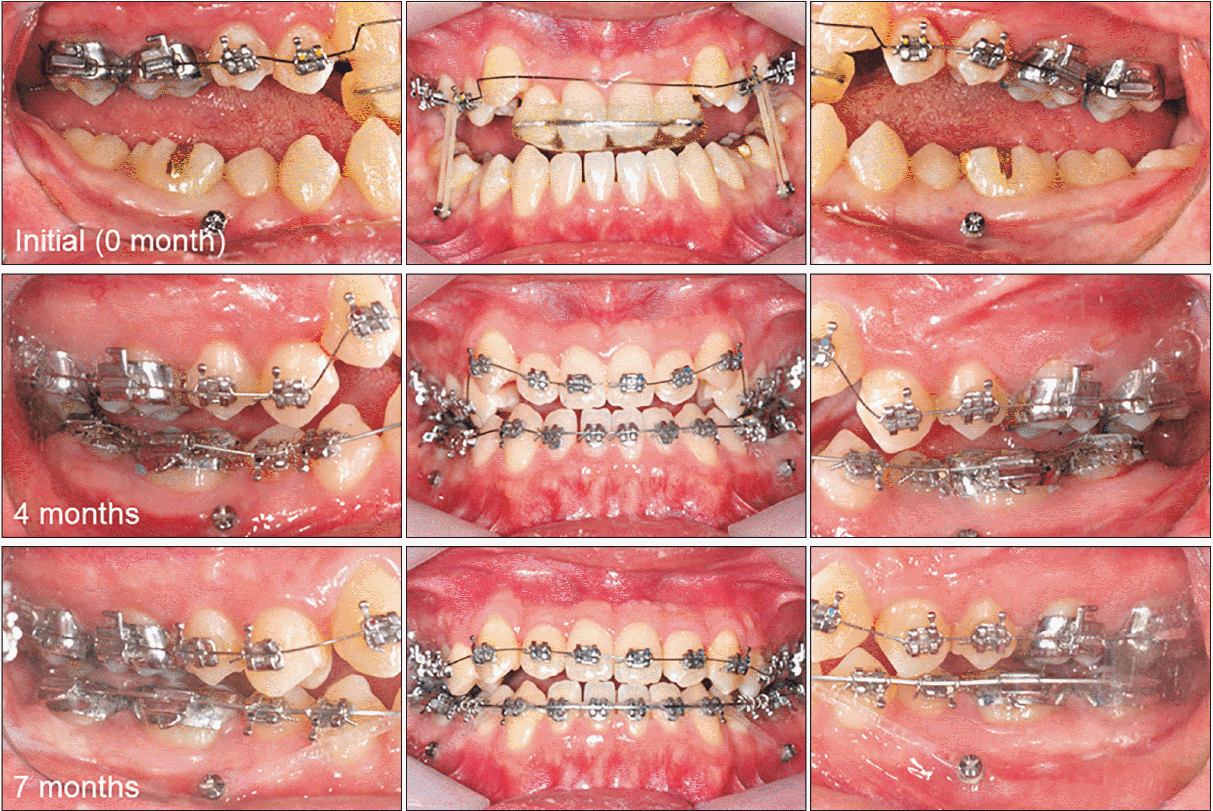
Preadjusted edgewise appliances with 0.022 × 0.028-inch slots and an MBT prescription (Victory Series; 3M Unitek, Monrovia, CA, USA) were bonded, and a 0.014-inch stainless steel archwire was used to align the maxillary posterior teeth. Intermaxillary elastics were used to extrude the posterior teeth from the maxillary first premolars to the miniscrews placed between the mandibular second premolars and first molars (Figure 5). A transpalatal arch was inserted into the first molars to maintain the arch width and buccopalatal inclination of the crown. The patient was instructed to wear elastics and a removable anterior bite plate at all times except during meals.
The anterior overjet and overbite were improved to an edge-to-edge bite at 4 months of after treatment. Therefore, the use of intermaxillary elastics and the anterior bite plate was discontinued. The brackets were bonded to the maxillary anterior and mandibular teeth. Then, 0.012-inch and 0.014-inch nickel-titanium (NiTi) archwires were applied to the maxillary and mandibular teeth, respectively (Figure 5). Two weeks before aligning the mandibular dentition, the mandibular third molars were extracted by an oral and maxillofacial surgeon. A 0.019 × 0.025-inch NiTi archwire was inserted into the mandibular dentition at seven months, and the mandibular teeth were retracted using miniscrews to correct the Class III molar relationship (Figure 5).
The fixed appliances were removed 17 months following the orthodontic treatment. Circumferential wraparound retainers and canine-to-canine fixed lingual retainers were used to retain the maxillary and mandibular dentition, respectively.
RESULTS
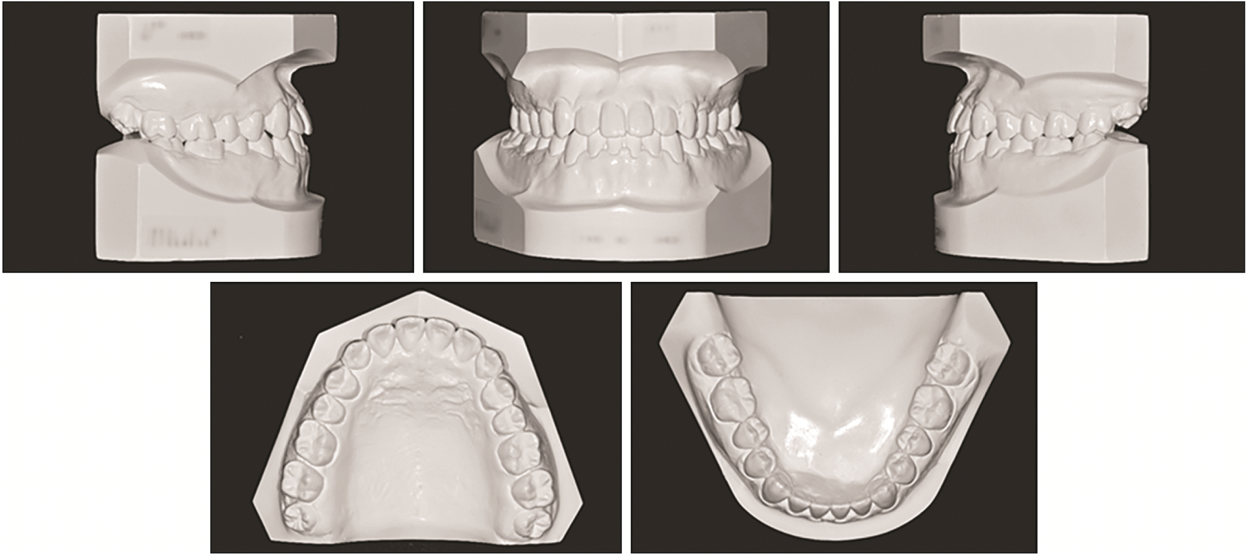
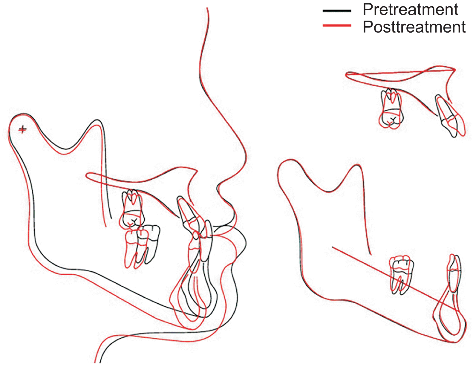
Figure 6 shows the improvement of the facial profiles and dental occlusion after the orthodontic treatment. The clockwise rotation of the mandible significantly reduced the facial concavity and prognathic mandible. The height/width ratio had also been improved by increasing the lower anterior facial height. The maxillary and mandibular dentitions were completely aligned (Figures 6 and 7). Normality in the anterior overjet and the overbite, and a Class I molar relationship, were achieved, although the dental midline did not coincide. A panoramic radiograph showed an absence of root resorption and good root parallelism. Cephalometric analysis revealed that mandibular rotations backward (sella-nasion-B point [SNB], from 88.2° to 85.5°) and downward (Y-axis, from 56.4° to 59.5°) had occurred following the orthodontic treatment and improved the maxillomandibular relationship (ANB, from –5.0° to –2.3°) (Figures 8, 9 and Table 1). The lower anterior facial height (anterior nasal spine [ANS]-Me) had increased by 6.0 mm with extrusion of the posterior teeth. The anterior crossbite had been corrected by the labial and lingual tipping of the maxillary and mandibular incisors, respectively, and by the clockwise rotation of the mandible. The patient was very satisfied with the overall treatment results.
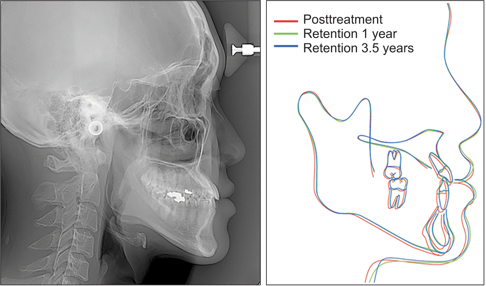
At 3.5 years of retention, the patient exhibited a harmonious facial profile, and the occlusion remained stable (Figure 10). However, cephalometric analysis during the retention period showed a mild rebounding in the mandibular position following debonding (Figure 11). The Y-axis angle decreased by 1.0°, and the lower anterior facial height (ANS-Me) decreased by 1.4 mm during the first year of the retention period (Table 1). However, no further relapse had occurred between the first year of retention and 3.5 years of retention.
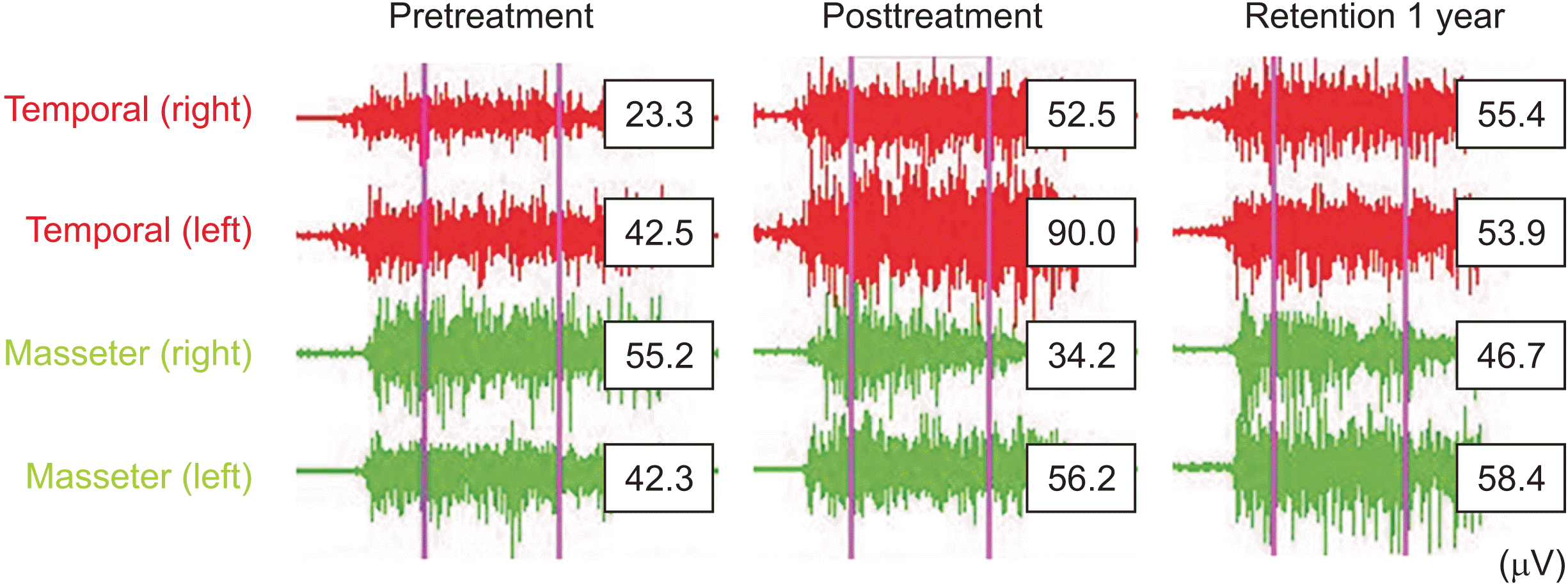
Figure 12 shows the electromyography of the masticatory muscle activities during maximum clenching at pretreatment, posttreatment, and 1-year of retention. The mean muscle activity was 32.9 μV for the temporal muscle and 48.8 μV for the masseter muscle. The overall temporal/masseter ratio (T/M ratio) was 0.67 at pretreatment, and it increased to 1.58 at posttreatment. A balance between the temporal and the masseter muscles was observed at one year following debonding (T/M ratio: 1.04).
DISCUSSION
Skeletal Class III malocclusion can be corrected using either orthognathic surgery or orthodontic camouflage. In general, the choice of treatment depends on the severity of the malocclusion and the patient’s chief complaint. Had there were no medical problems, the present patient would have undergone orthognathic surgery to improve his facial esthetics and achieve a normal occlusion. Eslami et al.10 investigated 65 patients who had received camouflage treatment or orthognathic surgery, and found that the Holdaway H angle and Wits appraisal can be used as critical diagnostic parameters for determining the treatment modality in borderline Class III cases. They concluded that Class III cases with a Wits appraisal greater than –5.8 mm could be corrected effectively by camouflage, but those with a Wits appraisal less than –5.8 mm must be treated by surgery. The present patient had a Wits appraisal of –9.7 mm at pretreatment and had no CO-MI discrepancy, indicating that surgery was the more suitable option. However, because the patient had a medical history of aplastic anemia, the patient selected orthodontic camouflage treatment instead of orthognathic surgery; this avoided possible surgical complications, such as bleeding and infection. His platelet count was 68,000 (normal range: 130,000–400,000 in males) when the mandibular third molars were extracted. Although orthognathic surgery is associated with a higher potential risk than tooth extraction, no complications occurred after the tooth extraction.
The combination of an anterior bite plate and posterior vertical elastics facilitated the correction of the deep overbite and severe Class III malocclusion in the present patient. Orthodontic tooth movement and significant rotations of the mandible downward (∆Y-axis, +3.1°) and backward (∆SNB, –2.7°; ∆ANB, +2.72) contributed to successful treatment outcomes. In the literature, the mandible is reported to be rotated significantly in a Class III elastic group (∆Y-axis, +0.6°; ∆ANB, +0.4°).11 However, the amount of mandibular rotation does not seem to be clinically significant. In contrast, in the present patient, the use of intermaxillary elastics with an anterior bite plate for molar extrusion contributed to a significant clockwise rotation of the mandible.
This case report demonstrates that backward rotation of the mandible by orthodontic molar extrusion can be a very effective and efficient treatment strategy to correct Class III malocclusion. However, a careful diagnosis is important before performing a backward mandibular rotation by orthodontic tooth extrusion. The authors suggest the following conditions as good indication for this treatment modality: (1) a Class III malocclusion with a deep overbite, (2) a concave facial profile showing chin protrusion, (3) a reduced lower anterior facial height, (4) lip redundancy at maximum occlusion, (5) elimination of lip redundancy and improvement of facial profile at the mandibular resting position (MRP), (6) improvement of the anterior overjet and overbite at MRP, (7) no discomfort in the temporomandibular joint (TMJ) or masticatory muscles at MRP, and (8) no airway disturbance at MRP.
In the present case, the freeway space was approximately 7 mm. The elimination of lip redundancy through increasing the vertical dimension increased the nasolabial angle and upper lip length, resulting in an improved lip profile and lower anterior facial height. However, in patients with a long lower anterior facial height, mandibular clockwise rotation could aggravate facial esthetics and cause lip incompetence.12
The main treatment in the present patient was the clockwise rotation of the mandible. Cephalometric superimposition showed that the center of rotation of the mandible was located at the mandibular condyle, and that there were no displacements of the condyle. However, three-dimensional angular changes of the condyle, and alteration of the vertical dimension, may lead to the development of TMJ disease (TMD).13-17 This is especially the case in adult patients, who have a lower ability for physiological adaptation than growing patients do. Therefore, a careful monitoring for TMJ was conducted in the present patient during the period in which he wore an anterior bite plate. The patient showed no signs or symptoms of TMD during or after the orthodontic treatment. In the relationship between altering the vertical dimension and development of TMD, Moreno-Hay and Okeson18 have concluded that the stomatognathic system has the ability to adapt rapidly to moderate vertical changes (< 5 mm).
What is the effect of increasing the vertical dimension on long-term stability? Maxillofacial positional interrelationships are known to be maintained by the balance of functional stresses exhibited by the neuromuscular and soft tissues of the stomatognathic system.19 Therefore, changes in the vertical muscular balance could result in an eventual relapse. However, it has been postulated that positional and structural changes in the stomatognathic system are quickly established, which may allow for alterations in the vertical dimension.20-23 It has been reported that an increase in the vertical dimension in adult patients, as expressed by a change in the Y-axis angle, relapsed 24.7% after orthodontic treatment, leaving a significant amount of treatment changes remain intact even after 5.6 years of posttreatment period.24 The present patient showed a 3.1° increase in Y-axis angle after the orthodontic treatment; however, the Y-axis angle decreased by 1.0° (32.3% relapse) after 1 year of retention. It has remained stable at 3.5 years of retention period. The improvement in the muscle balance that was observed at 1 year of retention might have affected the subsequent stability.
Neither increased labial gingival recession or tooth mobility were observed in this patient. However, a larger extrusion of the molars, or excessive proclination of the incisors, may have an adverse effect on periodontal support. Therefore, clinicians should be cautious when performing camouflage treatment, including molar extrusion or incisal proclination, for patients with gingival recession or a thin gingival biotype. In addition, patients should be followed-up for periodontal health after camouflage treatment with greater dental compensation. Mandibular molar distalization can be used to correct the Class III molar relationship and anterior crossbite, instead of proclination of the upper anterior teeth. The posterior anatomic limit appears to be the lingual cortex of the mandibular body,25 which is shorter in patients with hyperdivergent patterns.26 Therefore, computed tomography scans are recommended for patients who require significant mandibular molar distalization.
CONCLUSIONS
Backward rotation of the mandible by orthodontic molar extrusion can correct a Class III malocclusion and improve facial esthetics. This treatment modality can be an effective and efficient camouflage strategy for Class III patients with low mandibular plane angles.
SUPPLEMENTAL VIDEO
A video presentation of this article is available at https://youtu.be/mFyaQL6Nlio or www.e-kjo.org.
 XML Download