

 PDF
PDF Citation
 Print
Print


INTRODUCTION
Skeletal Class III malocclusion is one of the most complex conditions encountered in the orthodontic clinic. The global prevalence of this condition in the mixed and permanent dentition is 3.98% and 5.93%, respectively.1 The pathogenesis of this form of malocclusion includes recession of the upper jaw, protrusion of the lower jaw, or heteroplasia of both jaws. Almost two-thirds of cases with skeletal Class III malocclusion are accompanied by maxillary dysplasia.2 Growth modification is the major purpose of orthodontic treatment for children with mild Class III malocclusion. Maxillary protraction is the preferred form of treatment in clinical practice, and it aims to reduce the possibility of orthognathic surgery in adulthood.3
Traditional maxillary protraction, which is also referred to as tooth-anchored maxillary protraction, transmits an orthopedic force to the maxilla indirectly through the dentition by using intraoral retention devices, including banded4 or bonded5 rapid maxillary expansion (RME) devices, to stimulate the growth of the maxillary sutures. This approach can effectively promote maxillary growth and inhibit mandibular growth but is associated with side effects such as labial proclination of the maxillary incisors, clockwise rotation of the mandible, and an increase in the height of the lower third of the face due to the loss of anchorage.6
With the increasing use of miniscrews and miniplates in orthodontic clinical practice, the application of maxillary protraction with miniplates or miniscrews has developed rapidly and can be primarily categorized into two types: miniplates placed into the zygomatic buttress areas that protract the facemask (FM),7 or protraction with miniscrews placed between the mandibular lateral incisors and canines using Class III intermaxillary elastic.8 In a previous evidence-based study, Shi et al.9 demonstrated that bone-anchored maxillary protraction presented less labial proclination of the upper incisors in comparison with the tooth-anchored type, thus indicating that skeletal anchorage could reduce the incidence of side effects.
Different types of modified skeletal anchorage devices have been applied for maxillary protraction in the orthodontic clinic,10,11 including the Hybrid Hyrax RME appliance.12 However, these studies have not clarified whether appliances that combine tooth and bone anchorage provide better treatment effects than those using bone anchorage alone. Furthermore, previous studies have not clarified whether devices with stronger anchorage only induce undesirable dental compensation or provide more skeletal effects at the same time.
In contrast to a pairwise meta-analysis, network meta-analysis (NMA) provides the option to compare the effects of numbers of interventions.13 In this study, we aimed to compare the clinical effects of different types of bone-anchored maxillary protraction techniques with tooth-anchored maxillary protraction techniques through NMA and rank the recommended sequence of these interventions to provide practical clinical reference guidelines.
MATERIALS AND METHODS
Registration and literature search
This systematic review and NMA were registered in the International Prospective Register of Systematic Reviews (ID: CRD42021243210). Searches were performed using seven databases: MEDLINE (PubMed), Embase, Cochrane, Web of Science, Scopus, China National Knowledge Infrastructure, and the Wanfang Database. Gray literature was searched through Google Scholar. The search terms (Supplementary Table 1) combined subject terms and free terms without language limitations. The search date was up to May 15th, 2021.
The English subject terms included extraoral traction appliances, malocclusion, angle Class III, and orthodontic anchorage. The free terms included maxillary protraction, reverse headgear, anterior crossbite, and skeletal Class III.
Inclusion and exclusion criteria
The inclusion criteria were as follows: (1) Population: children with Class III malocclusion, ANB (the angle composed by the points subspinale-nasion-supramentale) < 0° edge-to-edge or reverse anterior bite, or inability to retract the mandible. (2) Intervention: a bone-anchored device for the experimental group and a tooth-anchored device or blank for the control group, or different types of bone-anchored devices for the experimental and control groups. (3) Outcome: primary outcomes included the SNA (the angle composed by the points sella-nasion-subspinale), SNB (the angle composed by the points sella-nasion-supramentale), and ANB. (4) Study design: randomized controlled trial (RCT) or controlled clinical trial (CCT).
The exclusion criteria were as follows: (1) history of surgery, orthodontic treatment, or orthognathic treatment; (2) presence of cleft lip and palate or other maxillofacial deformities; (3) presence of other genetic or systemic diseases.
Literature selection, data extraction, and quality assessment
After undergoing pre-experimental training, two researchers completed the literature selection, data extraction, and quality assessment independently according to the specific inclusion and exclusion criteria. In case of discrepancies between the assessments performed by the two researchers, another researcher made the final decision by cross-checking the results.
The data extraction mainly involved the collection of publication-related information and basic information about the study subjects, interventions, and outcomes. The quality assessment of the included RCTs was performed using the Risk of Bias (ROB) tool recommended by Cochrane. The same assessment of CCTs was performed using the Newcastle–Ottawa Scale.
Statistical analysis
The data were analyzed by R statistical software (R version 3.6.3) using the GeMTC package (version 1.0-1) based on the Bayesian generalized linear model, setting the number of pre-iterations to 10,000, the number of iterations to 50,000, the number of Markov chains to 3, and the step size to 1. The weighted mean difference (MD) was chosen as the effect size with the 95% confidence interval.
A network plot was drawn to depict the direct relationships among interventions. One point presented one type of intervention, and the size of the point represented the number of patients included in this intervention. The line segment between two points demonstrated the existence of a direct comparison between these two interventions. A closed-loop in the network plot indicated the presence of both direct and indirect comparisons; therefore, a consistency analysis would be applied. Consistency analysis between the direct and indirect comparison results was performed via node splitting. If p > 0.05, the consistency model was chosen. The heterogeneity of the included studies was analyzed according to the I2 calculation. p < 0.05 or I2 > 50% indicated the presence of significant heterogeneity and suggested that sensitivity analysis would be carried out to identify the source of heterogeneity. The random-effects model was applied for each indicator to take inter-study differences into account.
Potential scale reduction factors (PSRFs) were used to judge the degree of convergence of the model. The closer the PSRF was to 1, the more stable the model and the more credible the results were. When PSRF was > 1.1, the number of simulations was increased until the PSRF was closer to 1.
The ranking of the interventions was based on the surface under the cumulative ranking (SUCRA) values obtained by Stata (version 14.0; Stata Corp., College Station, TX, USA). The SUCRA values ranges from 0 to 100. The closer to 100 the value is, the larger the probability of the intervention being the most optimal. Publication bias was also tested using the funnel plot as generated by Stata.
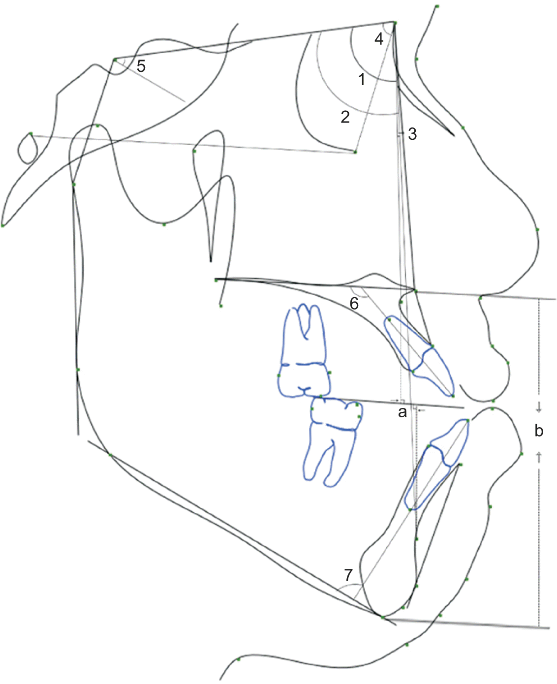
Our study involved a total of eleven outcomes: SNA, SNB, ANB, Wits (the distance between the perpendicular of the subspinale and supramentale points to the occlusal plane), SNOr (the angle composed by the points sella-nasion-orbitale), SN/MP (the angle composed by the sella-nasion plane and the mandibular plane), ANS-Me (the distance between the perpendicular of the anterior nasal spine and menton to the Frankel plane), U1/PP (the angle composed by the axis of the upper incisors and the palatal plane), IMPA (the angle composed by the axis of the lower incisors and the mandibular plane), overjet, and overbite (Figure 1).
RESULTS
Literature searches
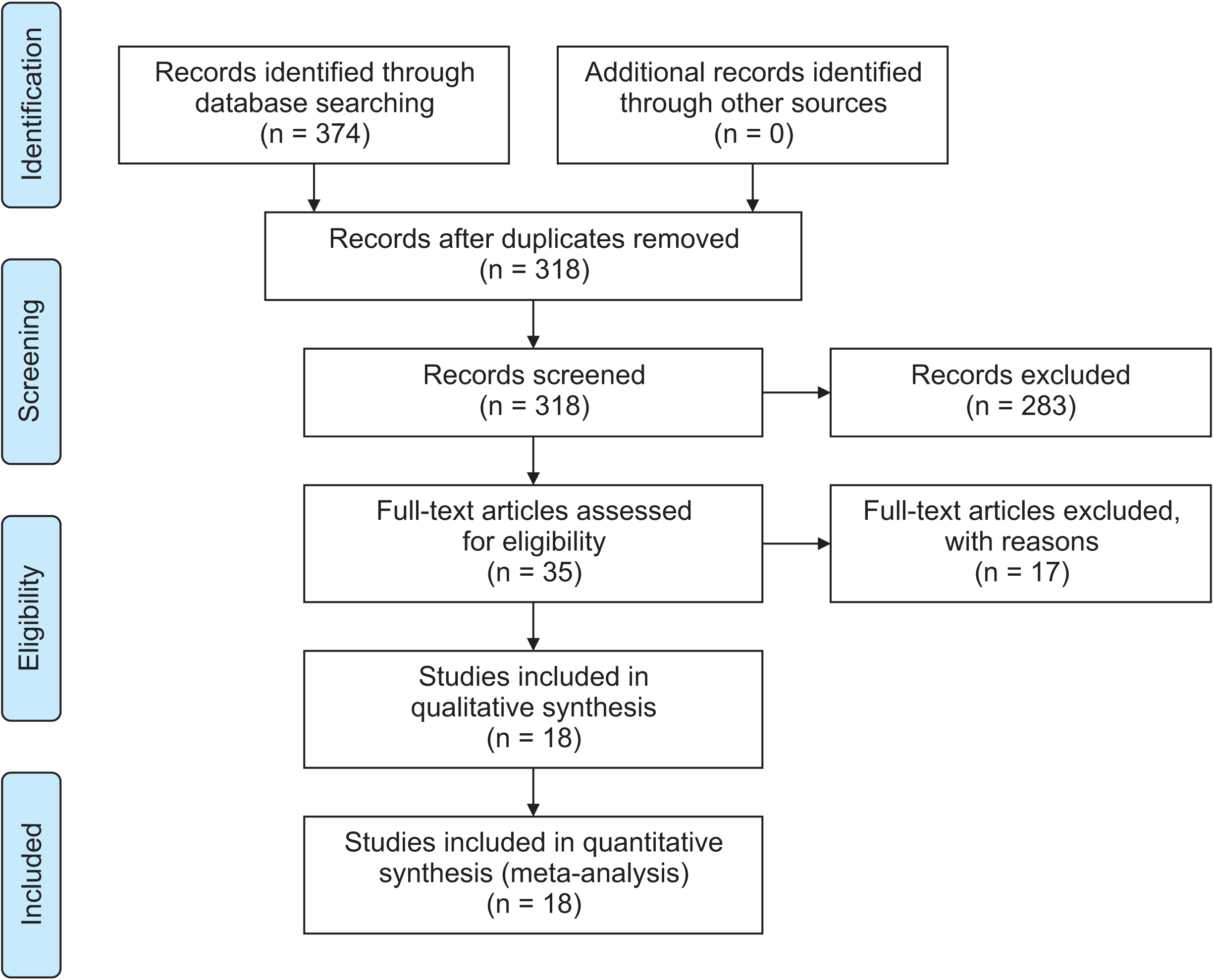
According to the search formula shown in Supplementary Table 1, a total of 1,374 relevant studies were retrieved from all search sources. Fifty-six duplicate studies were removed using EndNote X9 (Clarivate, Philadelphia, PA, USA). Two hundred and eighty-three studies that did not correspond with the inclusion criteria were excluded based on the title and abstract. After reading the full text of the 35 studies, 18 studies (four RCTs11,14-16 and 14 CCTs4,7,8,10,12,17-25) involving a total of 667 patients were finally included. The PRISMA (Preferred Reporting Items for Systematic Reviews and Meta-Analyses) flow diagram is given in Figure 2.
Study characteristics
Table 1 shows the essential characteristics of the included studies. In this study, interventions that only used bone anchorage such as miniplates or miniscrews were classified as the bone-anchored group, including bone anchorage with facemask appliance (BAFM) and bone anchorage with intermaxillary protraction (BAIP). Interventions that combined bone anchorage with bonds or bands were classified as the mixed-anchored group, including mixed anchorage with a facemask (MAFM) and mixed anchorage with intermaxillary protraction (MAIP). Detailed descriptions of the interventions are listed in Table 2 and Supplementary Table 2.
Risk of bias within studies
Results arising from our assessments of methodological quality are shown in Supplementary Table 3 and Supplementary Figure 1. The quality of the RCTs ranged from low to unclear: two studies11,14 were graded as having low ROB and two studies15,16 were graded as having unclear ROB. Quality assessment of the CCTs ranged from good to satisfactory: ten studies4,8,10,17-20,22,24,25 were graded as good and four studies7,12,21,23 were graded as satisfactory. The bias was mainly related to non-random study designs.
Network meta-analysis results
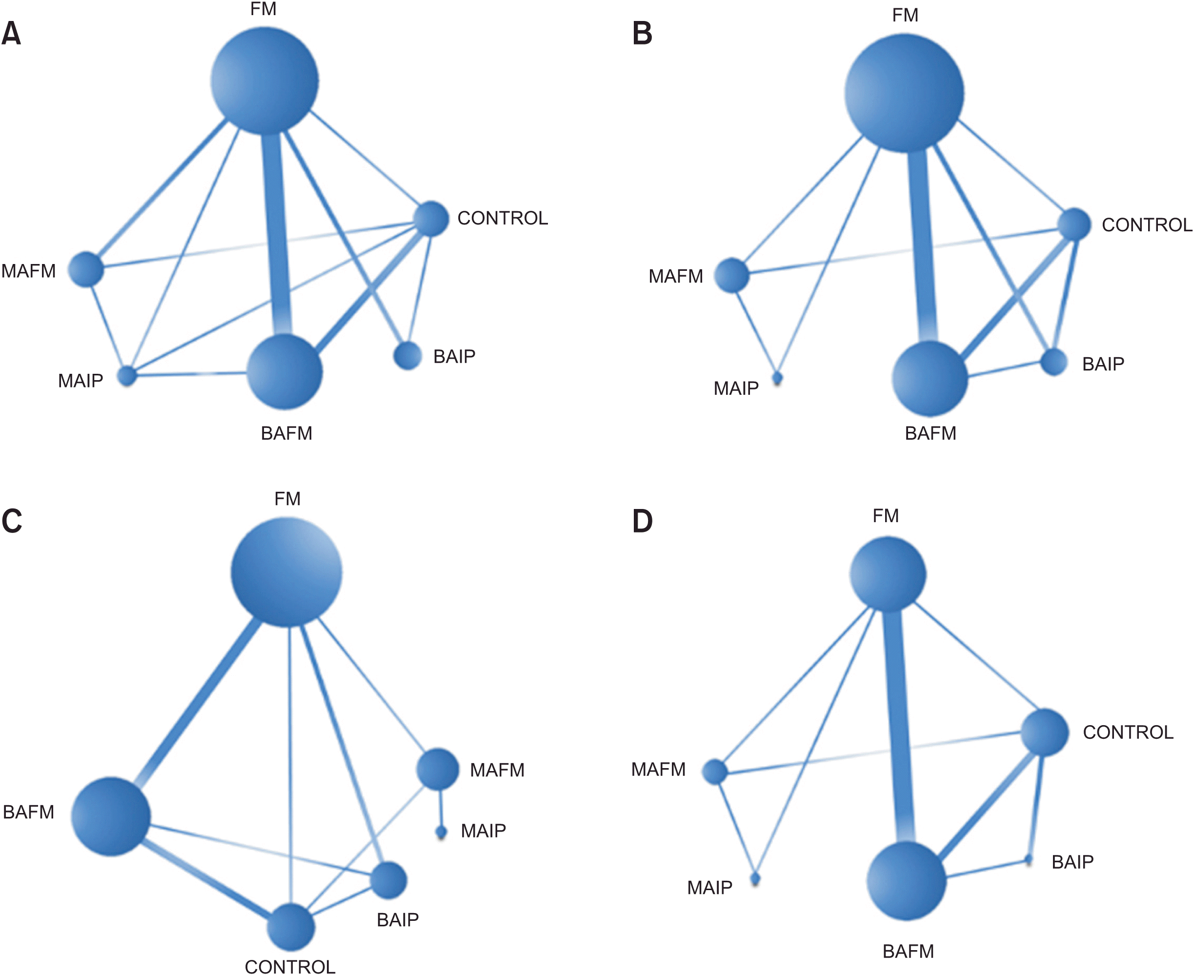
The network plot of each indicator had closed loops (Figure 3). Pairwise results are shown in Supplementary Table 4. The major results of the NMA are shown in Figure 4.
Skeletal changes
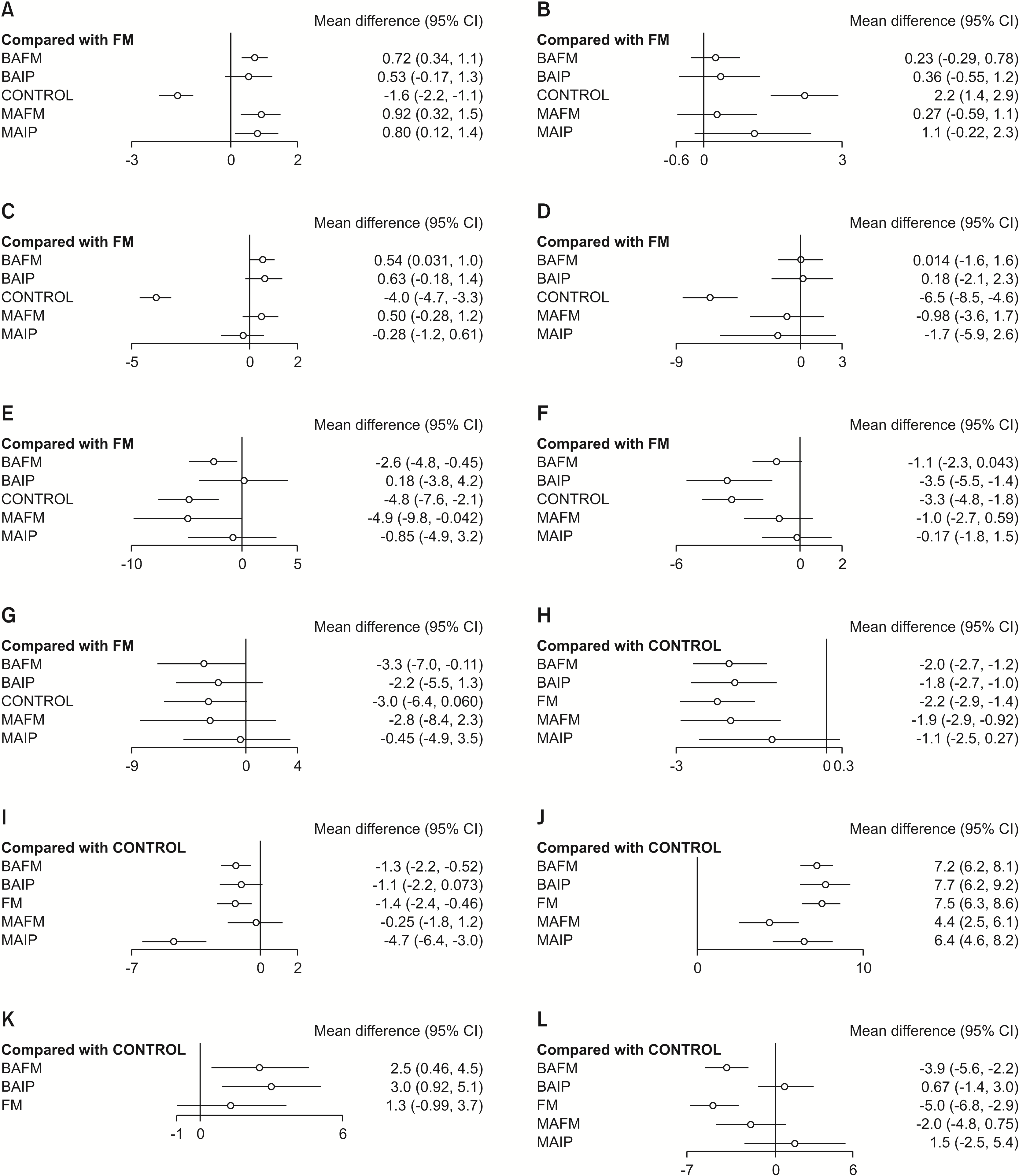
We identified 18 trials that included 667 patients and adopted SNA, SNB, and ANB as endpoints, and 11 trials that included 384 patients and adopted Wits as the endpoint. In comparison with the FM group, SNA (Figure 4A), ANB (Figure 4C), and Wits (Figure 4D) in the bone-anchored groups exhibited more advancement than those in the mixed-anchored groups. The SUCRA value (Table 3) showed that BAFM caused the greatest advancement of the A point and that BAIP had the best effect in terms of improving ANB and Wits. The increase in SNB (Figure 4H) was significantly inhibited in the treatment group in comparison with the control group, and the effects between the treatment groups showed no significant differences (Figure 4B).
Five studies that included 193 patients adopted SNOr as the outcome. In comparison with the control group, SNOr (Figure 4K) was higher in the treated groups. These changes were most apparent in the bone-anchored group than in the FM group. SUCRA values also indicated that BAIP resulted in the best effect in terms of increasing the SNOr.
Fourteen studies that included 493 patients adopted SN/MP as the outcome. In comparison with the FM group, SN/MP (Figure 4F) in the bone-anchored groups showed a lower increase than the mixed-anchored groups. SUCRA values implied that BAIP might be the best option for inhibiting the increase in SN/MP.
Seven studies that included 284 patients adopted ANS-Me as the outcome. ANS-Me (Figure 4E) increased in each treated group in comparison with the control group, and the changes caused by BAFM and MAFM were significantly less extensive than those caused by FM.
Dental changes
Nine studies that included 284 patients adopted U1/PP as the outcome indicator. In comparison with the FM group, the bone- and mixed-anchored groups showed a lower increase in U1/PP (Figure 4G; MDBAFM = –3.3, MDBAIP = –2.2, MDMAFM = –2.8, MDMAIP = –0.45). The SUCRA indicated that BAIP was the most effective intervention for controlling the labial proclination of the maxillary incisors.
Fifteen studies that included 534 patients adopted IMPA as the outcome indicator. In comparison with the control group, IMPA (Figure 4L) was lower in groups that utilized a FM (MDFM = –5.0, MDBAFM = –3.9, MDMAFM = –2.0), while a different degree of increase was observed in the groups that were facilitated by intermaxillary traction (MDBAIP = 0.67, MDMAIP = 1.5).
Heterogeneity tests, inconsistency tests, and other results
The heterogeneity test showed statistical heterogeneity between the included studies for the outcome indicators (I2 > 50%); therefore, the random-effects model was applied. Node-splitting methods showed inconsistencies for SNA, SNB, ANB, Wits, SN/MP, and IMPA (p < 0.05). Sensitivity analysis was applied to identify the source of the inconsistency. This inconsistency was removed after excluding two studies14,17 from the analysis of SNA and SN/MP, respectively. Inconsistencies also existed in SNB, ANB, Wits, and IMPA; this inconsistency was also eliminated after excluding the study by Sar et al.19 (p > 0.05). The results of the sensitivity analysis corresponded to the results of the NMA, indicating the stability of our statistical results. Furthermore, the funnel plots (Supplementary Figure 2) were symmetrical, indicating that the level of publication bias was acceptable. PSRF values for all indicators were equal to 1, indicating that our results were stable.
Supplementary data is available at https://doi.org/10.4041/kjod.21.264.
DISCUSSION
Maxillary protraction has been reported to present obvious effects in correcting the maxillary deficiency in skeletal Class III children.6 To reduce the adverse effects of the loss of anchorage, clinicians have used a variety of different methods in combination with skeletal anchorage. Thus, evaluation of the clinical effects of these approaches and identification of the preferred device for clinical practice is vital. Due to the diversity of clinical cases, such as cases with palatal transversal disharmony, designing trials that involve only maxillary protraction devices is difficult. Both Foersch et al.26 and Lee et al.27 evaluated the effect of maxillary protraction with or without RME over short- and long-terms but failed to identify any significant differences. Therefore, we did not perform subgrouping based on the usage of an RME appliance in our analysis. Our study instead investigated the influence of different anchorage strengths on the effects of maxillary protraction via evidence-based medical methods.
The results of our study showed that more strongly anchored maxillary protraction could promote greater anterior movement of the maxilla and significantly correct the intermaxillary Class III relationship better than a traditional tooth-anchored appliance. Bone-anchored devices that transmit orthodontic force to the maxilla directly could offer the best clinical effects; this was consistent with the findings of previous studies.4,8,9 BAFM may be the best way to promote maxillary growth and BAIP could be the best method to correct the Class III intermaxillary relationship. Furthermore, we found that maxillary protraction improved the advanced movement of the orbitale point; this was consistent with the results reported by Elnagar et al.14 and Lee et al.18 This is the first study to prove the effect of maxillary protraction on improving the growth of the upper half of the midface and correcting the concave profile of Class III patients through evidence-based medical methods.
Our results showed that maxillary protraction inhibited the sagittal growth of the mandible in the short-term. We also found no significant difference between different anchorage groups; this indicated that the restraining effects caused by the counterforce of the orthopedic force acting on the mandible and the mental region could only be influenced by the value of the protraction force rather than anchorage strength. Furthermore, mandibular growth has been generally considered to be difficult to inhibit,28 and overgrowth has been suggested to relapse after the end of maxillary protraction.3 Thus, maxillary protraction cannot be an ideal technique for inhibiting the mandible. The bone-anchored groups showed significantly less rotation because bone anchorage reduced elongation of the maxillary molars and the drop in the posterior palatal plane. Thus, the height of the lower face showed less increase.
The labial proclination of the maxillary incisors was significantly reduced after strengthening the anchorage; this was consistent with the results of a previous study.4 When stronger anchorage was applied, the dentition and the maxilla moved together rather than undergoing clockwise rotation, which was caused by a reduction in the labial force. Moreover, bone-anchored interventions led to greater maxillary growth, providing more space to relieve the congestion, with less labial proclination of the incisors than others.
Previous studies have presented different opinions relating to the changes in mandibular incisors. Ito et al.29 found that during the treatment of skeletal Class III malocclusion in animal experiments, the lower incisors tended to decompensate for the anterior growth of the maxillary position. However, a clinical study by Tripathi et al.23 concluded that a chin-cup appliance caused lingual inclination of the mandibular incisors. We found that groups using FM all showed more pronounced lingual inclination of the lower incisors, while groups using intermaxillary elastics showed labial proclination. These results suggest that the lingual compensation of the lower incisors could be predominantly related to the chin-cup30 instead of the undesired force produced by protraction.
The correction of overjet caused by maxillary protraction involved both dental and skeletal effects. According to our results, the proportion of skeletal effects was 57.87% in group FM, and the proportion increased to more than 90% with BAIP; groups with intermaxillary protraction showed greater skeletal effects than those with FM. Class III intermaxillary elastics could help correct the jaw position. This could be the reason for the presence of greater skeletal effects. The stability of the changes in jaw position depends on adaptive reconstruction of the temporomandibular joint. If the articular fossa cannot be modified in a timely manner, compulsive mandibular retrusion may lead to compression of the bilaminar region, which could be a hidden danger in temporomandibular disorders. However, this process can also increase the possibility of relapse. Dentists should pay more attention to the symptoms of the temporomandibular joint and prolong the follow-up time when applying maxillary protraction with intermaxillary elastics.
This study had some limitations that require consideration. Studies by Elnagar et al.14 in 2016 and Sar et al.19 in 2014 were designed as three-arm trials; all of the other included studies were two-arm trials. Thus, variations in study designs could be a source of the observed inconsistencies in SNA, SNB, ANB, Wits, and IMPA. Ağlarcı et al.17 applied bonded intraoral devices in their study; this controlled the extrusion of the maxillary molars and regulated the rotation of the mandibular plane. This could be the underlying reason for the appearance of inconsistency in SN/MP.
Furthermore, the included studies lacked sufficient and uniform outcome indicators, and accurate comparison of the initial malocclusion severity of the participants was difficult. In addition, the lack of randomization, the differences in treatment protocols, and the inconsistent development stages in children of the same age could all induce heterogeneity. More high-quality and well-designed RCTs are required to eliminate the effects of these variables and establish the true efficacy of the treatment.
CONCLUSIONS
Our analysis showed that bone-anchored maxillary protraction could significantly promote the forward movement of the maxilla and correct the Class III intermaxillary relationship. Strengthening the anchorage could yield more bone effects in terms of maxillary protraction and reduce dental compensations and side effects, including the labial proclination of the upper incisors, clockwise rotation of the mandibular bone, and an increase in the height of the lower face. Among the maxillary protraction interventions described in this study, BAIP yielded the best treatment effect.
The lingual inclination of mandibular incisors after maxillary protraction is mainly related to compression by the chin-cup. Maxillary protraction could also promote the forward growth of the middle-third of the face and improve the concave profile in an effective manner.
 XML Download