Journal List > Korean J Orthod > v.52(5) > 1516079860

Ku, Han, Kim, Oh, Kook, and Kim: Common dental anomalies in Korean orthodontic patients: An update

Abstract

Objective

The aim of this study was 1) to investigate the prevalence and pattern of dental anomalies (DAs), 2) to compare DAs according to the type of malocclusion, and 3) to investigate the correlation between tooth impaction and other DAs in the Korean orthodontic population.

Methods

A total of 3,240 orthodontic patients were classified as Class I, Class II, or Class III malocclusion groups. The presence and location of common DAs, including impaction, microdontia, agenesis, supernumerary tooth, transposition, and fusion, were identified by examining diagnostic records. Furthermore, samples were classified as Group 1 without impaction or Group 2 with impaction. The prevalence of other DAs concurrent with impaction was investigated and compared to Group 1.

Results

Impaction was the most prevalent DA, followed by microdontia, agenesis, and supernumerary. Class I and Class III groups showed the same order of prevalence, but agenesis was more frequent than microdontia in the Class II group. The prevalence of the four DAs was lowest in the Class III group. Overall, 8.6% of patients were classified into Group 2. The incidence of DAs other than impaction and the prevalence of multiple concurrent DAs were significantly higher in Group 2. Impaction showed a significant relationship with supernumerary tooth, transposition, and fusion.

Conclusions

The prevalence and pattern of DAs varied depending on the type of malocclusion. As there was a higher risk of other DAs in patients with impacted teeth, early detection of the impacted tooth and a detailed diagnosis of other possible DAs may be essential.

INTRODUCTION

Dental anomalies (DAs) are defined as changes in the numbers, shapes, structures, exfoliations, and eruptions of teeth during dental development.1,2 Tooth impaction, microdontia, tooth agenesis, and supernumerary tooth are the most common DAs encountered in clinical practice.3,4 By affecting the number and size of teeth and the discrepancy of the arch length, these congenital anomalies can deteriorate both esthetics and function.5 Furthermore, DAs of orthodontic patients tend to be more frequently compared to non-orthodontic patients.6,7 Early diagnosis of DAs enables optimal planning of orthodontic treatment, reducing side effects, and the complexity of treatment.8
Extensive literature has been reported on the prevalence and distribution of DAs in different populations.3-9 In contrast, relatively few studies have demonstrated the association between the presence of DAs and the malocclusion type.10-12 Fernandez et al.10 found that DAs were most prevalent in Class III malocclusion and microdontia was significantly more frequent in Class III malocclusion. Basdra et al.11 reported that Class III malocclusion showed significantly higher rates of DAs compared to Class II division 1 malocclusion. Uslu et al.12 demonstrated that tooth impaction had a significantly lower prevalence in Class II and Class II division 2 malocclusion. The types of DAs investigated and diagnostic criteria for each DA vary in the literature, showing inconsistent results. Furthermore, due to differences in ethnicity and environmental factors, there are discrepancies in the prevalence of DAs in previous studies.9,13 For the Korean population, there is little literature on the frequency and pattern of DAs in relation to the malocclusion type or skeletal feature. To date, only a few studies have reported the prevalence of hypodontia according to the type of malocclusion.14,15
The prevalence of tooth impaction is known to be the highest among the different types of DAs even when excluding the impaction of third molars, and ranges from 3.1% to 13.7% depending on the characteristics of the population.4,8,16 Laganà et al.8 reported that the most frequently impacted teeth were the maxillary canine, followed by maxillary lateral incisors, and maxillary central incisors. The prevalence pattern may be different in Korean population.
In patients with impacted teeth, a concurrent supernumerary tooth, or a small-sized tooth, is often encountered. Baccetti17 demonstrated the existence of associations among different DAs and emphasized the importance of early diagnosis of one anomaly as it is an indicator of a higher risk of other DAs. Peck et al.18 discovered that patients with a palatally displaced canine (PDC) tooth have a higher incidence of permanent tooth agenesis and small lateral incisors. Sigler et al.19 also showed that subjects with PDC exhibited a significantly higher prevalence of small lateral incisors and distoangulation of the second mandibular premolars. However, these studies investigated only individuals affected by DAs and a comparison with a control group was absent.18-20 For the Korean population, there have been few studies on the association between tooth impaction and other common DAs.
Therefore, the purpose of this study was 1) to investigate the prevalence and pattern of DAs, 2) to compare DAs according to the type of malocclusion, and 3) to investigate the correlation between tooth impaction and other DAs in the Korean orthodontic population.

MATERIALS AND METHODS

Study population

The subjects of the present study included 3,753 patients who visited the Department of Orthodontics at Seoul St. Mary’s Hospital, the Catholic University of Korea and underwent a diagnostic examination for orthodontic treatment between September 2002 and October 2019. The minimum age of the sample was 6 years, and any patient with a history of orthodontic treatment, multiple dental prosthesis, tooth loss, craniofacial disorders, incomplete records, or a tooth (or teeth) whose identification was unclear in the records was excluded. Furthermore, if there was uncertainty in the diagnosis of DAs, it was confirmed through follow-up panoramic radiographs or excluded from the sample. A final sample consisting of 3,240 patients (mean age, 22.2 ± 11.6 years) was included in the study. The presence and location of common DAs; tooth impaction, microdontia, tooth agenesis, supernumerary tooth, transposition, and fusion were identified by examination of their initial diagnostic records. This study was approved by the institutional review board of the Catholic University of Korea (KC20RISI0442 and KC21RISI0109).

Characterization of malocclusion type

The type of malocclusion in this study was defined according to the anteroposterior relationship of the maxilla and the mandible. The type of malocclusion was classified according to the value of the A point–Nasion–B point (ANB) angle measured in the lateral cephalograms, as follows.
  • - Class I (ANB angle with values between 0° and 4°)

  • - Class II (ANB angle with values > 4°)

  • - Class III (ANB angle with values < 0°)

Diagnosis of dental anomalies

Pretreatment diagnostic records including clinical photographs, panoramic radiographs, lateral cephalograms and cone-beam computed tomography images, if any, were examined to identify the following DAs:
  1. Tooth impaction: a tooth that was buried in the bone or gingiva obstructed on its normal eruption path and failed to erupt after the normal eruption period, except for the third molar.21

  2. Microdontia: a tooth smaller than the normal tooth size on the opposite side when comparing the mesiodistal width of the crown.22

  3. Tooth agenesis: the developmental absence of at least one permanent tooth, also known as hypodontia and congenitally missing tooth, except for the third molar.23 To exclude uncertainty, the central or lateral mandibular incisors were counted together.

  4. Supernumerary tooth: an excess of the regular number of teeth, with an additional tooth, which may be erupted or unerupted8,21

  5. Transposition: an unusual type of ectopic eruption or positional interchange of two teeth8

  6. Fusion: the union between the dentin or enamel of two or more separated tooth germs24

Group classification according to the presence or absence of an impacted tooth

The subjects were further classified as Group 1 without an impacted tooth (control group) or Group 2 with an impacted tooth (impaction group). The inclusion criteria for Group 2 were the presence of one or more permanent impacted teeth, excluding the impacted third molars. An impacted supernumerary tooth or an impacted third molar was not included in Group 2. The following DAs were detected; tooth impaction, agenesis, supernumerary tooth, microdontia, transposition, and fusion.

Statistical analysis

All measurements were examined by the same investigator and were repeated after two weeks. The systemic intraexaminer error between the two measurements was evaluated with a paired t-test. The extent of the measurement error between the first and second evaluations was also assessed by calculating the intraclass correlation coefficient. Patients and the DAs rates were stratified by sex and type of malocclusion. To determine the statistical significance of DAs by sex and malocclusion type, the chi-square test (sex) and chi-square test with Bonferroni correction (malocclusion type) were conducted. In addition, the chi-square test was used to compare the prevalence of impacted teeth and their relationship with other DAs in each group. The level of significance in all tests was set at 5% (p < 0.05). The Phi correlation coefficient was calculated to assess the correlation between the DAs. A Phi coefficient of 0.1 to 0.3 was interpreted as a weak correlation, 0.3 to 0.5 as moderate, and 0.5 to 1.0 as a strong correlation. All statistical analyses were performed using SPSS ver. 20.0 (IBM Corp., Armonk, NY, USA).

RESULTS

Sample composition and prevalence of dental anomalies

The total sample (3,240 patients) was composed of 1,310 male (40.4%) and 1,930 female (59.6%) patients. The most common DA was tooth impaction, which showed a prevalence of 8.6%, followed by microdontia (6.8%), tooth agenesis (6.5%), and supernumerary tooth (2.2%) as shown in Table 1. There was no significant sex-based difference with regard to the prevalence of DAs, except for the presence of the supernumerary tooth which was more frequent in male (3.3%) than in female (1.5%, p < 0.001) patients. For each DA, the mean number of affected teeth per individual was less than two. However, only for the tooth agenesis, two or more (2.18 ± 0.45) teeth were affected on average. Regarding the reliability of the measurements, no significant difference was observed in the measurements of the first and the second evaluations.
Table 1 compares the prevalence of DAs according to the type of malocclusion. There were 1,430 (44.1%) patients in the Class III group, followed by 1,178 (36.4%) Class II patients, and 632 (19.5%) Class I patients. On comparing the prevalence of each DA among the types of malocclusions, tooth impaction was found to be more prevalent in the Class I group (12.2%) than in the Class III group (6.7%, p < 0.001). Conversely, tooth agenesis was detected more frequently in the Class II group (8.1%) than in the Class III group (5.3%, p < 0.05). A supernumerary tooth was more common in the Class I group (3.8%) than in the Class II group (1.8%, p < 0.05) or in the Class III group (1.8%, p < 0.05).

Maxillomandibular distribution of dental anomalies

The maxillary and mandibular distribution of DAs was further investigated in each malocclusion type (Table 2). Most DAs, except for tooth agenesis, were more common in the maxilla. Microdontia (> 90%) generally occurred in the maxilla and there was no significant difference between the types of malocclusions. However, the distribution of tooth impaction, tooth agenesis, and supernumerary tooth differed according to the type of malocclusion. An impacted tooth was more likely to exist in the maxilla in the Class III group than in the Class I (p < 0.001) or Class II group (p < 0.01). Similarly, the incidence rate of tooth agenesis in the maxilla was higher in the Class III group than in the Class I group (p < 0.05) or the Class II group (p < 0.001). The supernumerary tooth fraction found in the maxilla was 93.1% in the Class I group and was significantly higher than 64.3% in the Class II group (p < 0.05).

The most affected teeth for each dental anomaly

Figure 1 shows that the tooth most frequently impacted was the maxillary canine (36.5%), followed by the second maxillary premolar (14.6%), the second mandibular molar (12.4%), and the second mandibular premolar (7.3%). Microdontia was by far the most prevalent in the maxillary lateral incisor (86.6%) followed by the maxillary second premolar (6.9%). For tooth agenesis, the mandibular incisor (27.4%) and the second premolar (25.5%) were the most frequently affected teeth. The most common site for the supernumerary tooth was the premaxillary region (U1–3, 69.4%), followed by the mandibular premolar area (L4, L5; 11.1%).
Table 3 describes the most frequently affected teeth (areas) of DAs according to the type of malocclusion. Regarding tooth impaction, the total sample and the Class III group showed the same order of prevalence, which was as follows: the maxillary canine, the second premolar of the maxillary and the second mandibular molar. However, in Class I and Class II groups, the second mandibular molar was impacted more frequently than the second maxillary premolar. The percentage of maxillary canine impaction was highest in the Class III group. Concerning tooth agenesis, the mandibular incisor was the most frequently affected tooth, while the mandibular second premolar was the most affected only in the Class I group. In any type of malocclusion, microdontia and supernumerary tooth were the most common in the lateral maxillary incisor and premaxillary region, respectively.

Prevalence of multiple dental anomalies

The overall frequency of any of the four DAs in Class III (17.3%) malocclusions was significantly lower than that of Class I (23.4%, p < 0.05) and Class II (21.4%, p < 0.01) malocclusions (Table 4). In addition, the prevalence of multiple (two or more) types of DAs in Class III (2.9%) was lower than that in Class I (5.4%, p < 0.05) malocclusion.

Group classification according to the presence or absence of an impacted tooth

Among a total of 3,240 patients, 280 patients (8.6%) presented with at least one impacted tooth and were grouped as Group 2. The other patients with no impacted teeth were grouped as Group 1. Group 1 (2,960 patients; mean age, 22.8 ± 11.7 years) consisted of 1,183 male (40.0%) and 1,777 female (60.0%) patients, and Group 2 (280 patients; mean age, 15.4 ± 8.2 years) consisted of 126 male (45.0%) and 154 female (55.0%) patients. No statistically significant sex-based differences were found in either group (p > 0.05).

The prevalence of dental anomalies other than tooth impaction in Group 1 and Group 2

The prevalence of the five DAs other than tooth impaction in Group 1, Group 2, and in the total sample is shown in Table 5 and Figure 2. The five DAs were found more frequently in Group 2 (p < 0.01), and the difference in prevalence between Group 1 and Group 2 was statistically significant (p < 0.001). Microdontia was the most frequently observed DA in both Group 1 (6.3%) and Group 2 (12.1%). The prevalence of tooth agenesis was 6.3% in Group 1 and 9.6% in Group 2. The supernumerary tooth was also detected more frequently in Group 2 (11.8%), while it occurred significantly less often in Group 1 (1.3%). The incidence of transposition and fusion was very low in both groups.
The prevalence of multiple concurrent DAs other than tooth impaction was also significantly higher in Group 2 than in Group 1 (p < 0.001, Table 6 and Figure 3). No patient had more than four DAs.

Correlation between tooth impaction and other dental anomalies

Table 7 shows that tooth impaction had a significant relationship with supernumerary tooth, transposition, and fusion (weak correlation, 0.1 ≤ Phi < 0.3). Furthermore, microdontia and tooth agenesis was correlated with supernumerary tooth, transposition, and fusion.

DISCUSSION

Dental anomalies can be classified into various types in relation to the number, shape, size and location of teeth.25 Some are observed relatively often and should be considered in advance during orthodontic treatment planning because they affect function, aesthetics, or occlusion.12 Tooth impaction, microdontia, tooth agenesis, and supernumerary tooth, which are known as the four most common DAs,3,4 were mainly investigated here. As in many other studies, tooth impaction showed the highest prevalence. Additionally, the prevalence of the four DAs was similar or greater than that of previous reports.4,10,11 Investigations of the general population or general dental patients showed a lower prevalence, while studies focusing on orthodontic patients at university-affiliated general hospitals indicated similar prevalence rates with this study. In other words, orthodontic patients in general hospitals are more likely to be referred due to different DAs as well as skeletal discrepancies.6,26
With regard to sex-based differences, there was a significant difference only in the prevalence of supernumerary tooth. The supernumerary tooth is known to occur twice as often in males than in females and the prevalence is 1.5–3.5% in the permanent dentition.27,28 The results of this study showed the same prevalence range and males were more frequently affected than females at a ratio of 2.2:1. Furthermore, the average number of affected teeth per individual was investigated, and only tooth agenesis affected more than two teeth, which is consistent with a previous study by Fernandez et al.10
The prevalence of the four DAs was highest in Class I (23.4%), followed by Class II (21.4%) and Class III (17.3%) regardless of the type of DA (Table 4). However, for each DA, significant differences among different malocclusion types were found for tooth impaction, tooth agenesis, and supernumerary tooth (Table 1). Tooth agenesis was detected more frequently in Class II (8.1%) malocclusion than in Class III (5.3%) malocclusion in this study. However, Celikoglu et al.29 found that hypodontia was significantly less frequent among patients with skeletal Class II malocclusion, and Chung et al.14 showed that hypodontia was associated with skeletal Class III malocclusion.
Maxillary teeth were more often affected by DAs than mandibular teeth, except for tooth agenesis (Table 2). In Class III malocclusion, both tooth impaction and tooth agenesis were found to be more common in the maxilla than in Class I or Class II malocclusion. Therefore, it may be postulated that the relative size of the maxilla and mandible appears to be related to the distribution of these two DAs, i.e., tooth impaction and tooth agenesis.
To our knowledge, no study has examined the most frequently affected tooth among DAs according to the type of malocclusion. For tooth agenesis, the most frequently affected teeth were, in order, the mandibular incisors, the second mandibular premolars, and the second maxillary premolars, reflecting the same results as Chung et al.14 The proportion of missing mandibular incisors was particularly high (33.1%) in the Class II group. This result may indicate an increase in the difficulty of treatment in Class II patients with missing mandibular incisors. This high prevalence of mandibular incisor agenesis was suggested as a characteristic of the hypodontia pattern of ‘mongoloids' by Kim.15
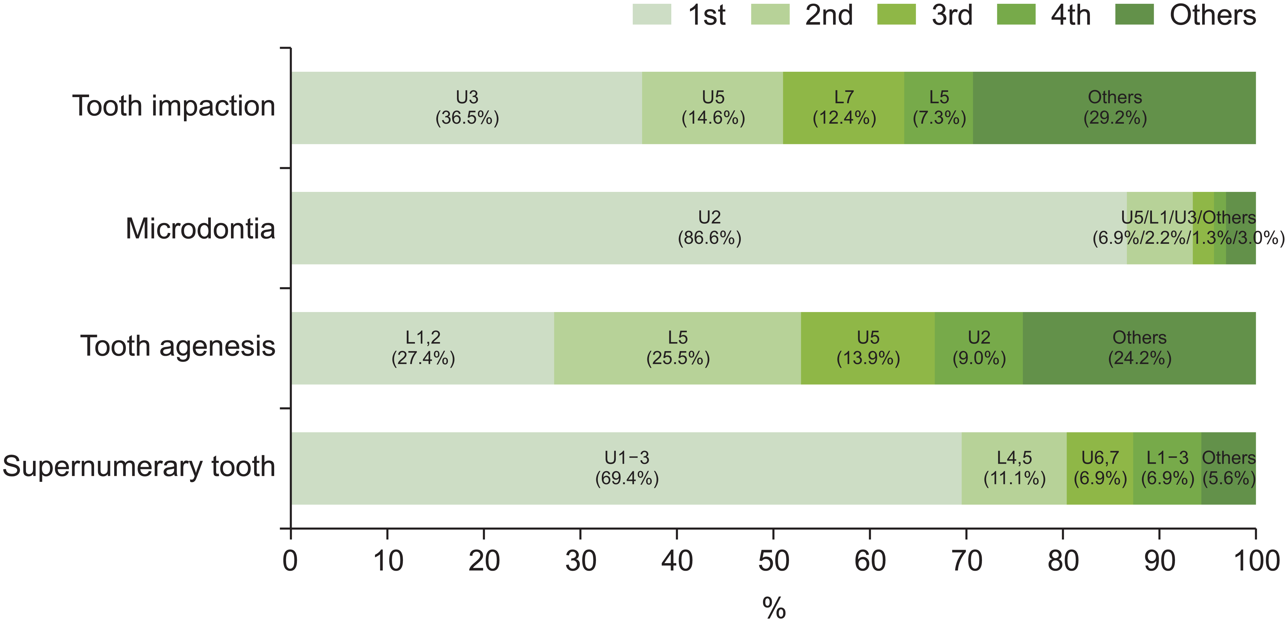
The most frequently affected teeth by microdontia were the maxillary lateral incisors, known as peg lateralis. In addition, the majority of supernumerary tooth occurred in the premaxilla, for example, in the mesiodens (Figure 1). The same results have already been reported in various studies.8-10 The most commonly impacted tooth was the maxillary canine among all malocclusion types (Table 3). The second most impacted tooth was the mandibular second molar in Class I and Class II malocclusion, and the maxillary second premolar in Class III malocclusion and in the total sample. Since Class III patients (n = 1,430, 44.1%) accounted for a large proportion of the total sample, the overall trend would have been the same as that of the Class III group. The impaction of the maxillary second premolar may induce a decrease in the maxillary arch length, making it more difficult to treat Class III malocclusion already complicated with anteroposterior jaw disharmony.
Since tooth impaction was the most common DA and may lead to severe side effects such as root resorption, tooth loss, and gingival problems,30 the total sample was grouped according to the presence or absence of an impacted tooth. The prevalence of an impacted tooth (Group 2) was 8.6% (n = 280), which was higher than the 3.09% reported by Lee et al.4 In another study by Fardi et al.16 evaluating the Northern Greek population, the incidence of impacted teeth was 13.7%. One of the reasons for this may be methodological differences, for example, the definition of impacted tooth and selection of the sample. They considered an unerupted supernumerary tooth as an impacted tooth,16 while in this study, the supernumerary tooth as well as the impacted third molars were considered as additional teeth and were not included as an impacted tooth.
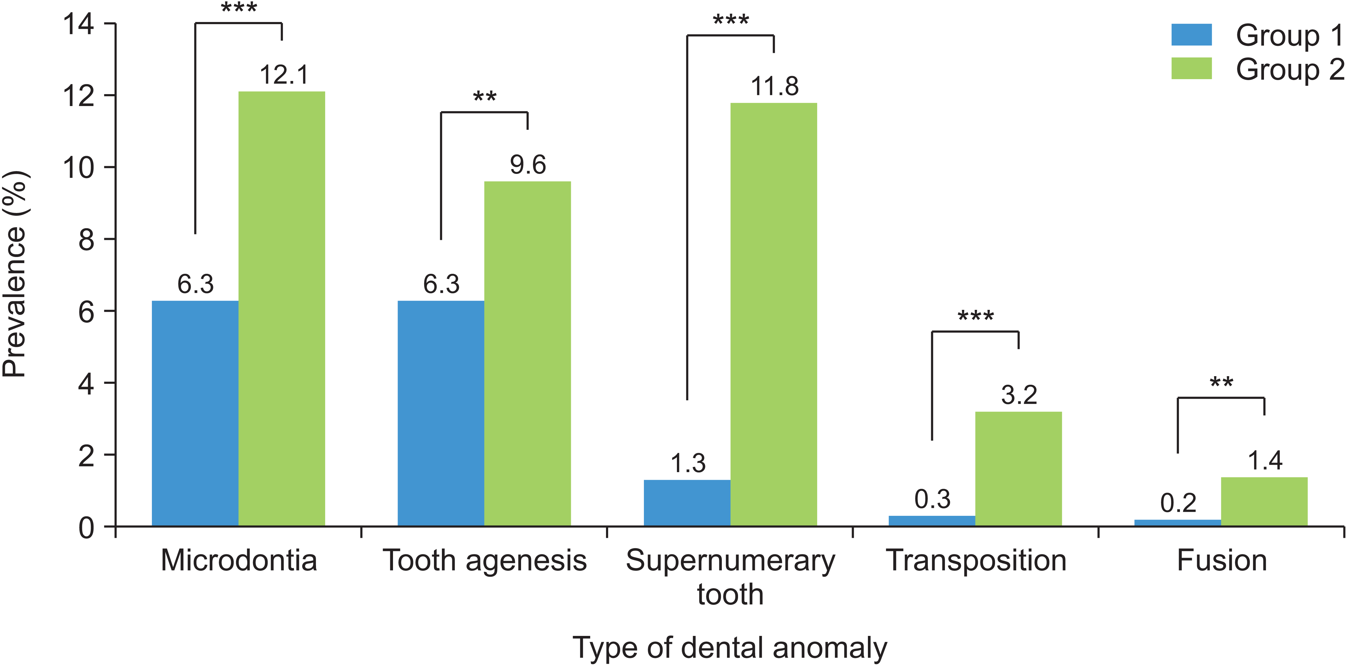
When the prevalence of all other DAs except the impacted tooth was compared between the groups (Table 5 and 
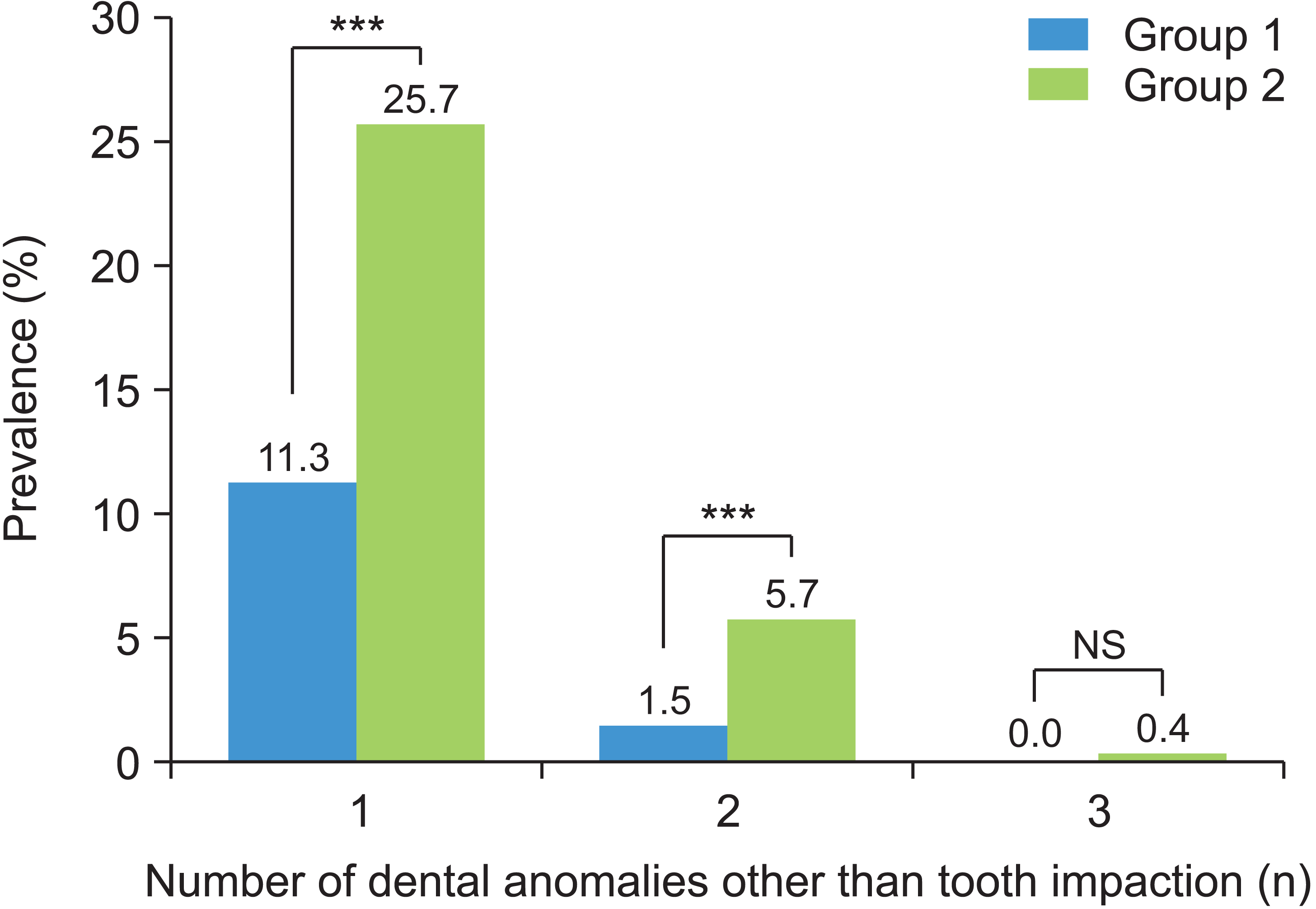
Figure 2), the prevalence was significantly higher in the impaction group (Group 2) than in the control group (Group 1). In particular, the prevalence of a supernumerary tooth and an impacted tooth in Group 2 (11.8%) was approximately ten times higher than the solitary presence of a supernumerary tooth in Group 1 (1.3%). Furthermore, the prevalence of multiple concurrent DAs such as simultaneous tooth agenesis, microdontia, supernumerary tooth or teeth was also significantly higher in Group 2 than in Group 1 (Table 6 and 
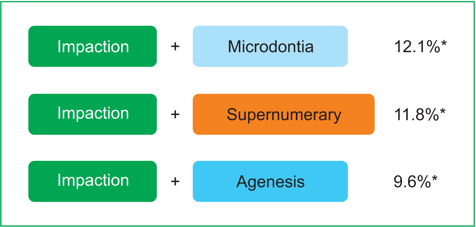
Figure 3). The most common patterns of concurrent DAs with tooth impaction in our study have been summarized in Figure 4, since understanding these common patterns would be clinically useful.
Regarding the associations between impacted teeth and other DAs, tooth impaction had a significant relationship with supernumerary teeth, transposition, and fusion (0.1 ≤ Phi < 0.3) (Table 7). Additionally, microdontia and tooth agenesis showed a correlation with supernumerary tooth, transposition, and fusion. The associations between tooth impaction and supernumerary/transposition and between tooth agenesis and transposition have also been reported by Laganà et al.8 Baccetti17 also demonstrated significant reciprocal associations between the agenesis of the second premolars, the peg lateralis, and the palatal displacement of maxillary canines, suggesting their common heredity. In this study, tooth agenesis was often discovered as multiple missing teeth state. Most supernumerary teeth were located in the maxillary anterior region, including mesiodens located between the two maxillary central incisors. Since maxillary canines, excluding third molars, were also the most commonly impacted teeth in many previous reports,4,16,21 a number of investigations dealing only with impacted maxillary canines have been described. Several previous reports have demonstrated a statistically significant association between PDC and other tooth impactions, agenesis, transposition, and peg-shaped maxillary lateral incisors.
According to this study, there was a greater possibility that patients with tooth impaction would have other DAs at the same time. Additionally, significant relationships among DAs suggest that an anomaly may present a potential risk of other anomalies. Therefore, a more thorough examination and follow-up would be required for patients with tooth impaction. In addition, early detection and accurate diagnosis of other possible DAs can decrease the complexity of treatment and provide optimal treatment for orthodontic patients.

CONCLUSIONS

In our study of the Korean orthodontic population: (1) the prevalence and pattern of DAs varied depending on the type of malocclusion; (2) the overall prevalence of the four common DAs was lower in Class III malocclusion than in Classes I and II; (3) patients with impacted teeth had a significantly higher incidence among other DAs; and (4) the prevalence of multiple concurrent DAs was also significantly higher in patients with impacted teeth. Therefore, early detection of impacted teeth and detailed diagnosis of other DAs is essential to provide better treatment.

SUPPLEMENTAL VIDEO

A video presentation of this article is available at https://youtu.be/18u4z_QBcn0 or www.e-kjo.org.

ACKNOWLEDGEMENTS

This study was supported in part by the Alumni Fund of the Department of Dentistry and Graduate School of Clinical Dental Science, The Catholic University of Korea. We would like to thank Mrs. YJ Lee for the meticulous data collection.

Notes

CONFLICTS OF INTEREST

No potential conflict of interest relevant to this article was reported.

REFERENCES

1. Brook AH. 2009; Multilevel complex interactions between genetic, epigenetic and environmental factors in the aetiology of anomalies of dental development. Arch Oral Biol. 54(Suppl 1):S3–17. DOI: 10.1016/j.archoralbio.2009.09.005. PMID: 19913215. PMCID: PMC2981858. PMID: https://www.scopus.com/inward/record.uri?partnerID=HzOxMe3b&scp=71549118817&origin=inward.
2. Baron C, Houchmand-Cuny M, Enkel B, Lopez-Cazaux S. 2018; Prevalence of dental anomalies in French orthodontic patients: a retrospective study. Arch Pediatr. 25:426–30. DOI: 10.1016/j.arcped.2018.07.002. PMID: 30249487. PMID: https://www.scopus.com/inward/record.uri?partnerID=HzOxMe3b&scp=85053683621&origin=inward.
3. Patil S, Doni B, Kaswan S, Rahman F. 2013; Prevalence of dental anomalies in Indian population. J Clin Exp Dent. 5:e183–6. DOI: 10.4317/jced.51119. PMID: 24455078. PMCID: PMC3892239. PMID: https://www.scopus.com/inward/record.uri?partnerID=HzOxMe3b&scp=84885168141&origin=inward.
4. Lee JH, Yang BH, Lee SM, Kim YH, Shim HW, Chung HS. 2011; A study on the prevalence of dental anomalies in Korean dental-patients. Korean J Orthod. 41:346–53. DOI: 10.4041/kjod.2011.41.5.346. PMID: https://www.scopus.com/inward/record.uri?partnerID=HzOxMe3b&scp=80055057248&origin=inward.
5. Guttal KS, Naikmasur VG, Bhargava P, Bathi RJ. 2010; Frequency of developmental dental anomalies in the Indian population. Eur J Dent. 4:263–9. DOI: 10.1055/s-0039-1697838. PMID: 20613914. PMCID: PMC2897859.
6. Thongudomporn U, Freer TJ. 1998; Prevalence of dental anomalies in orthodontic patients. Aust Dent J. 43:395–8. PMID: 9973708. PMID: https://www.scopus.com/inward/record.uri?partnerID=HzOxMe3b&scp=0032233633&origin=inward.
7. Al-Jabaa AH, Aldrees AM. 2013; Prevalence of dental anomalies in Saudi orthodontic patients. J Contemp Dent Pract. 14:724–30. DOI: 10.5005/jp-journals-10024-1391. PMID: 24309355. PMID: https://www.scopus.com/inward/record.uri?partnerID=HzOxMe3b&scp=84893407069&origin=inward.
8. Laganà G, Venza N, Borzabadi-Farahani A, Fabi F, Danesi C, Cozza P. 2017; Dental anomalies: prevalence and associations between them in a large sample of non-orthodontic subjects, a cross-sectional study. BMC Oral Health. 17:62. DOI: 10.1186/s12903-017-0352-y. PMID: 28284207. PMCID: PMC5346249. PMID: https://www.scopus.com/inward/record.uri?partnerID=HzOxMe3b&scp=85014841562&origin=inward.
9. Altug-Atac AT, Erdem D. 2007; Prevalence and distribution of dental anomalies in orthodontic patients. Am J Orthod Dentofacial Orthop. 131:510–4. DOI: 10.1016/j.ajodo.2005.06.027. PMID: 17418718. PMID: https://www.scopus.com/inward/record.uri?partnerID=HzOxMe3b&scp=34047127502&origin=inward.
10. Fernandez CCA, Pereira CVCA, Luiz RR, Vieira AR, De Castro Costa M. 2018; Dental anomalies in different growth and skeletal malocclusion patterns. Angle Orthod. 88:195–201. DOI: 10.2319/071917-482.1. PMID: 29215300. PMCID: PMC8312537. PMID: https://www.scopus.com/inward/record.uri?partnerID=HzOxMe3b&scp=85043574845&origin=inward.
11. Basdra EK, Kiokpasoglou MN, Komposch G. 2001; Congenital tooth anomalies and malocclusions: a genetic link? Eur J Orthod. 23:145–51. DOI: 10.1093/ejo/23.2.145. PMID: 11398552. PMID: https://www.scopus.com/inward/record.uri?partnerID=HzOxMe3b&scp=0035317373&origin=inward.
12. Uslu O, Akcam MO, Evirgen S, Cebeci I. 2009; Prevalence of dental anomalies in various malocclusions. Am J Orthod Dentofacial Orthop. 135:328–35. DOI: 10.1016/j.ajodo.2007.03.030. PMID: 19268831. PMID: https://www.scopus.com/inward/record.uri?partnerID=HzOxMe3b&scp=61349115073&origin=inward.
13. Dang HQ, Constantine S, Anderson PJ. 2017; The prevalence of dental anomalies in an Australian population. Aust Dent J. 62:161–4. DOI: 10.1111/adj.12443. PMID: 27471093. PMID: https://www.scopus.com/inward/record.uri?partnerID=HzOxMe3b&scp=85013042356&origin=inward.
14. Chung CJ, Han JH, Kim KH. 2008; The pattern and prevalence of hypodontia in Koreans. Oral Dis. 14:620–5. DOI: 10.1111/j.1601-0825.2007.01434.x. PMID: 18248591. PMID: https://www.scopus.com/inward/record.uri?partnerID=HzOxMe3b&scp=53149132810&origin=inward.
15. Kim YH. 2011; Investigation of hypodontia as clinically related dental anomaly: prevalence and characteristics. ISRN Dent. 2011:246135. DOI: 10.5402/2011/246135. PMID: 21991459. PMCID: PMC3170076.
16. Fardi A, Kondylidou-Sidira A, Bachour Z, Parisis N, Tsirlis A. 2011; Incidence of impacted and supernumerary teeth-a radiographic study in a North Greek population. Med Oral Patol Oral Cir Bucal. 16:e56–61. DOI: 10.4317/medoral.16.e56. PMID: 20711166. PMID: https://www.scopus.com/inward/record.uri?partnerID=HzOxMe3b&scp=78751662159&origin=inward.
17. Baccetti T. 1998; A controlled study of associated dental anomalies. Angle Orthod. 68:267–74. DOI: 10.1043/0003-3219(1998)068<0267:ACSOAD>2.3.CO;2. PMID: 9622764.
18. Peck S, Peck L, Kataja M. 1996; Prevalence of tooth agenesis and peg-shaped maxillary lateral incisor associated with palatally displaced canine (PDC) anomaly. Am J Orthod Dentofacial Orthop. 110:441–3. DOI: 10.1016/S0889-5406(96)70048-3. PMID: 8876497. PMID: https://www.scopus.com/inward/record.uri?partnerID=HzOxMe3b&scp=0030265939&origin=inward.
19. Sigler LM, Baccetti T, McNamara JA Jr. 2011; Effect of rapid maxillary expansion and transpalatal arch treatment associated with deciduous canine extraction on the eruption of palatally displaced canines: a 2-center prospective study. Am J Orthod Dentofacial Orthop. 139:e235–44. DOI: 10.1016/j.ajodo.2009.07.015. PMID: 21392667. PMID: https://www.scopus.com/inward/record.uri?partnerID=HzOxMe3b&scp=79952468852&origin=inward.
20. Peck S, Peck L, Kataja M. 2002; Concomitant occurrence of canine malposition and tooth agenesis: evidence of orofacial genetic fields. Am J Orthod Dentofacial Orthop. 122:657–60. DOI: 10.1067/mod.2002.129915. PMID: 12490878. PMID: https://www.scopus.com/inward/record.uri?partnerID=HzOxMe3b&scp=0036884421&origin=inward.
21. Patil S, Maheshwari S. 2014; Prevalence of impacted and supernumerary teeth in the North Indian population. J Clin Exp Dent. 6:e116–20. DOI: 10.4317/jced.51284. PMID: 24790709. PMCID: PMC4002339. PMID: https://www.scopus.com/inward/record.uri?partnerID=HzOxMe3b&scp=84909595776&origin=inward.
22. Agurto-S P, Nicholson C, del Sol M. 2019; Proposal of anatomical terms for alterations in tooth size: "Microdontia and macrodontia.". Int J Morphol. 37:375–8. DOI: 10.4067/S0717-95022019000100375.
23. Al-Ani AH, Antoun JS, Thomson WM, Merriman TR, Farella M. 2017; Hypodontia: an update on its etiology, classification, and clinical management. Biomed Res Int. 2017:9378325. DOI: 10.1155/2017/9378325. PMID: 28401166. PMCID: PMC5376450. PMID: https://www.scopus.com/inward/record.uri?partnerID=HzOxMe3b&scp=85017171204&origin=inward.
24. Tailor M. 2012; Tooth fusion- a rare dental anomaly: analysis of six cases. Int J Oral Maxillofac Pathol. 4:50–3. PMID: https://www.scopus.com/inward/record.uri?partnerID=HzOxMe3b&scp=0030265939&origin=inward.
25. Schulze C. Gorlin RJ, Goldman HM, editors. 1970. Developmental abnormalities of the teeth and jaws. Thoma's oral pathology. 6th ed. CV Mosby;St Louis: PMID: https://www.scopus.com/inward/record.uri?partnerID=HzOxMe3b&scp=0030265939&origin=inward.
26. Silverman NE, Ackerman JL. 1979; Oligodontia: a study of its prevalence and variation in 4032 children. ASDC J Dent Child. 46:470–7. PMID: 290641. PMID: https://www.scopus.com/inward/record.uri?partnerID=HzOxMe3b&scp=0018546853&origin=inward.
27. Garvey MT, Barry HJ, Blake M. 1999; Supernumerary teeth--an overview of classification, diagnosis and management. J Can Dent Assoc. 65:612–6. PMID: 10658390. PMID: https://www.scopus.com/inward/record.uri?partnerID=HzOxMe3b&scp=0033253662&origin=inward.
28. Kinirons MJ. 1982; Unerupted premaxillary supernumerary teeth. A study of their occurrence in males and females. Br Dent J. 153:110. DOI: 10.1038/sj.bdj.4804863. PMID: 6957208. PMID: https://www.scopus.com/inward/record.uri?partnerID=HzOxMe3b&scp=0020474191&origin=inward.
29. Celikoglu M, Kazanci F, Miloglu O, Oztek O, Kamak H, Ceylan I. 2010; Frequency and characteristics of tooth agenesis among an orthodontic patient population. Med Oral Patol Oral Cir Bucal. 15:e797–801. DOI: 10.4317/medoral.15.e797. PMID: 20383097. PMID: https://www.scopus.com/inward/record.uri?partnerID=HzOxMe3b&scp=77956405148&origin=inward.
30. Bedoya MM, Park JH. 2009; A review of the diagnosis and management of impacted maxillary canines. J Am Dent Assoc. 140:1485–93. DOI: 10.14219/jada.archive.2009.0099. PMID: 19955066. PMID: https://www.scopus.com/inward/record.uri?partnerID=HzOxMe3b&scp=75249101074&origin=inward.

Figure 1
Most frequently affected teeth (areas) for each dental anomaly in the total sample.
U, maxillary teeth; L, mandibular teeth; 1, central incisor; 2, lateral incisor; 3, canine; 4, first premolar; 5, second premolar; 6, first molar; 7, second molar.
kjod-52-5-324-f1.tif
Figure 2
Comparison of the prevalence of dental anomalies other than tooth impaction in Group 1 and Group 2. The prevalence of all the dental anomalies was significantly higher in Group 2 than in Group 1. A chi-squared test was performed.
Group 1, without an impacted tooth; Group 2, with an impacted tooth.
**p < 0.01; ***p < 0.001.
kjod-52-5-324-f2.tif
Figure 3
Comparison of the prevalence of multiple dental anomalies other than tooth impaction in Group 1 and Group 2. The prevalence of the concurrent two dental anomalies in Group 2 was significantly higher than Group 1 (p < 0.0001). No patient had more than four dental anomalies. A chi-squared test was performed.
NS indicates no statistical significance between groups.
Group 1, without an impacted tooth; Group 2, with an impacted tooth.
***p < 0.0001.
kjod-52-5-324-f3.tif
Figure 4
The most common patterns of concurrent dental anomaly with impacted teeth in Group 2. Impacted tooth and microdontia were the most common patterns of concurrent dental anomaly found in Group 2, followed by impacted tooth and supernumerary tooth, impacted tooth and tooth agenesis.
*The numbers indicate their prevalence in Group 2.
kjod-52-5-324-f4.tif
Table 1
Prevalence of the four most common dental anomalies according to malocclusion type
Type of dental anomalies Total
(n = 3,240)
No. of affected
teeth per
individual
Class I
(n = 632)
Class II
(n = 1,178)
Class III
(n = 1,430)
p-value
Tooth impaction 279 (8.6) 1.31 ± 0.74 77 (12.2) 106 (9.0) 96 (6.7) 0.0003*** (I > III)
Microdontia 221 (6.8) 1.75 ± 0.62 47 (7.4) 81 (6.9) 93 (6.5) NS
Tooth agenesis 212 (6.5) 2.18 ± 0.45 40 (6.3) 96 (8.1) 76 (5.3) 0.0111* (II > III)
Supernumerary tooth 71 (2.2) 1.27 ± 0.38 24 (3.8) 21 (1.8) 26 (1.8) 0.0261* (I > II), 0.0213* (I > III)

Values are presented as number (%) or mean ± standard deviation.

p-values are based on chi-square test and adjusted using the Bonferroni correction.

NS indicates no statistical significance between groups.

*p < 0.05; ***p < 0.001.

Table 2
Comparison of maxillary and mandibular distribution of dental anomalies according to malocclusion type
Type of dental anomaly Total Class I Class II Class III p-value
Maxilla Mandible Maxilla Mandible Maxilla Mandible Maxilla Mandible
Tooth
impaction
261 (70.7) 108 (29.3) 64 (60.4) 42 (39.6) 87 (66.9) 43 (33.1) 110 (82.7) 23 (17.3) 0.0003*** (III – I), 0.0096** (III – II)
Microdontia 373 (96.6) 13 (3.4) 72 (98.6) 1 (1.4) 144 (98.0) 3 (2.0) 157 (94.6) 9 (5.4) NS
Tooth
agenesis
189 (40.9) 273 (59.1) 32 (36.8) 55 (63.2) 68 (33.0) 138 (67.0) 89 (52.7) 80 (47.3) 0.0477* (III – I), 0.0003*** (III – II)
Supernumerary
tooth
70 (77.8) 20 (22.2) 27 (93.1) 2 (6.9) 18 (64.3) 10 (35.7) 25 (75.8) 8 (24.2) 0.0228* (I – II)

Values are presented as number (%).

p-values are based on chi-square test and adjusted using the Bonferroni correction.

NS indicates no statistical significance between groups.

*p < 0.05; **p < 0.01; ***p < 0.001.

Table 3
Most frequently affected teeth (areas) for each dental anomaly according to malocclusion type
Type of dental anomaly Total Class I Class II Class III
Tooth
(area)
n % Tooth (area) n % Tooth
(area)
n % Tooth (area) n %
Tooth impaction U3 115 36.5 U3 26 28.6 U3 41 35.0 U3 48 44.9
U5 46 14.6 L7 15 16.5 L7 16 13.7 U5 19 17.8
L7 39 12.4 U5 12 13.2 U5 15 12.8 L7 8 7.5
Others 115 36.5 Others 38 41.8 Others 45 38.5 Others 32 29.9
Microdontia U2 201 86.6 U2 43 89.6 U2 74 87.1 U2 84 84.8
U5 16 6.9 U5 4 8.3 U5 6 7.1 U5 6 6.1
L1 5 2.2 L1 1 2.1 U4 2 2.4 L1 3 3.0
Others 10 4.3 Others 0 0 Others 3 3.5 Others 6 6.1
Tooth agenesis L1,2 85 27.4 L5 21 35.6 L1,2 45 33.1 L1,2 26 22.6
L5 79 25.5 L1,2 14 23.7 L5 34 25.0 L5 24 20.9
U5 43 13.9 U5 9 15.3 U5 14 10.3 U5 20 17.4
Others 103 33.2 Others 15 25.4 Others 43 31.6 Others 45 39.1
Supernumerary tooth U1–3 50 69.4 U1–3 18 72.0 U1–3 10 47.6 U1–3 22 84.6
L4,5 8 11.1 U6,7 2 8.0 L4,5 4 19.0 L4,5 3 11.5
U6,7 5 6.9 U4,5 2 8.0 U6,7/L1–3 3 14.3 L1–3 1 3.8
Others 9 12.5 Others 3 12.0 Others 4 19.0 Others 0 0

U, maxillary teeth; L, mandibular teeth; 1, central incisor; 2, lateral incisor; 3, canine; 4, first premolar; 5, second premolar; 6, first molar; 7, second molar.

Table 4
Prevalence of multiple dental anomalies according to malocclusion type
No. of dental anomalies Total Class I Class II Class III p-value
None 2,593 (80.0) 484 (76.6) 926 (78.6) 1,183 (82.7)
Yes (≥ 1) 647 (20.0) 148 (23.4) 252 (21.4) 247 (17.3) 0.0234* (I > III), 0.0033** (II > III)
One 524 (16.2) 114 (18.0) 205 (17.4) 205 (14.3) NS
Two 111 (3.4) 29 (4.6) 42 (3.6) 40 (2.8) NS
Multiple (≥ 2) 123 (3.8) 34 (5.4) 47 (4.0) 42 (2.9) 0.0198* (I > III)

Values are presented as number (%).

p-values are based on the chi-squared test and adjusted using the Bonferroni correction.

NS indicates no statistical significance between groups.

*p < 0.05; **p < 0.01.

Table 5
Comparison of the prevalence of dental anomalies other than tooth impaction in Group 1 and Group 2
Type of dental anomaly Total sample (%) Group 1 (Control) Group 2 (Impaction) p-value
Microdontia 6.8 188 (6.3) 34 (12.1) < 0.0001***
Tooth agenesis 6.5 186 (6.3) 27 (9.6) 0.0064**
Supernumerary tooth 2.2 38 (1.3) 33 (11.8) < 0.0001***
Transposition 0.5 8 (0.3) 9 (3.2) < 0.0001***
Fusion 0.3 6 (0.2) 4 (1.4) 0.0026**

Values are presented as number (%).

Chi-square test was performed.

**p < 0.01; ***p < 0.001.

Table 6
Comparison of the prevalence of multiple dental anomalies other than tooth impaction in Group 1 and Group 2
No. of concurrent
dental anomalies
Group 1 (Control) Group 2 (Impaction) p-value
1 335 (11.3) 72 (25.7) < 0.001***
2 44 (1.5) 16 (5.7) < 0.001***
3 1 (0.0) 1 (0.4) NS

Values are presented as numbers (%).

NS indicates no statistical significance between groups.

A chi-squared test was performed.

***p < 0.001.

Table 7
Correlation between impaction and other dental anomalies in Group 2
ϕ coefficient Tooth
impaction
Microdontia Tooth
agenesis
Supernumerary tooth Transposition Fusion
Tooth impaction - 0.039 0.016 0.161* 0.198* 0.211*
Microdontia - 0.023 0.127* 0.167* 0.182*
Tooth agenesis - 0.147* 0.185* 0.199*
Supernumerary tooth - 0.055 0.080
Transposition - 0.032
Fusion -

Phi (ϕ) correlation analysis was performed.

*0.1 ≤ ϕ < 0.3 (small).

TOOLS
Similar articles