Journal List > Korean J Orthod > v.52(5) > 1516079859

Huang, Han, and Yang: Effect and stability of miniscrew-assisted rapid palatal expansion: A systematic review and meta-analysis

Abstract

Objective

This study aimed to systematically analyze the effect and stability of miniscrew-assisted rapid palatal expansion (MARPE) to provide a reference for the clinical treatment of patients with maxillary transverse deficiency (MTD).

Methods

We searched PubMed, Science Direct, Web of Science, Embase, Cochrane Library, CNKI, and Wanfang Database for relevant studies published before February 18, 2021 and selected them according to the eligibility criteria. The Cochrane Handbook for Systematic Reviews (version 5.1.0) criteria were used for the quality assessment of randomized controlled trials, while the scoring protocol of the methodological index for non-randomized studies was used for non-randomized controlled trials. Statistical analysis was performed using the RevMan5.3 software.

Results

All the included studies showed a relatively high success rate of expansion. The changes in both the intermolar and alveolar widths after MARPE were statistically significant. MARPE exhibited greater skeletal expansion effects than did conventional RPE. The midpalatal suture was opened in parallel after MARPE. A small amount of relapse was observed 1 year after expansion. MARPE caused tooth inclination and a decrease in alveolar height, but it was less significant than in conventional RPE.

Conclusions

MARPE may be an effective treatment modality for patients with MTD. It causes great transverse skeletal expansion in late adolescence. In comparison to conventional RPE, MARPE has lower detrimental periodontal effects and has certain clinical advantages.

INTRODUCTION

Maxillary transverse deficiency (MTD) is a common malocclusion that is accompanied by high vault, unilateral or bilateral posterior cross-bite, excessive buccal corridor space, and crowding.1 Rapid palatal expansion (RPE) is a useful treatment to correct transverse maxillary deficiency and posterior cross-bite. It was introduced by Angell2 in 1860 and gained popularity in the late 1900s. The most common application of RPE is as a tooth-borne expander. However, it is difficult for late adolescents to achieve orthopedic expansion due to the bony interdigitation of the midpalatal suture.3,4 Some detrimental periodontal effects of the conventional RPE have also been reported, such as restricted skeletal movement, tooth inclination, root resorption, and gingival recession.5
An alternative method is surgically assisted rapid palatal expansion (SARPE). It has been used to release closed sutures that resist expansion in adults, and it helps to reduce the adverse effects of conventional RPE.6,7 However, SARPE is costly, traumatic, and often accompanied by complications. Furthermore, for patients with maxillary constriction accompanied by severe anteroposterior discrepancies, SARPE is not a good alternative because of the requirement for phase 2 surgery.8
Miniscrew-assisted rapid palatal expansion (MARPE) was developed with the purpose of achieving maxillary expansion without surgical intervention. The rigid element of the MARPE appliance connects to the miniscrews, which are implanted into the para-midpalatal area.9 Accordingly, the appliance is able to directly deliver the expansion force to the palatal surface of the maxilla and therefore maximize the skeletal effect.10 Several studies on MARPE have reported its expansion effects and stability.8,10-20 However, the types, frequency, measurement methods, measurement indexes, and retention time vary among studies; therefore, there is a lack of comparability among different studies. A comprehensive and systematic analysis is necessary to accurately assess the efficacy of MARPE.
This study aimed to systematically analyze the short- and long-term effects of MARPE on teeth, alveolar bone, maxilla, and circummaxillary structures and to evaluate its efficacy and stability to ultimately provide a reference for the clinical treatment of MTD patients.

MATERIALS AND METHODS

Literature search

We searched PubMed, Science Direct, Web of Science, Embase, Cochrane Library, CNKI, and Wanfang Database for relevant studies published before February 18, 2021. The search strategy used for PubMed was as follows: #1= (miniscrew OR micro-implant OR implant) AND rapid AND (palatal OR maxillary OR maxilla) AND (expansion OR expanded). #2=MARPE[Title/Abstract] OR MARME[Title/Abstract]. The articles cited in the reference lists of the included studies were also reviewed.

Eligibility criteria

The inclusion criteria were randomized controlled trials (RCTs) or non-randomized controlled trials (NRCTs) performed in patients with MTD who underwent MARPE, holding accurate and useful data, or has the possibility of obtaining data from the primary data. Excluded studies were as follows; studies with less than 8 samples; studies that included patients with cleft lip, craniofacial syndrome, or orthodontic treatment or maxillofacial surgery histories; and studies where other interventions were performed.

Study selection

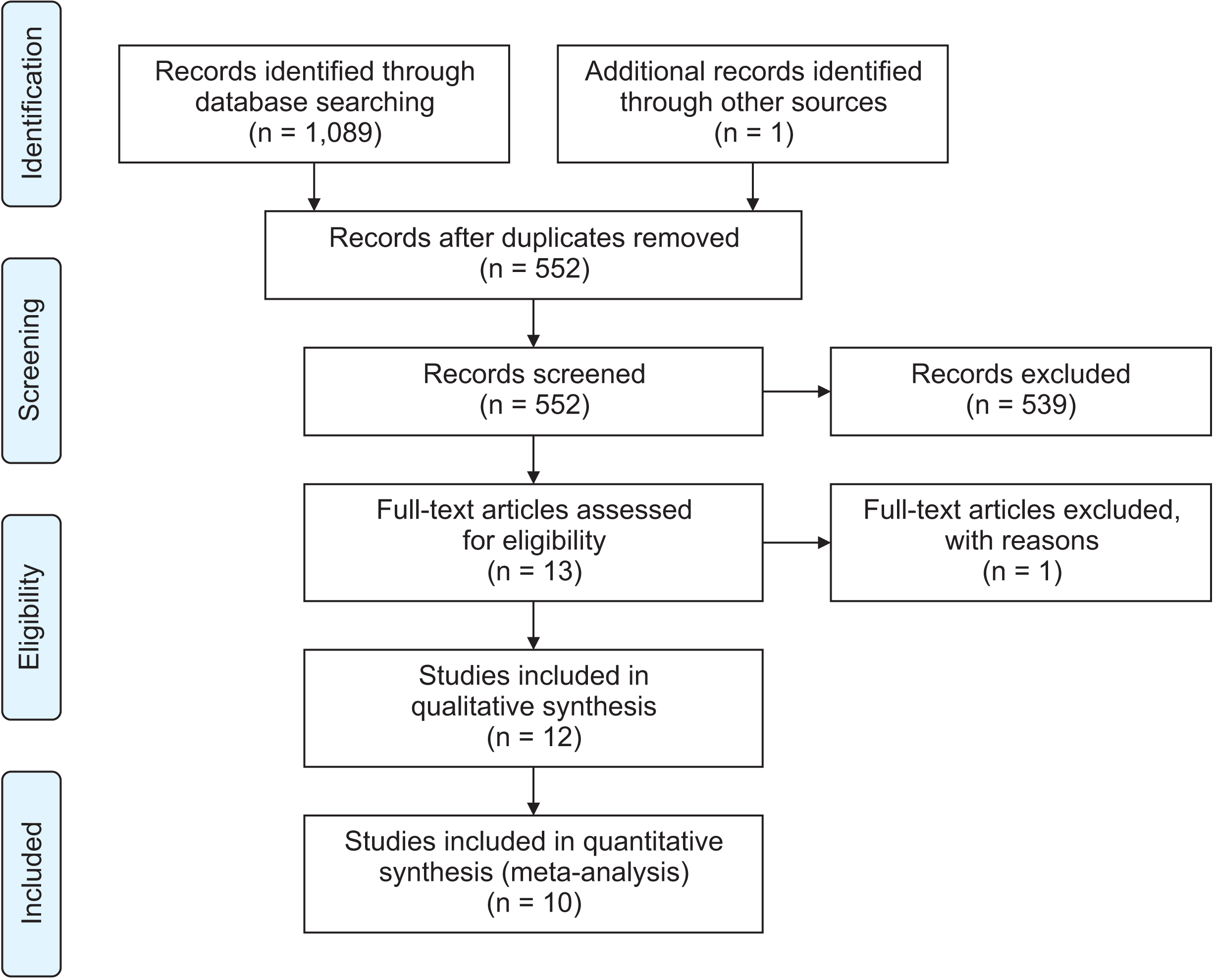
A flow diagram of the study selection process is shown in Figure 1. A total of 1,090 studies were identified through database searches and other sources. Of these, 552 studies were reviewed after duplicates were removed, and 13 full-text articles were assessed for eligibility. Finally, 12 studies were included in the qualitative synthesis and 10 studies were included in the meta-analysis.

Data extraction

Data were extracted by two reviewers using the data collection form. Data collection included size, sex (male/female), average age, appliance, activation protocol, retention duration, measurement methods, and follow-up period. Additionally, the following clinical data were collected to analyze the effects and stability of MARPE: success rate, intermolar width, alveolar width, suture expansion at the anterior and posterior nasal spine, inclination of the first molars, and alveolar height. Three measurement periods, that is, before MARPE, after MARPE, and 1 year follow-up, were selected for the meta-analysis. The definitions of these parameters are presented in Table 1. Relevant characteristics of the selected studies were also recorded.

Quality assessment

Two reviewers assessed the study quality. The quality assessment criteria introduced in the Cochrane Handbook for Systematic Reviews (version5.1.0) were used for RCTs, while the scoring protocol of the methodological index for non-randomized studies (MINORS) was used for NRCTs.

Statistical analysis

Data were presented as the mean difference with 95% confidence interval. I2 statistic and p-value were used to assess heterogeneity. No or low heterogeneity was defined as I2 ≤ 50% and p ≥ 0.1, and a fixed-effects model was applied. Moderate heterogeneity was defined as I2 > 50% and p < 0.1, and subgroup analysis or the random effects model was used. High heterogeneity was defined as I2 > 75% and p < 0.1, and descriptive analysis was used. All statistical analyses were performed using the RevMan5.3 software.

RESULTS

Study characteristics and quality

The characteristics of the selected studies are summarized in Table 2. Table 3 reported the risk of bias assessments. All 3 RCTs had low risk of bias. Among the 9 NRCTs, 3 and 6 trials were of high and moderate quality, respectively, based on the MINORS score (Table 4).

Synthesis of results

Success rate

Oliveira et al.11 reported a negative correlation between the age and MARPE success rate. The success rates of MARPE among individuals aged 15–19 years, 20–29 years, and 30–37 years were 83.3%, 81.8%, and 20%, respectively. In the study by Choi et al.,8 the success rate was 86.96%. Among the 69 patients with MTD, opening of the midpalatal suture failed in 9 patients. Lim et al.10 reported that among 38 patients diagnosed with MTD and treated with MARPE, midpalatal suture opening was not observed in five patients, and the success rate was 86.8%. In the study by Park et al.,12 the success rate was 84.2% in 19 patients with MTD. In the other included studies, the midpalatal suture was successfully opened with MARPE in all patients.

Intermolar width

Choi et al.,8 Park et al.,12 Jia et al.,13 Ngan et al.,14 and Yılmaz et al.15 reported the intermolar width before and after MARPE, and the heterogeneity test showed high homogeneity (I2 = 5%, p = 0.38); therefore, we applied a fixed-effects model to synthesize the results. The results showed that the change in the intermolar width before and after MARPE was statistically significant, with an average increase of 6.48 mm (Figure 2A).

Alveolar width

Choi et al.,8 Park et al.12 and Jia et al.13 reported the alveolar width before and after MARPE. The heterogeneity test showed high homogeneity (I2 = 0%, p = 0.59), and thus, the fixed-effects model was used for the analysis. The result showed that the change in the alveolar width before and after MARPE was statistically significant, with an average increase of 3.23 mm (Figure 2B).

Expansion pattern

Cantarella et al.16 reported that the transverse width of the suture was 4.75 mm at the anterior nasal spine (ANS) and 4.33 mm at the posterior nasal spine (PNS). Further, the amount of suture expansion at PNS was 90% of that at the ANS, indicating that the midpalatal suture was opened in parallel. In the study by Yılmaz et al.,15 the intermolar and intercanine width increased by 5.56 mm and 5.20 mm, respectively. This similar increases indicated a parallel expansion in the anteroposterior direction. Lin et al.17 also reported that the expansion pattern was parallel in the MARPE group.

Long-term stability

Lim et al.10 and Lagravère et al.18 reported changes in the intermolar width and alveolar width immediately after MARPE and 1 year later. Both heterogeneity test results showed high homogeneity (I2 = 0%, p = 0.46 for the changes of intermolar width and I2 = 0%, p = 0.74 for the changes of alveolar width). As such, a fixed-effects model was applied for the synthesis, and the results showed that the changes in the intermolar width and alveolar width were both statistically significant 1 year after expansion. Intermolar width decreased by 1.56 mm, while alveolar width decreased by 0.55 mm (Figure 2C and 2D).

Tooth inclination

Jia et al.,13 Ngan et al.,14 Lin et al.,17 Lagravère et al.,18 and Akin et al.19 reported the inclination of maxillary first molars on both sides after MARPE. The heterogeneity test showed high homogeneity (I2 = 0%, p = 0.63), and thus, the fixed-effects model was used. The results showed no statistical significance in tooth inclination between the right and left maxillary first molars. This indicated that although various degrees of inclination can be observed after MARPE, there was no significant difference between both sides (Figure 2E).

Alveolar height

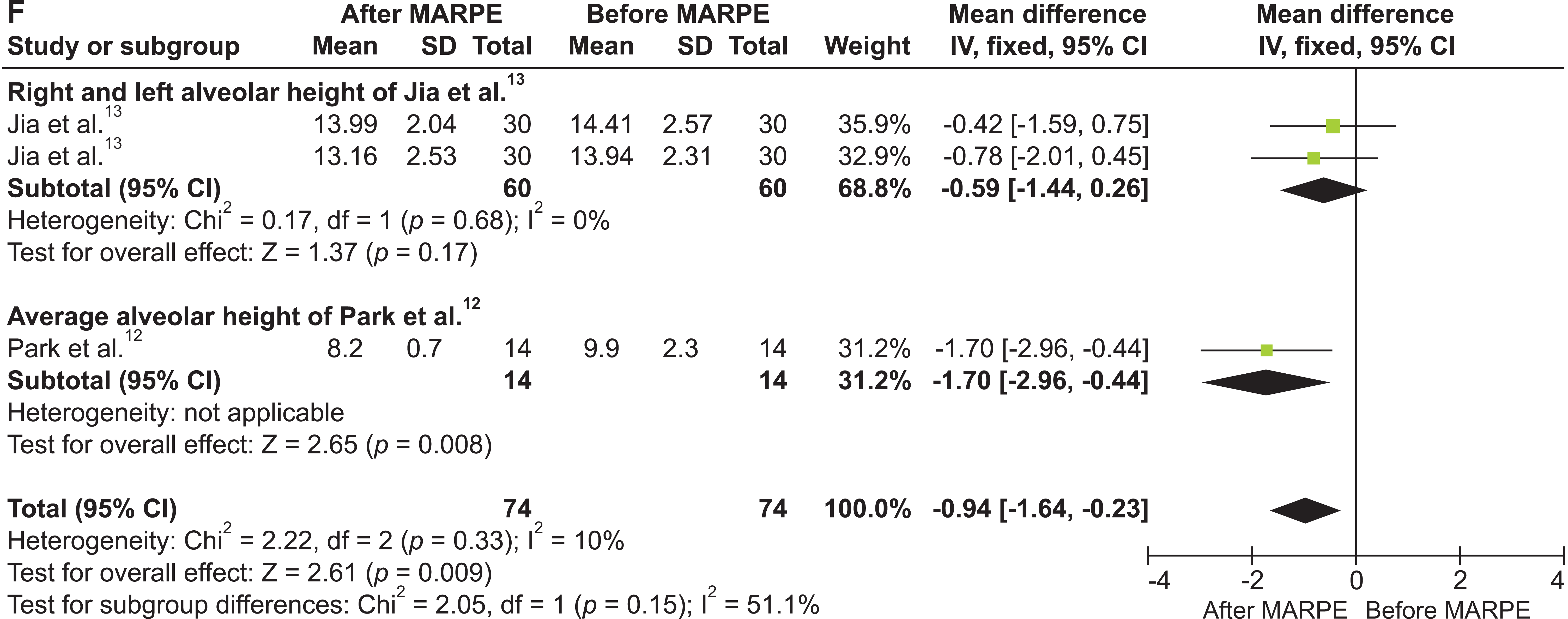
Jia et al.13 reported the buccal alveolar height of the right and left maxillary first molars, and the heterogeneity result was I2 = 0% (p = 0.68). After synthesis, the results showed no statistically significant difference between the right and left maxillary first molars, indicating a similar decrease in both sides. Park et al.12 reported change of the distance from the mesiobuccal cusp tip to the buccal alveolar crest of the maxillary first molar, indicating decrease of the buccal alveolar height. We synthesized the results of Park et al.12 and the study above and found high homogeneity (I2 = 10%, p = 0.33). The fixed-effects model was applied, and the results showed significant changes in alveolar height, with an average decrease of 0.94 (Figure 2F). Lin et al.17 also reported a statistically significant decrease of the alveolar height in the MARPE group; however, the actual amount of bone loss was not clinically significant.

Comparison with conventional RPE

Celenk-Koca et al.20 reported similar increase of the interdental width between the MARPE group and the conventional RPE group, but there were significant intergroup skeletodental differences. The miniscrew-supported expander increased the midpalatal suture 2.5 times more than the conventional tooth-borne expander. In a study by Lin et al.,17 the ratio of skeletal expansion to dental crown expansion in the C-expander group was approximately 57.5% to 77.0%, while that in the hyrax group was only 25.6% to 42.9%. Meanwhile, Yılmaz et al.15 reported similar maxillary intermolar width and palatal width in the MARPE group, while the intermolar width increased more than the palatal width in the banded expansion group, demonstrating that skeletal expansion was more significant in the MARPE group.
For tooth inclination, Celenk-Koca et al.20 reported 1.3° lingual and 3.9° buccal crown tipping of the maxillary first molars in the MARPE and conventional RPE groups, respectively. Buccal inclination in the MARPE group was significantly lower than the conventional RPE group. In the study by Yılmaz et al.,15 the MARPE group showed a significantly smaller amount of tooth inclination than the banded and bonded RPE groups. Lagravère et al.18 also reported a more significant dental inclination after expansion in the conventional RPE group.
Regarding the bone loss, Lin et al.17 reported a vertical buccal bone loss of 0.91 ± 0.68 mm on the right maxillary first molar and 0.59 ± 0.94 mm on the left side in the hyrax group. Meanwhile, in the MARPE group, it was 0.54 ± 0.49 mm and 0.27 ± 0.36 mm on the right and left sides, respectively. Similarly, Celenk-Koca et al.20 also showed less buccal bone loss in the MARPE group than in the conventional RPE group for both premolars and molars.

DISCUSSION

Age is as a significant influencing factor of the success rate of expansion.4,21 Age less than 13–15 years is considered to be optimal for conventional RPE treatment, and the effects may already be unsatisfactory in post-pubertal patients.22 In the current analysis, majority of patients in 8 of 12 studies were aged > 15 years,8,10-12,14,16,17 but they all reported a relatively high success rate of expansion, attributed to the suture structure.23 However, due to small number of studies and the differences in success rates, it is unclear whether the success rate of MARPE is superior to that of conventional RPE in late adolescents.
In addition, MARPE has greater skeletal expansion effects than conventional RPE. Handelman et al.24 evaluated the trans-arch width of molars and premolars in adults after using a Haas expander and reported that dental expansion may be the predominant effect in conventional RPE. In a study by Jia et al.,13 the ratio of skeletal to dental expansion in the MARPE group was nearly twice as high as the hyrax group. Similarly, Lin et al.17 revealed that the skeletal expansion ratio (hard palate expansion compared to the dental crown expansion) was nearly two-fold greater in the MARPE group than the hyrax group. Celenk-Koca et al.20 found that the miniscrew-supported expander had more than 2.5 times greater expansion than the tooth-anchored expander. By measuring the maxillary intermolar and palatal widths, Yılmaz et al.15 also revealed a more significant skeletal expansion in the MARPE group. These results may be attributed to the design of MARPE expanders. Additional miniscrews used as the main anchorage increase the skeletal anchorage force, helping overcome the expansion resistance.
In the study by Cantarella et al.,16 the suture showed a parallel expansion pattern at ANS and PNS. Lin et al.17 and Yılmaz et al.15 also confirm that the midpalatal suture was opened in parallel. However, Jia et al.13 and Akin et al.19 reported a V-shaped opening of the midpalatal suture, which differed from the parallel expansion pattern. This difference could be explained by the position of the device. According to Braun et al.,25 the expansion resistance is centered between the first and second molars in the sagittal plane. Jackscrews and miniscrews in the studies by Jia et al.13 and Akin et al.19 were located between the second premolars and first molars. In contrast, jackscrews and miniscrews in the study by Cantarella et al.16 were positioned between the maxillary first molars. Such a closer position to the expansion resistance center helps achieve a more parallel expansion pattern.
With respect to long-term stability, our meta-analysis showed a low rate of relapse 1 year after expansion. Although MARPE can effectively obtain skeletal expansion, it cannot avoid relapse, which also appears in SARPE and conventional RPE.26 The relapse can be explained by the elastic recovery of the soft tissue in the midpalatal suture and the shrinkage of transseptal fibers after the mechanical force is removed.27,28 Furthermore, the immaturity of the newly formed bone tissue may lead to bone resorption under a recurrence force, contributing to relapse.29,30
Tooth inclination is a typical adverse effect of RPE. Our meta-analysis indicates that various degrees of inclination can be observed after MARPE, but there was no significant difference between right and left sides. According to Lim et al.10 and Lin et al.,17 the change of tooth axis is mainly caused by bending of the alveolar bone, and absolute tooth inclination is negligible. The mechanical force of expansion appliances may cause shrinking of the periodontal membrane, bending of the alveolar bone, and inclination of the anchored teeth. Therefore, inclination is inevitable in RPE treatment. The studies by Celenk-Koca et al.,20 Yılmaz et al.,15 and Lagravère et al.18 revealed that tooth inclination was significantly lower in the MARPE group than in the conventional RPE group.
The force produced during conventional RPE was delivered to the para-midpalatal bone tissue through the anchored teeth, thereby causing buccal rotation of the alveolar bone.31-34 However, the MARPE appliance can directly deliver the force to the bone tissue and reduce the buccal force acting on the teeth, decreasing tooth inclination. Seong et al.35 analyzed the stress distribution of different types of expanders and found that the stress distribution was relatively even in MARPE and that the stress on the buccal plate of the anchored teeth was decreased. The additional miniscrews in the MARPE appliance may move the vector of the expansion force closer to the center of resistance,36 consequently, it can secure sufficient expansion with decreased tooth inclination.
Tipping movements of teeth can cause changes in the alveolar bone,37-39 resulting decrease in alveolar bone thickness and height. As shown in our meta-analysis, the buccal alveolar height decreased by an average of 0.94 mm after MARPE. According to Lin et al.17 and Celenk-Koca et al.,20 the extent of buccal bone loss in MARPE is lesser than that in conventional RPE. This could be explained by the reduced tooth inclination in MARPE, which may be correlated with the initial bone quantity. Lim et al.10 showed that changes in alveolar bone thickness and alveolar height were negatively correlated with the initial thickness and height of the alveolar bone, that is, the greater the initial bone quantity, the lesser the extent of alveolar bone loss.39,40
The findings of our meta-analysis revealed an overall beneficial effect of MARPE in treating patients with MTD. Compared with conventional RPE, MARPE had greater skeletal expansion and fewer adverse effects, including tooth inclination and alveolar height decrease. In addition, the MARPE appliance allows the use of fixed orthodontic appliances, eliminating the retention period and shortening the total treatment duration. Moreover, MARPE is more effective for patients with high angle, lacking anchorage teeth, or severe MTD.
There are some limitations to our study, the primary is the small number of articles included. Some articles were excluded from the meta-analysis due to incomplete data. Consequently, the number of included patients was insufficient, and as such, grouping and comparative analysis of different designs or placements could not be performed. This may have affected the heterogeneity of the meta-analysis. Furthermore, some methodological limitations exist because of the retrospective nature of the meta-analysis. Additional studies of high quality and large sample sizes in this field are needed to further evaluate MARPE.

CONCLUSIONS

MARPE can be an effective treatment modality for patients with MTD, with a relatively high success rate in opening the midpalatal suture in late adolescents. MARPE also has certain clinical advantages. In comparison to conventional RPE, MARPE produces greater transverse skeletal expansion and is associated with fewer detrimental periodontal effects such as tooth inclination and alveolar height decrease.

ACKNOWLEDGEMENTS

This study was supported by the National Natural Science Foundation of China (Grant Nos. 81800926).

Notes

CONFLICTS OF INTEREST

No potential conflict of interest relevant to this article was reported.

REFERENCES

1. Proffit WR, Fields HW Jr, Moray LJ. 1998; Prevalence of malocclusion and orthodontic treatment need in the United States: estimates from the NHANES III survey. Int J Adult Orthodon Orthognath Surg. 13:97–106. PMID: 9743642.
2. Angell EH. 1860; Treatment of irregularity of the permanent or adult teeth. Dental Cosmos. 1:540–4.
3. Melsen B, Melsen F. 1982; The postnatal development of the palatomaxillary region studied on human autopsy material. Am J Orthod. 82:329–42. DOI: 10.1016/0002-9416(82)90467-5. PMID: 6961805. PMID: https://www.scopus.com/inward/record.uri?partnerID=HzOxMe3b&scp=0020374694&origin=inward.
4. Persson M, Thilander B. 1977; Palatal suture closure in man from 15 to 35 years of age. Am J Orthod. 72:42–52. DOI: 10.1016/0002-9416(77)90123-3. PMID: 267435. PMID: https://www.scopus.com/inward/record.uri?partnerID=HzOxMe3b&scp=0017379719&origin=inward.
5. Cao Y, Zhou Y, Song Y, Vanarsdall RL Jr. 2009; Cephalometric study of slow maxillary expansion in adults. Am J Orthod Dentofacial Orthop. 136:348–54. Erratum in: Am J Orthod Dentofacial Orthop 2009;136:760. DOI: 10.1016/j.ajodo.2008.03.017. PMID: 19732668. PMID: https://www.scopus.com/inward/record.uri?partnerID=HzOxMe3b&scp=69249245460&origin=inward.
6. Betts NJ, Vanarsdall RL, Barber HD, Higgins-Barber K, Fonseca RJ. 1995; Diagnosis and treatment of transverse maxillary deficiency. Int J Adult Orthodon Orthognath Surg. 10:75–96. PMID: 9082002. PMID: https://www.scopus.com/inward/record.uri?partnerID=HzOxMe3b&scp=0029422049&origin=inward.
7. Cureton SL, Cuenin M. 1999; Surgically assisted rapid palatal expansion: orthodontic preparation for clinical success. Am J Orthod Dentofacial Orthop. 116:46–59. DOI: 10.1016/S0889-5406(99)70302-1. PMID: 10393580. PMID: https://www.scopus.com/inward/record.uri?partnerID=HzOxMe3b&scp=0033158865&origin=inward.
8. Choi SH, Shi KK, Cha JY, Park YC, Lee KJ. 2016; Nonsurgical miniscrew-assisted rapid maxillary expansion results in acceptable stability in young adults. Angle Orthod. 86:713–20. DOI: 10.2319/101415-689.1. PMID: 26938955. PMCID: PMC8600851. PMID: https://www.scopus.com/inward/record.uri?partnerID=HzOxMe3b&scp=84984698676&origin=inward.
9. Lee SR, Lee JW, Chung DH, Lee SM. 2020; Short-term impact of microimplant-assisted rapid palatal expansion on the nasal soft tissues in adults: a three-dimensional stereophotogrammetry study. Korean J Orthod. 50:75–85. DOI: 10.4041/kjod.2020.50.2.75. PMID: 32257933. PMCID: PMC7093666. PMID: https://www.scopus.com/inward/record.uri?partnerID=HzOxMe3b&scp=85083220475&origin=inward.
10. Lim HM, Park YC, Lee KJ, Kim KH, Choi YJ. 2017; Stability of dental, alveolar, and skeletal changes after miniscrew-assisted rapid palatal expansion. Korean J Orthod. 47:313–22. DOI: 10.4041/kjod.2017.47.5.313. PMID: 28861393. PMCID: PMC5548712. PMID: https://www.scopus.com/inward/record.uri?partnerID=HzOxMe3b&scp=85027020683&origin=inward.
11. Oliveira CB, Ayub P, Angelieri F, Murata WH, Suzuki SS, Ravelli DB, et al. 2021; Evaluation of factors related to the success of miniscrew-assisted rapid palatal expansion. Angle Orthod. 91:187–94. DOI: 10.2319/051420-436.1. PMID: 33351888. PMCID: PMC8028491. PMID: https://www.scopus.com/inward/record.uri?partnerID=HzOxMe3b&scp=85102965531&origin=inward.
12. Park JJ, Park YC, Lee KJ, Cha JY, Tahk JH, Choi YJ. 2017; Skeletal and dentoalveolar changes after miniscrew-assisted rapid palatal expansion in young adults: a cone-beam computed tomography study. Korean J Orthod. 47:77–86. DOI: 10.4041/kjod.2017.47.2.77. PMID: 28337417. PMCID: PMC5359634. PMID: https://www.scopus.com/inward/record.uri?partnerID=HzOxMe3b&scp=85016124277&origin=inward.
13. Jia H, Zhuang L, Zhang N, Bian Y, Li S. 2021; Comparison of skeletal maxillary transverse deficiency treated by microimplant-assisted rapid palatal expansion and tooth-borne expansion during the post-pubertal growth spurt stage. Angle Orthod. 91:36–45. DOI: 10.2319/041920-332.1. PMID: 33289835. PMCID: PMC8032284. PMID: https://www.scopus.com/inward/record.uri?partnerID=HzOxMe3b&scp=85098713317&origin=inward.
14. Ngan P, Nguyen UK, Nguyen T, Tremont T, Martin C. 2018; Skeletal, dentoalveolar, and periodontal changes of skeletally matured patients with maxillary deficiency treated with microimplant-assisted rapid palatal expansion appliances: a pilot study. APOS Trends Orthod. 8:71–85. DOI: 10.4103/apos.apos_27_18.
15. Yılmaz A, Arman-Özçırpıcı A, Erken S, Polat-Özsoy Ö. 2015; Comparison of short-term effects of mini-implant-supported maxillary expansion appliance with two conventional expansion protocols. Eur J Orthod. 37:556–64. DOI: 10.1093/ejo/cju094. PMID: 25564504. PMID: https://www.scopus.com/inward/record.uri?partnerID=HzOxMe3b&scp=84943409917&origin=inward.
16. Cantarella D, Dominguez-Mompell R, Mallya SM, Moschik C, Pan HC, Miller J, et al. 2017; Changes in the midpalatal and pterygopalatine sutures induced by micro-implant-supported skeletal expander, analyzed with a novel 3D method based on CBCT imaging. Prog Orthod. 18:34. DOI: 10.1186/s40510-017-0188-7. PMID: 29090368. PMCID: PMC5663987. PMID: https://www.scopus.com/inward/record.uri?partnerID=HzOxMe3b&scp=85032944341&origin=inward.
17. Lin L, Ahn HW, Kim SJ, Moon SC, Kim SH, Nelson G. 2015; Tooth-borne vs bone-borne rapid maxillary expanders in late adolescence. Angle Orthod. 85:253–62. DOI: 10.2319/030514-156.1. PMID: 25490552. PMCID: PMC8631879. PMID: https://www.scopus.com/inward/record.uri?partnerID=HzOxMe3b&scp=84924918691&origin=inward.
18. Lagravère MO, Carey J, Heo G, Toogood RW, Major PW. 2010; Transverse, vertical, and anteroposterior changes from bone-anchored maxillary expansion vs traditional rapid maxillary expansion: a randomized clinical trial. Am J Orthod Dentofacial Orthop. 137:304.e1–12. discussion 304–5. DOI: 10.1016/j.ajodo.2009.09.016. PMID: 20197161.
19. Akin M, Akgul YE, Ileri Z, Basciftci FA. 2016; Three-dimensional evaluation of hybrid expander appliances: a pilot study. Angle Orthod. 86:81–6. DOI: 10.2319/121214-902.1. PMID: 25923245. PMCID: PMC8603971. PMID: https://www.scopus.com/inward/record.uri?partnerID=HzOxMe3b&scp=85000401648&origin=inward.
20. Celenk-Koca T, Erdinc AE, Hazar S, Harris L, English JD, Akyalcin S. 2018; Evaluation of miniscrew-supported rapid maxillary expansion in adolescents: a prospective randomized clinical trial. Angle Orthod. 88:702–9. DOI: 10.2319/011518-42.1. PMID: 30102085. PMCID: PMC8174075. PMID: https://www.scopus.com/inward/record.uri?partnerID=HzOxMe3b&scp=85055816459&origin=inward.
21. Stuart DA, Wiltshire WA. 2003; Rapid palatal expansion in the young adult: time for a paradigm shift? J Can Dent Assoc. 69:374–7. PMID: 12787474. PMID: https://www.scopus.com/inward/record.uri?partnerID=HzOxMe3b&scp=0037530291&origin=inward.
22. Bishara SE, Staley RN. 1987; Maxillary expansion: clinical implications. Am J Orthod Dentofacial Orthop. 91:3–14. DOI: 10.1016/0889-5406(87)90202-2. PMID: 3541577. PMID: https://www.scopus.com/inward/record.uri?partnerID=HzOxMe3b&scp=0023175086&origin=inward.
23. Cohen MM Jr. 1993; Sutural biology and the correlates of craniosynostosis. Am J Med Genet. 47:581–616. DOI: 10.1002/ajmg.1320470507. PMID: 8266985. PMID: https://www.scopus.com/inward/record.uri?partnerID=HzOxMe3b&scp=0027383692&origin=inward.
24. Handelman CS, Wang L, BeGole EA, Haas AJ. 2000; Nonsurgical rapid maxillary expansion in adults: report on 47 cases using the Haas expander. Angle Orthod. 70:129–44. DOI: 10.1043/0003-3219(2000)070<0129:NRMEIA>2.0.CO;2. PMID: 10833001.
25. Braun S, Bottrel JA, Lee KG, Lunazzi JJ, Legan HL. 2000; The biomechanics of rapid maxillary sutural expansion. Am J Orthod Dentofacial Orthop. 118:257–61. DOI: 10.1067/mod.2000.108254. PMID: 10982925. PMID: https://www.scopus.com/inward/record.uri?partnerID=HzOxMe3b&scp=0034264360&origin=inward.
26. Gauthier C, Voyer R, Paquette M, Rompré P, Papadakis A. 2011; Periodontal effects of surgically assisted rapid palatal expansion evaluated clinically and with cone-beam computerized tomography: 6-month preliminary results. Am J Orthod Dentofacial Orthop. 139(4 Suppl):S117–28. DOI: 10.1016/j.ajodo.2010.06.022. PMID: 21435529. PMID: https://www.scopus.com/inward/record.uri?partnerID=HzOxMe3b&scp=79952981513&origin=inward.
27. Kanzaki R, Daimaruya T, Takahashi I, Mitani H, Sugawara J. 2007; Remodeling of alveolar bone crest after molar intrusion with skeletal anchorage system in dogs. Am J Orthod Dentofacial Orthop. 131:343–51. DOI: 10.1016/j.ajodo.2006.04.025. PMID: 17346589. PMID: https://www.scopus.com/inward/record.uri?partnerID=HzOxMe3b&scp=33847418932&origin=inward.
28. Storey E. 1973; Tissue response to the movement of bones. Am J Orthod. 64:229–47. DOI: 10.1016/0002-9416(73)90017-1. PMID: 4199007. PMID: https://www.scopus.com/inward/record.uri?partnerID=HzOxMe3b&scp=0015819709&origin=inward.
29. Hou B, Fukai N, Olsen BR. 2007; Mechanical force-induced midpalatal suture remodeling in mice. Bone. 40:1483–93. DOI: 10.1016/j.bone.2007.01.019. PMID: 17398175. PMCID: PMC1939974. PMID: https://www.scopus.com/inward/record.uri?partnerID=HzOxMe3b&scp=34248566783&origin=inward.
30. Ma J, Wu Y, Zhang W, Smales RJ, Huang Y, Pan Y, et al. 2008; Up-regulation of multiple proteins and biological processes during maxillary expansion in rats. BMC Musculoskelet Disord. 9:37. DOI: 10.1186/1471-2474-9-37. PMID: 18366685. PMCID: PMC2291465. PMID: https://www.scopus.com/inward/record.uri?partnerID=HzOxMe3b&scp=42049120035&origin=inward.
31. Garib DG, Henriques JF, Janson G, Freitas MR, Coelho RA. 2005; Rapid maxillary expansion--tooth tissue-borne versus tooth-borne expanders: a computed tomography evaluation of dentoskeletal effects. Angle Orthod. 75:548–57. DOI: 10.1043/0003-3219(2005)75[548:RMETVT]2.0.CO;2. PMID: 16097223.
32. Garrett BJ, Caruso JM, Rungcharassaeng K, Farrage JR, Kim JS, Taylor GD. 2008; Skeletal effects to the maxilla after rapid maxillary expansion assessed with cone-beam computed tomography. Am J Orthod Dentofacial Orthop. 134:8–9. DOI: 10.1016/j.ajodo.2008.06.004. PMID: 18617096.
33. Lione R, Ballanti F, Franchi L, Baccetti T, Cozza P. 2008; Treatment and posttreatment skeletal effects of rapid maxillary expansion studied with low-dose computed tomography in growing subjects. Am J Orthod Dentofacial Orthop. 134:389–92. DOI: 10.1016/j.ajodo.2008.05.011. PMID: 18774085. PMID: https://www.scopus.com/inward/record.uri?partnerID=HzOxMe3b&scp=50849144400&origin=inward.
34. Weissheimer A, de Menezes LM, Mezomo M, Dias DM, de Lima EM, Rizzatto SM. 2011; Immediate effects of rapid maxillary expansion with Haas-type and hyrax-type expanders: a randomized clinical trial. Am J Orthod Dentofacial Orthop. 140:366–76. DOI: 10.1016/j.ajodo.2010.07.025. PMID: 21889081. PMID: https://www.scopus.com/inward/record.uri?partnerID=HzOxMe3b&scp=80052350652&origin=inward.
35. Seong EH, Choi SH, Kim HJ, Yu HS, Park YC, Lee KJ. 2018; Evaluation of the effects of miniscrew incorporation in palatal expanders for young adults using finite element analysis. Korean J Orthod. 48:81–9. DOI: 10.4041/kjod.2018.48.2.81. PMID: 29564217. PMCID: PMC5854885. PMID: https://www.scopus.com/inward/record.uri?partnerID=HzOxMe3b&scp=85044119408&origin=inward.
36. Koo YJ, Choi SH, Keum BT, Yu HS, Hwang CJ, Melsen B, et al. 2017; Maxillomandibular arch width differences at estimated centers of resistance: comparison between normal occlusion and skeletal Class III malocclusion. Korean J Orthod. 47:167–75. DOI: 10.4041/kjod.2017.47.3.167. PMID: 28523243. PMCID: PMC5432438. PMID: https://www.scopus.com/inward/record.uri?partnerID=HzOxMe3b&scp=85031101300&origin=inward.
37. Thilander B, Nyman S, Karring T, Magnusson I. 1983; Bone regeneration in alveolar bone dehiscences related to orthodontic tooth movements. Eur J Orthod. 5:105–14. DOI: 10.1093/ejo/5.2.105. PMID: 6574916. PMID: https://www.scopus.com/inward/record.uri?partnerID=HzOxMe3b&scp=0020757717&origin=inward.
38. Tian YL, Liu F, Sun HJ, Lv P, Cao YM, Yu M, et al. 2015; Alveolar bone thickness around maxillary central incisors of different inclination assessed with cone-beam computed tomography. Korean J Orthod. 45:245–52. DOI: 10.4041/kjod.2015.45.5.245. PMID: 26445719. PMCID: PMC4593869. PMID: https://www.scopus.com/inward/record.uri?partnerID=HzOxMe3b&scp=84942318935&origin=inward.
39. Garib DG, Henriques JF, Janson G, de Freitas MR, Fernandes AY. 2006; Periodontal effects of rapid maxillary expansion with tooth-tissue-borne and tooth-borne expanders: a computed tomography evaluation. Am J Orthod Dentofacial Orthop. 129:749–58. DOI: 10.1016/j.ajodo.2006.02.021. PMID: 16769493. PMID: https://www.scopus.com/inward/record.uri?partnerID=HzOxMe3b&scp=33744918064&origin=inward.
40. Rungcharassaeng K, Caruso JM, Kan JY, Kim J, Taylor G. 2007; Factors affecting buccal bone changes of maxillary posterior teeth after rapid maxillary expansion. Am J Orthod Dentofacial Orthop. 132:428.e1–8. DOI: 10.1016/j.ajodo.2007.02.052. PMID: 17920493. PMID: https://www.scopus.com/inward/record.uri?partnerID=HzOxMe3b&scp=34848823758&origin=inward.

Figure 1
Flow diagram showing the study selection.
kjod-52-5-334-f1.tif
Figure 2
Forest plot of the effects and stability of miniscrew-assisted rapid palatal expansion (MARPE). The outcomes assessed are: (A) intermolar width before and after MARPE; (B) alveolar width before and after MARPE; (C) intermolar width immediately after and 1 year after MARPE; (D) alveolar width immediately after and 1 year after MARPE; (E) tooth inclination of the right and left maxillary first molars after MARPE; and (F) buccal alveolar height of the right and left maxillary first molars before and after MARPE.
CI, confidence interval; IV, inverse variance; SD, standard deviation.
kjod-52-5-334-f2a.tifkjod-52-5-334-f2b.tif
Table 1
Variable definition
Variable Definition
Intermolar width Linear distance between right and left maxillary first molars
Alveolar width Linear distance between right and left maxillary first molar alveolar bone
Suture expansion at ANS Transverse width between anterior nasal spine points
Suture expansion at PNS Transverse width between posterior nasal spine points
Tooth inclination Angle between the long axis of the maxillary first molar and the palatal plane
Alveolar height Distance from the most inferior alveolar point at mesiobucccal root of maxillary first molar to
baseline

Baseline: Line passing through the lowest point at the inferior inner contour of the nasal cavity on the same side, parallel to the axial plane.

Table 2
Characteristics of included studies
Author Year Design Group Size Sex (male/female) Average age (yr) Appliance Activation protocol Retention time Measurement methods Follow-up
Lagravère et al.18 2010 >RCT >MARPE 21 >8/13 14.24 ± 1.32 >Bone-borne >0.25 mm/d >6 months >CBCT >6 months, 1 year
>RPE 20 >5/15 14.05 ± 1.35 >Hyrax expander >0.5 mm/d >6 months
>Control 21 >6/15 12.86 ± 1.19
Lin et al.17 2015 >NRCT >MARPE 15 >0/15 18.1 ± 4.4 >C-expander >0.25 mm/d >CBCT >3 months
>RPE 13 >0/13 17.4 ± 3.4 >Hyrax expander
Yılmaz et al.15 2015 >NRCT >MARME 14 >8/6 13.2 ± 2.1 >Bone-borne expander 0.4 mm/d for first 7–10 d, 0.2 mm/3 d after the opening of the suture Cephalograms, posteroanterior radiographs, dental casts
>Bonded RME 14 >8/6 12.1 ± 2.1 >Bonded expander
>Banded RME 14 >6/8 13.4 ± 1.7 >Banded expander
Akin et al.19 2016 >NRCT >MARPE 9 >5/4 13.61 ± 0.72 >Hybrid expander 0.5 mm/d for the first week, then 0.25 mm/d for 3 weeks >CBCT
Choi et al.8 2016 >NRCT >MARPE 20 >10/10 20.9 ± 2.9 >Bone-borne expander >0.2 mm/2 d >3 months Posteroanterior cephalograms >30.2 ± 13.2 months
Cantarella et al.16 2017 >NRCT >MARPE 15 >6/9 17.2 ± 4.2 Maxillary skeletal expander 0.5 mm/d at first, then 0.25 mm/d after inter-incisal diastema appeared >More than 3 months >CBCT
Lim et al.10 2017 >NRCT >MARPE 24 >8/16 21.6 ± 3.1 >Bone-borne expander >0.2 mm/d >4 months >CBCT 1 year (14.17 ± 2.70 month)
Park et al.12 2017 >NRCT >MARPE 14 >9/5 20.1 ± 2.4 >Bone-borne expander >0.2 mm/d >CBCT
Celenk-Koca et al.20 2018 >RCT >Conventional-
RME
20 >8/12 13.84 ± 1.36 >Hyrax expander >2 turns/d >CBCT
>MARPE 20 >7/13 13.81 ± 1.23 Miniscrew-support expander
Ngan et al.14 2018 >NRCT >MARPE 8 >6/2 21.9 ± 1.5 Maxillary skeletal expander Varied with the severity of transverse discrepancy >CBCT
Oliveira et al.11 2020 >NRCT >MARPE 28 >10/18 15–37 Miniscrew-support expander >2/4 turn/d >4 months >CBCT >6 months
Jia et al.13 2021 >RCT >MARPE 30 >9/21 14.8 ± 1.5 >Hyrax expander >0.5 mm/d >3 months >CBCT
>Hyrax 30 >12/18 15.1 ± 1.6 Four-point MARPE expander

RCT, randomized controlled trial; NRCT, non-randomized controlled trial; MARPE, miniscrew-assisted rapid palatal expansion; RPE, rapid palatal expansion; RME, rapid maxillary expansion; CBCT, cone beam computed tomography.

Table 3
Risk of bias for randomized controlled trials based on Cochrane’s risk of bias tool
Author Year Random sequence generation Allocation concealment Blinding of participants and personnel Blinding of outcome assessment Incomplete outcome data Selective reporting Other sources of bias Overall bias
Lagravère et al.18 2010 Low risk Unclear Low risk Unclear Low risk Unclear Low risk Low risk
Celenk-Koca
et al.20
2018 Low risk Low risk Low risk Low risk Low risk Unclear Low risk Low risk
Jia et al.13 2021 Low risk Low risk Low risk Unclear Low risk Unclear Low risk Low risk
Table 4
Methodological index for non-randomized controlled trials based on MINORS
Author Year 1 2 3 4 5 6 7 8 9 10 11 12 Total
Lin et al.17 2015 2 1 2 2 1 1 2 0 2 1 1 2 17
Yılmaz et al.15 2015 2 1 2 2 2 0 2 2 2 2 2 2 21
Akin et al.19 2016 2 1 2 2 0 0 2 0 9
Choi et al.8 2016 2 2 2 2 0 2 0 2 12
Cantarella et al.16 2017 2 2 2 2 0 0 2 0 10
Lim et al.10 2017 2 2 2 2 0 2 2 2 14
Park et al.12 2017 2 2 2 2 0 0 2 0 10
Ngan et al.14 2018 2 2 2 2 0 0 2 0 10
Oliveira et al.11 2020 2 2 2 2 2 0 2 2 14

Items 1–12 represent the following: (1) a clearly stated aim; (2) inclusion criteria for consecutive patients; (3) prospective collection of data; (4) endpoints appropriate for the aim of this study; (5) unbiased assessment of the study endpoint; (6) follow-up period appropriate to the aim of the study; (7) loss to follow-up of less than 5%; (8) prospective calculation of study size; (9) adequate control group; (10) contemporary group; (11) baseline equivalence of groups; and (12) appropriate statistical analyses. A score of 0 means not mentioned, 1 means reported but inadequate, and 2 means reported and adequate. The total score is 24 for studies with control groups and 16 for studies without control groups. Quality is considered low (0–9 for studies with control groups, 0–7 for studies without control groups), moderate (10–20 for studies with control groups, 8–13 for studies without control groups), or high (20–24 for studies with control groups, 14–16 for studies without control groups) based on the total score.

MINORS, methodological index for non-randomized studies.

TOOLS
Similar articles