

 PDF
PDF Citation
 Print
Print


INTRODUCTION
Robin sequence (RS) is a rare congenital condition with remarkable phenotypic heterogeneity due to its association with various craniofacial malformations. A recent clinical consensus report defines the triad of RS as micrognathia, glossoptosis, and upper airway obstruction (UAO).1 A palatal cleft is present in 80–90% of infants with RS.2 Severe feeding and breathing difficulties are the two primary concerns in the management of neonates with RS. Discordance of the suck-swallow-breath mechanism in infants with RS often results from unsecured upper airway during swallowing due to tongue base obstruction.3 Interceptive treatment to separate the tongue base away from the posterior pharyngeal wall, therefore, can be an effective mechanism in reclaiming the pharyngeal airway space.
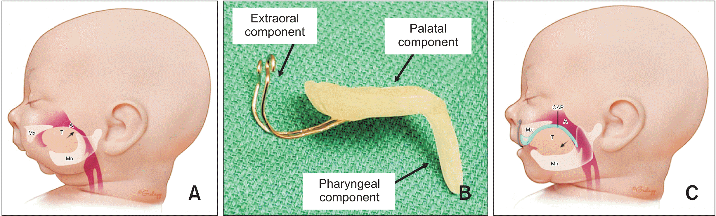
Orthodontic airway plate (OAP) treatment, first introduced in Europe in 1967,4 is a nonsurgical option among various surgical and nonsurgical treatments for neonates with RS with tongue-base UAO. Tübingen palatal plate (TPP) and pre-epiglottal baton plate (PEBP) are examples of conventional OAPs with varying iterations in design and application.5,6 Briefly, an OAP is comprised of three components: palatal, pharyngeal, and extraoral (Figure 1). The palatal component (palatal plate) blocks the tongue from passing through the palatal cleft, separates the nasal cavity and the oral cavity, and provides a solid surface for efficient bottle feeding. The pharyngeal component (velar spur) prevents the tongue from prolapsing, establishes a secure pharyngeal airway, and enables the anterior tongue positioning for bottle feeding. Mandibular catch-up growth of RS infants treated with the OAP has been suggested to be a positive association with the anterior positioning of the tongue by an OAP.7 The extraoral component (a pair of anterior extension wires) functions as hooks connecting to the facial tapes for additional retention of an OAP. Figure 1 illustrates an example of a conventional OAP and how it is situated inside a RS patient’s mouth to enlarge the airway.
Despite the proven efficacy, craniofacial orthodontists are often hesitant to initiate OAP treatment because the overall process is extremely time-consuming and labor-intensive requiring multiple periodic adjustments to accommodate a neonate’s rapid maxillary growth during several months of treatment. This article reports an innovative method of manufacturing and utilizing an OAP to increase efficiency over current clinical and procedural challenges.
SPLIT ORTHODONTIC AIRWAY PLATE
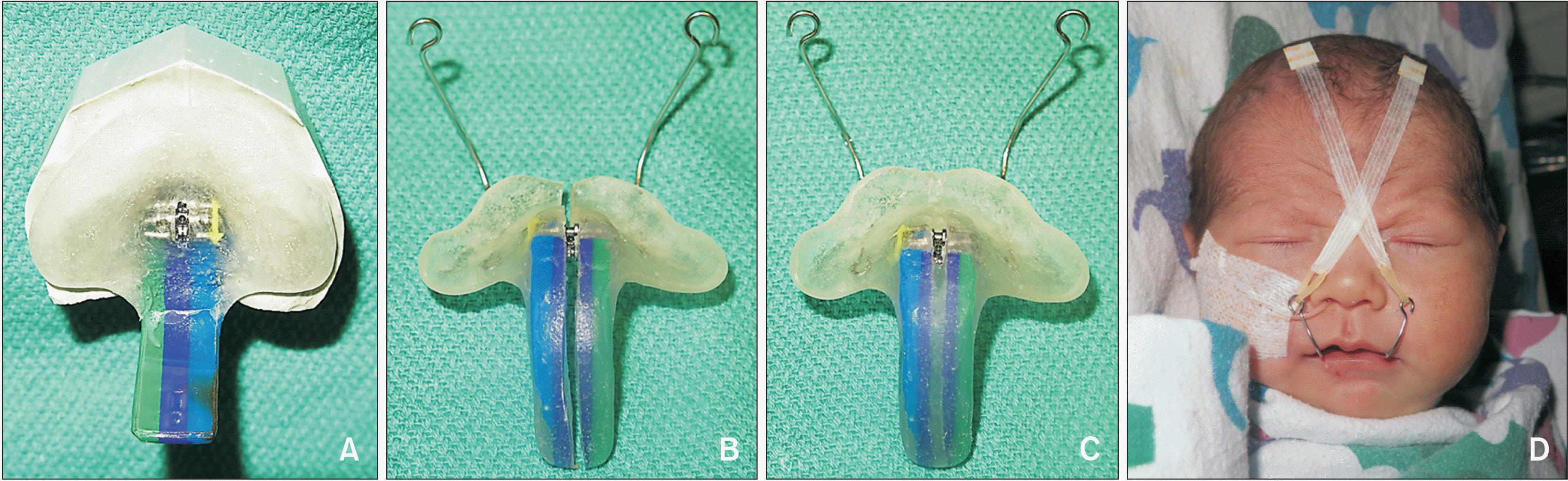
The innovation of split orthodontic airway plate (S-OAP) begins with an installation of a mini-expansion screw (Dentaurum, Ispringen, Germany) at the center of the palatal component of the OAP during fabrication in an orthodontic laboratory (Figure 2A).8,9 Only after placing an expansion screw is the splint acrylic (Great Lakes Orthodontics Ltd., Tonawanda, NY, USA) poured to build the palatal component, followed by connecting the pharyngeal component as previously described.10 A S-OAP is adjusted and installed inside a RS baby’s mouth following the same protocol as conventional OAP delivery.11 As the patient grows, an appearance of a linear continuous pressure mark on the labial slope of the maxillary alveolar ridge is an indication that the palatal plate is becoming too small for the growing maxillary arch. At this time, the S-OAP is split in half and slightly expanded by turning the embedded jackscrew (Figure 2B). The split is then re-sealed using fresh splint acrylic (Great Lakes Orthodontics Ltd.) to maintain the structural integrity and rigidity of the S-OAP prior to reinsertion inside the patient’s mouth (Figure 2C). Figure 2D shows a facial frontal view of an infant wearing the S-OAP. This enlargement mechanism is precise and quantifiable in contrast to the existing conventional method of arbitrarily grinding the palatal acrylic. The internal anatomy of the palatal plate is preserved, which continues to provide intimate and continuous contact between the palatal plate and alveolar ridge despite the enlargement of the palatal plate. The acrylic at areas where enlargement is not desirable (palatal cleft and lateral surfaces of the velar spur) must be ground off to maintain its original width. This cycle of split enlargement is repeated every 1–2 weeks at the neonatal intensive care unit (NICU) and every 3–4 weeks at the ambulatory craniofacial airway orthodontic clinic following the neonate’s hospital discharge.
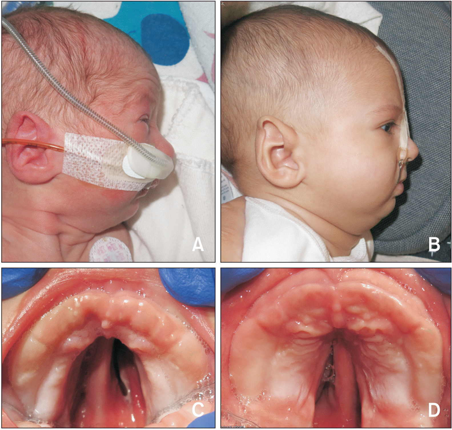
At our institution, using this S-OAP protocol, seven infants with RS were able to complete the treatment using just one device per patient; hence, the conventional requirement of re-impression, re-fabrication, and re-delivery of multiple OAPs as mid-course corrections was avoided. Figure 3 shows an exemplary patient with isolated RS treated using a S-OAP. As expected with any OAPs, this infant’s mandible demonstrated catch-up growth over 6 months of treatment. The palatal plate was periodically enlarged using the S-OAP protocol. Five turns of expansion screw activation were applied at a 4-week interval. A significant reduction of the size of palatal cleft was also noticeable.
DISCUSSION
The safety and effectiveness of nonsurgical OAP treatment in improving breathing and oral feeding difficulties of neonates with RS have been well documented in previous literature.11-14 Its pharyngeal component dictates the posterior limit of the tongue base and serves as the anterior border of the pharynx, thereby guaranteeing the patency of upper airway for infants with RS. Although clinical data suggest that the OAP treatment may stimulate the mandibular catch-up growth in infants with RS, it is only speculated that the growth may occur in the context of the Melvin Moss’ functional matrix theory postulating reactive nature of the bone growth to specific functional demands such as the guided anterior tongue movement.7,15
In conventional OAP treatments using PEBP or TPP, periodic adjustments to accommodate an infant’s rapid maxillary growth are achieved by subjectively grinding off acrylic layers from the tissue side of the palatal plate. Excessive grinding can compromise the intimate adaptation between the gingivopalatal tissue and the OAP, greatly reducing the OAP’s intraoral retention and comfort. Conversely, insufficient and uneven grinding may lead to restricted maxillary growth and can cause intraoral pressure marks. Inevitably, it is often required that the initial OAP be replaced by a new larger OAP every two to three months, which means obtaining a new maxillary impression, fabricating a new custom-fit device, and appliance delivery under awake nasopharyngoscopy in collaboration with pediatric otolaryngology at the NICU. All these repetitive procedures exhaust significant amounts of resources from the family, insurance payors, and healthcare professionals of the transdisciplinary high-risk care unit.5,6
In 2019, our institution adopted the OAP treatment for the first time in the United States and recognized the need for modifying the existing utilization protocol in order to reduce labor-intensive and time-consuming aspects of its application. The S-OAP is the product of our efforts to avoid repeated hospital admission, maxillary impression, fabrication, delivery, and adjustments of new OAPs in the NICU. The innovative concept was conceived following the philosophy of Biocreative Orthodontic Strategy with an emphasis on simplicity, efficacy, and comfort of orthodontic devices for patients.16 Installation of a mini-expansion screw on the mid-sagittal plane at the center of the palatal plate of a conventional OAP provides a mechanism for a quick and quantifiable enlargement of the palatal plate and eliminates guesswork of grinding the acrylic. The intimate but evenly spacious contact between the tissue and the S-OAP appears to be critical for providing optimal retention of the device and sufficient room for infant maxillary growth.
It is noteworthy that the palatal seal will leak at the expansion screw segment. However, nutritious suckling of infants with palatal cleft is accomplished by effective expression of milk against a solid palatal surface using a Dr. Brown's® specialty nipple/bottle system (DrBrowns, St. Louis, MO, USA) with an infant-paced feeding valve, which does not require complete palatal seal. All isolated RS babies (without associations with other anomalies) treated using a S-OAP at our institution showed successful oral feeding with excellent weight gain trajectory, achieving 100% oral feeding within a few months of wearing a S-OAP. Similar to conventional OAPs, a S-OAP also triggers a gagging reflex when the device is inserted inside the mouth. Although it no longer triggers a gagging reflex when the device remains inside the mouth, caregivers are recommended to perform the daily cleaning and reinsertion of the device approximately 30–60 minutes prior to the next scheduled feeding in order to avoid risks of emesis or aspiration.
With aggregate expertise in orthodontics, medicine, surgery, and engineering using health innovation technologies, our team continues to investigate modalities for creating more user-friendly and patient-centered procedural protocols in the implementation of OAP treatment for neonates with RS.
CONCLUSIONS
The OAP is a safe and effective nonsurgical treatment for RS. A craniofacial orthodontist’s leading role in a transdisciplinary management team for neonates and infants with RS is critical to the success of OAP treatments. Despite extensive evidence of the therapeutic efficacy, OAP treatments are still infrequently offered around the world. This may be due to demands of laborious and time-consuming adjustments and re-fabrication requirements for rapidly growing patients. Innovation of a S-OAP protocol may mitigate a craniofacial orthodontist’s apprehension to initiate OAP treatment and serve to remind our profession of the importance of craniofacial orthodontics in the management of growth and development of very young patients.
 XML Download