Abstract
Glossectomy combined with radiotherapy causes different levels of tongue function disorders and leads to severe malocclusion, with poor periodontal status in cancer survivors. Although affected patients require regular access to orthodontic care, special considerations are crucial for treatment planning. This case report describes the satisfactory orthodontic management for the correction of severe dental crowding in a 43-year-old female 6 years after treatment for tongue cancer with total glossectomy combined with radiotherapy, to envision the possibility of orthodontic care for oral cancer survivors. Extraction was performed to correct dental crowding and establish proper occlusion following alignment, after considering the possibility of osteoradionecrosis. Orthodontic mini-implants were used to provide skeletal anchorage required for closure of the extraction space and intrusion of the anterior teeth. The dental crowding was corrected, and Class I occlusal relationship was established after 36 months of treatment. The treatment outcome was sustained after 15 months of retention, and long-term follow-up was recommended.
Tongue squamous cell carcinoma (SCC) is the most common oral malignancy.1 It predominantly occurs in tobacco and alcohol users in the sixth and seventh decades of life.1,2 However, recent research from several countries has demonstrated a notable increase in the frequency of SCC among young patients, including non-smokers and non-drinkers.1-3 Systematic oral examinations are considered key for contributing to the early detection of oral cancer.3
Tongue SCC can be treated with surgery, radiotherapy, chemotherapy, or combinations of these approaches, depending on the individual case.4 Partial or total glossectomy results in tongue function disorders of varying intensity, which in turn causes uneven pressure distribution between the tongue and perioral muscles.5,6 This pressure imbalance plays a major role in the occurrence and development of malocclusion.7 Moreover, radiotherapy for malignant head and neck tumors has significant limitations owing to acute and late toxiciy. This irreversible damage to the oral tissues often leads to severe chronic periodontal problems, requiring complex interdisciplinary dental management before, during, and after cancer treatment.8,9
Cancer survivors are a specific patient population encountered by orthodontists in clinical practice.10,11 Previous studies suggest that since orthodontic treatment is commonly an elective procedure, it should be delayed until the patient has completed cancer treatment and is in long-term remission without potential sources of dental infection.10,11 However, reports of comprehensive orthodontic treatment in tongue SCC survivors with severe loss of tongue structure are limited.
Therefore, this case report described the orthodontic management for a female patient who had completed tongue SCC treatment with total glossectomy and radiotherapy 6 years previously, and envisioned the possibility of orthodontic care for oral cancer survivors.
A 43-year-old female with a chief complaint of dental crowding requested orthodontic treatment. She had been diagnosed with tongue SCC and finished cancer treatment via total glossectomy combined with radiotherapy (at a dose of < 50 Gy to the mandible for 5 weeks) 6 years earlier. She had received complex interdisciplinary dental management before, during, and after the cancer treatment until no periodontal or dental disease was noted. Before orthodontic treatment, no evidence of tumor recurrence was observed in the regular magnetic resonance imaging checkups in this patient; no xerostomia or potential sources of dental infection were noted, and chronic periodontitis was stably controlled by periodontists. Written informed consent was obtained from the patient.
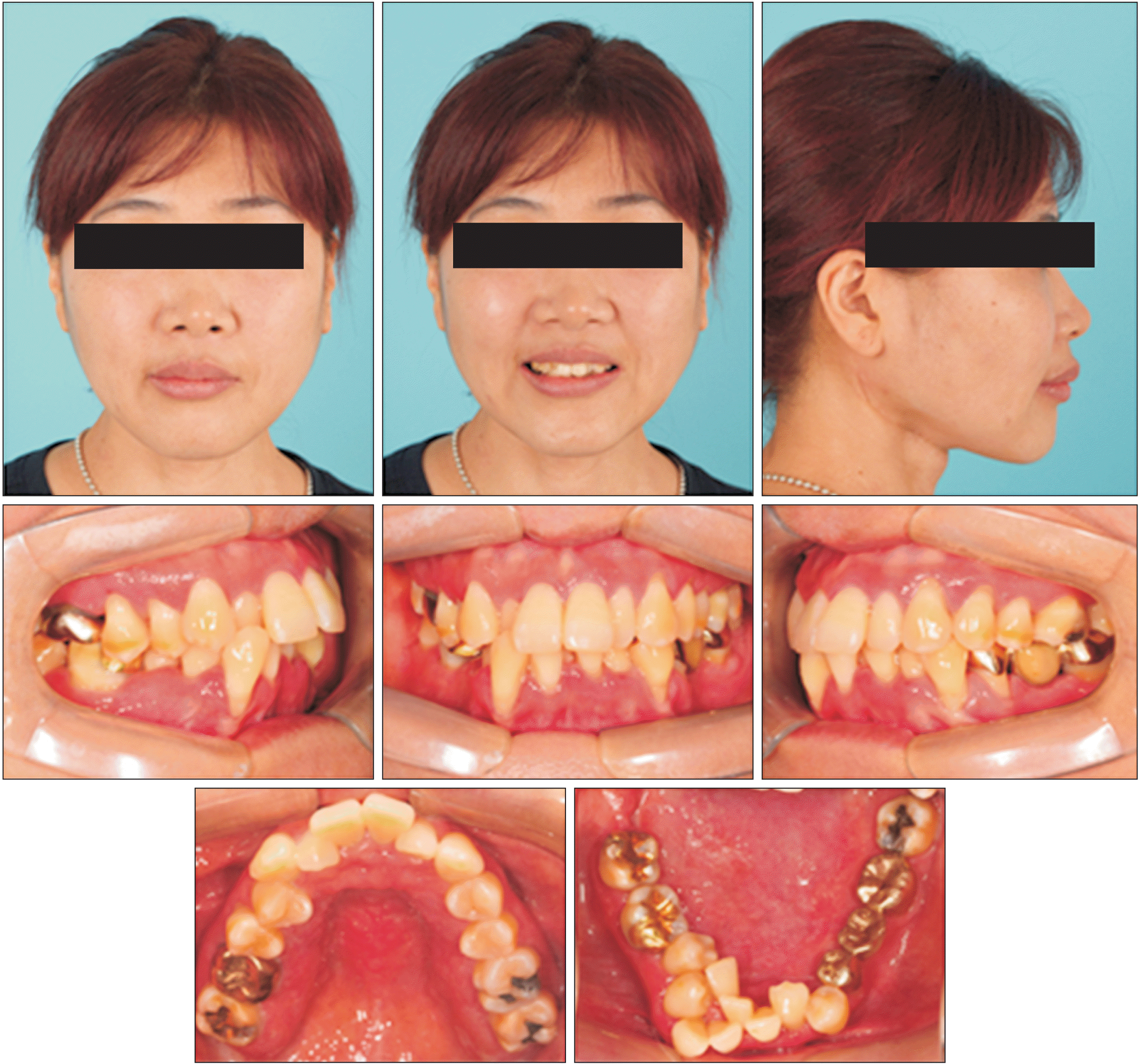
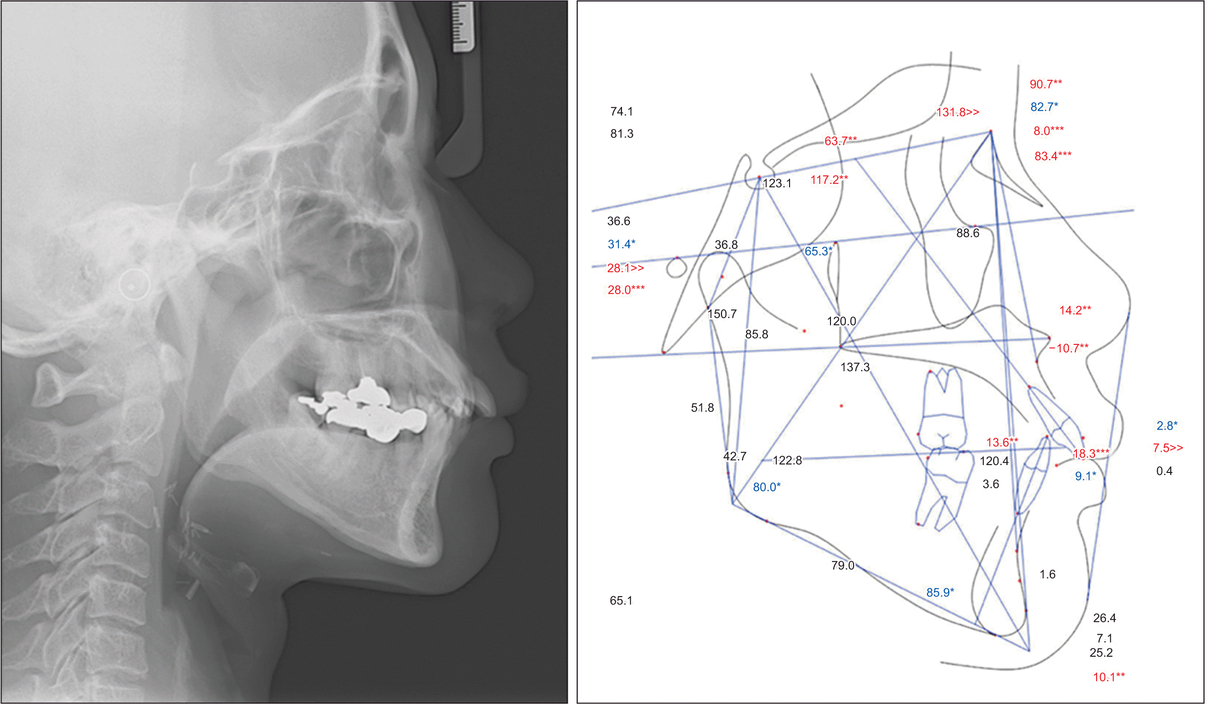
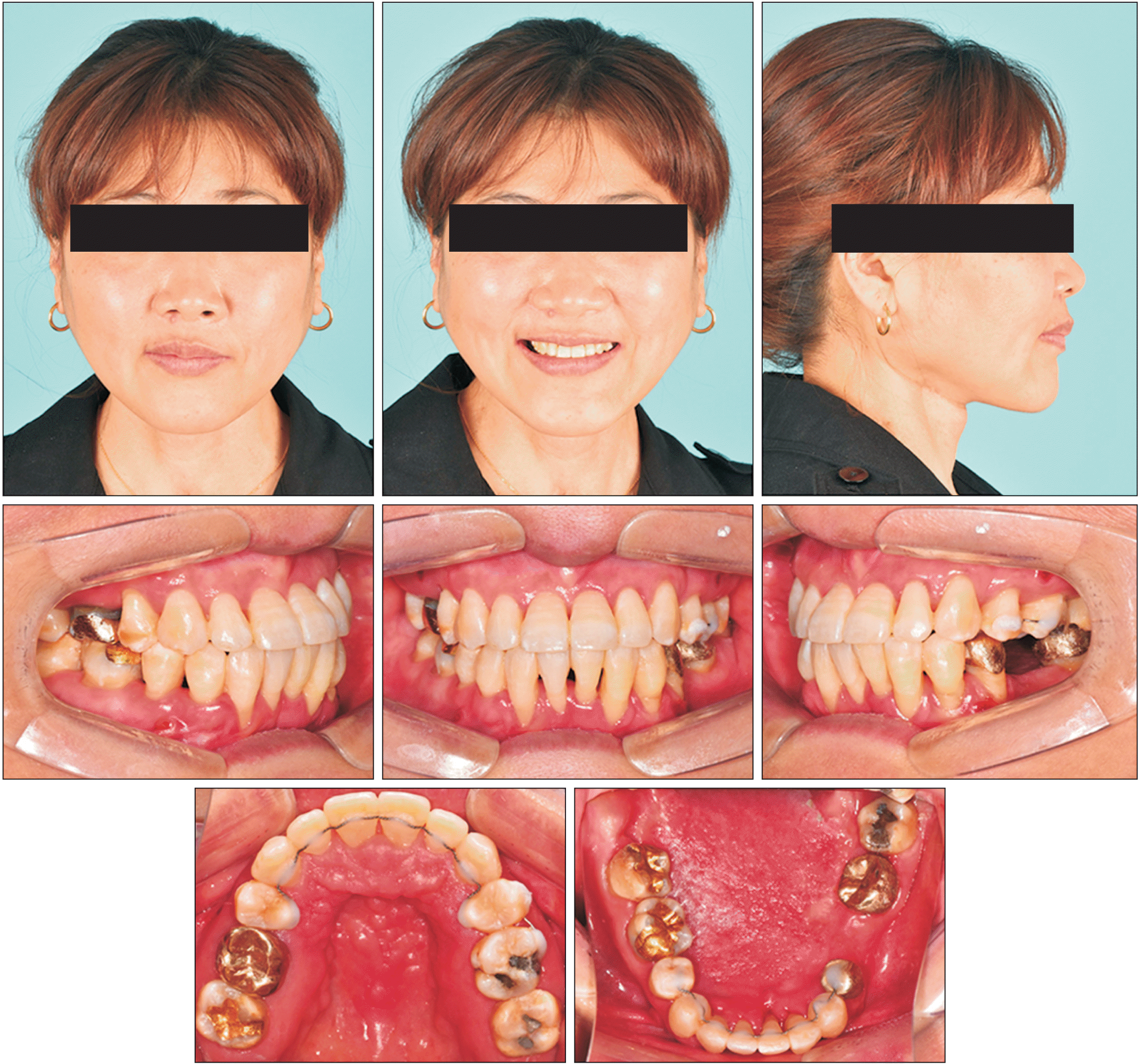
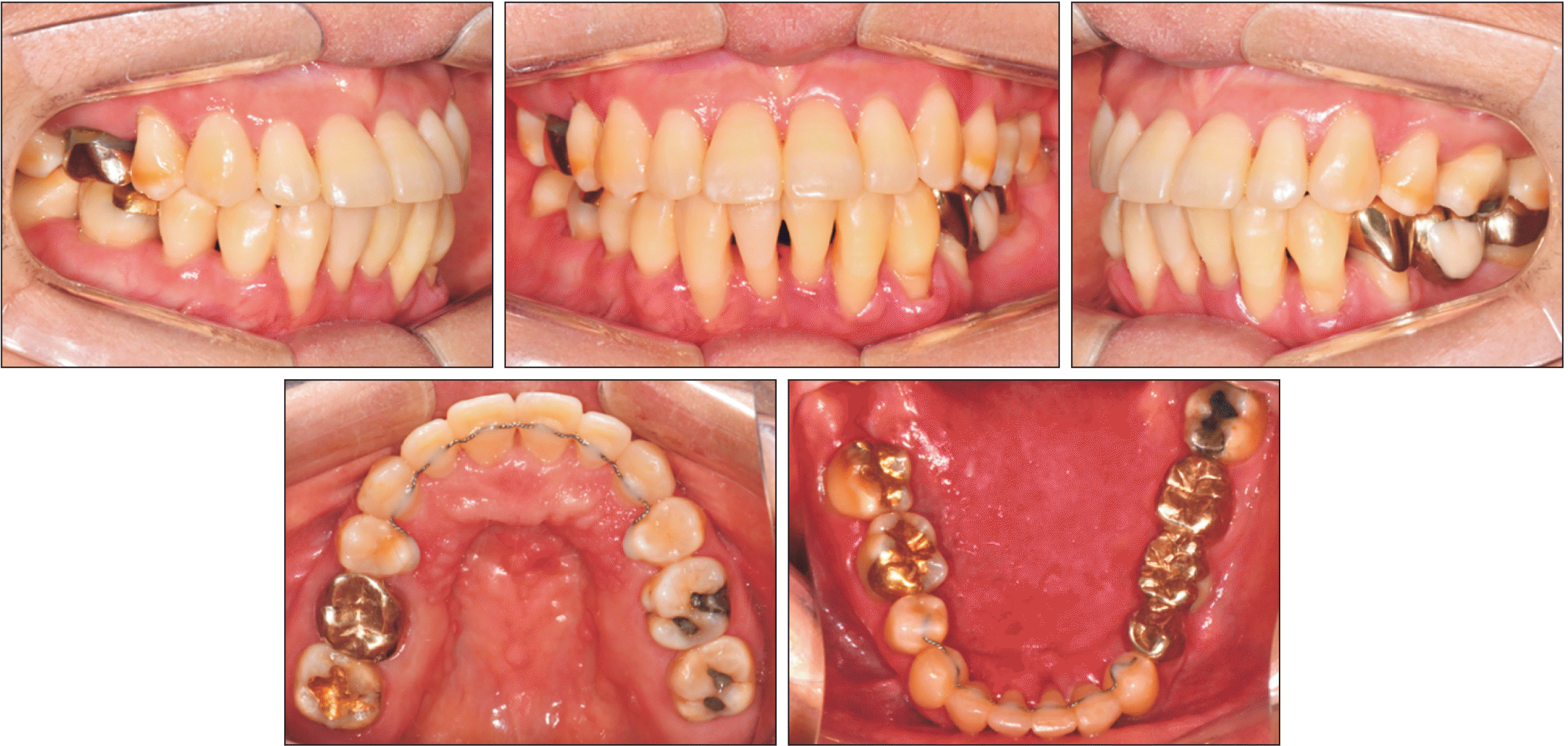
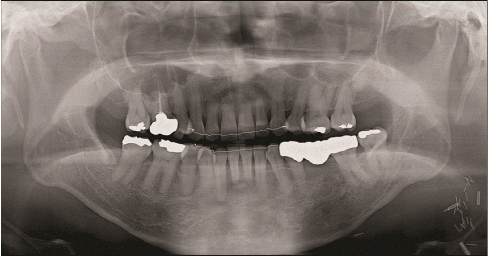
At the initial stage of orthodontic treatment, severe dental crowding was observed, especially in the mandibular anterior teeth due to absence of the tongue, with severe alveolar bone loss (Figure 1). The patient exhibited skeletal Class II with Class I occlusal relationship (Sella-Nasion-A point angle = 90.7°; Sella-Nasion-B point angle = 82.7°; A point-Nasion-B point angle = 8.0°; Figures 2 and 3). Other notable findings included crossbite with the right maxillary lateral incisor and scissor bite with the maxillary right second premolar and first molar. The patient had lost the mandibular left first molar, which was restored with a fixed prosthesis spanning the second premolar to the second molar; however, the bridge appeared to be mobile. The maxillary midline was concordant with the facial midline, and the facial profile was acceptable. The patient had good oral hygiene (Figures 2 and 3).
The treatment objectives included: (1) correction of dental crowding, and (2) establishment of proper occlusion.
Extraction was considered to achieve the treatment objectives; however, abnormal response of the alveolar bone and teeth (with respect to tooth movement) to the orthodontic forces were major concerns based on the patient’s medical history. Therefore, the reaction of the alveolar bone and teeth to orthodontic forces was monitored, and the final treatment plan was determined after attaining leveling and alignment.
The maxillary arch was first aligned with a self-ligating bracket system using a 0.018-inch Roth prescription (Tomy Inc., Tokyo, Japan) with 0.012-inch nickel-titanium as an initial wire. A maxillary posterior bite block was used to avoid interferences and correct the crossbite with the right maxillary lateral incisor. Maxillary arch alignment and crossbite correction were achieved after 5 months of treatment, followed by the initiation of treatment for the mandibular arch. The 0.016 × 22-inch stainless steel archwire was the working wire in the continuous wire sequence for the maxillary arch. The same bracket system was used for the mandibular arch, with 0.012-inch nickel-titanium archwire as the initial wire. The two out-of-arch and lingually placed mandibular incisors were left unbracketed. The fixed prosthesis in the mandibular left quadrant was removed because it was mobile, and space was retained for a pontic after orthodontic treatment. Intermaxillary elastics were used to correct the scissor bite. The 0.016 × 22-inch stainless steel archwire (working wire) was the ultimate in the wire sequence. Mandibular alignment was achieved after an additional 8 months of treatment, with correction of all instances of scissor bite. Subsequently, the maxillary posterior bite block was removed. The condition of the patient was re-evaluated to determine the final treatment plan (Figure 4A).
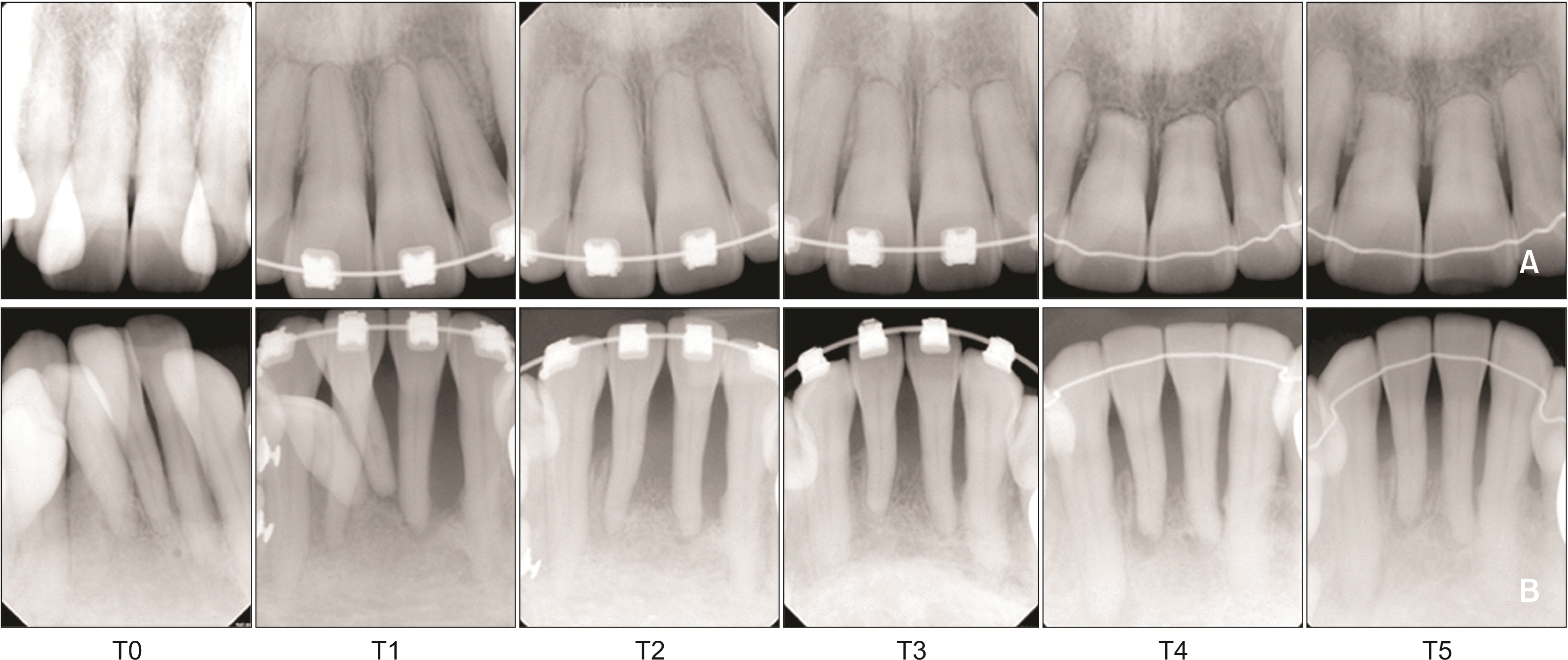
At this juncture, the patient exhibited a large overjet, Class II canine relationship, and black triangular spaces in the mandibular anterior region (Figure 4A). The right and left maxillary first premolars were extracted to reduce the overjet and establish Class I molar relationship, and the two periodontally hopeless mandibular incisors were extracted (Figure 4B). Orthodontic mini-implants (1.8 × 7 mm, BMK; Biomaterials Korea, Seoul, Korea) were used to provide skeletal anchorage. Two mini-implants were placed on the mesial side of the maxillary second premolars, and two mini-implants were also inserted in the inter-radicular space between the mandibular first and second premolars (Figure 4B). A retraction force used to close the extraction space in the maxillary arch, and an intrusive force used to control the torque over the anterior teeth and establish a proper overbite in the anterior segment. A light intrusive force was applied to the mandibular arch to improve alveolar bone support for the anterior teeth (Figure 4B). Interproximal reduction was performed for the mandibular anterior teeth during the treatment period to compensate for Bolton’s discrepancy and establish Class I occlusion. Trauma from the occlusion in the mandibular anterior teeth, alveolar bone and root status, and oral hygiene were examined regularly through follow-up appointments (Figure 5). However, root resorption was observed in the maxillary incisor teeth (Figure 5).
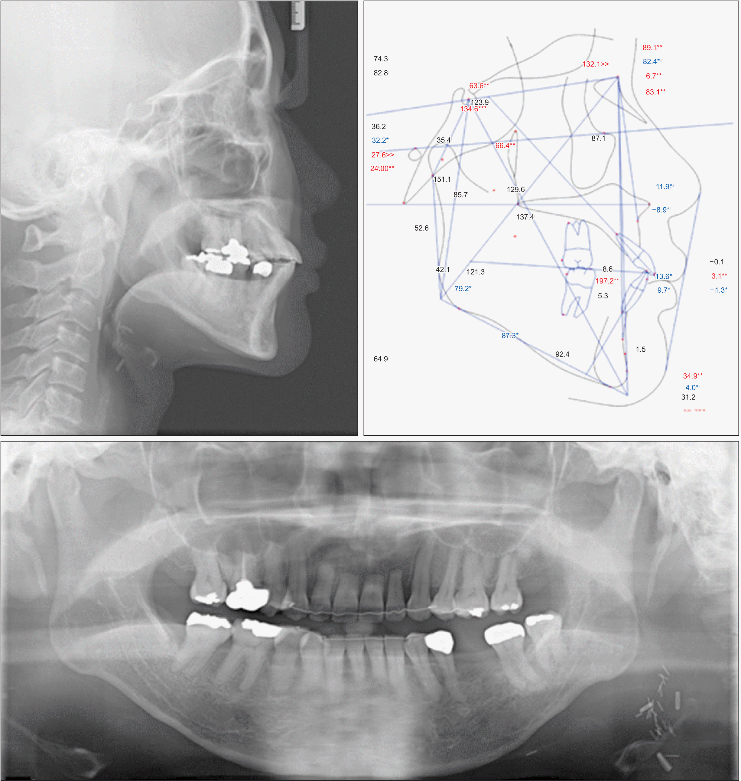
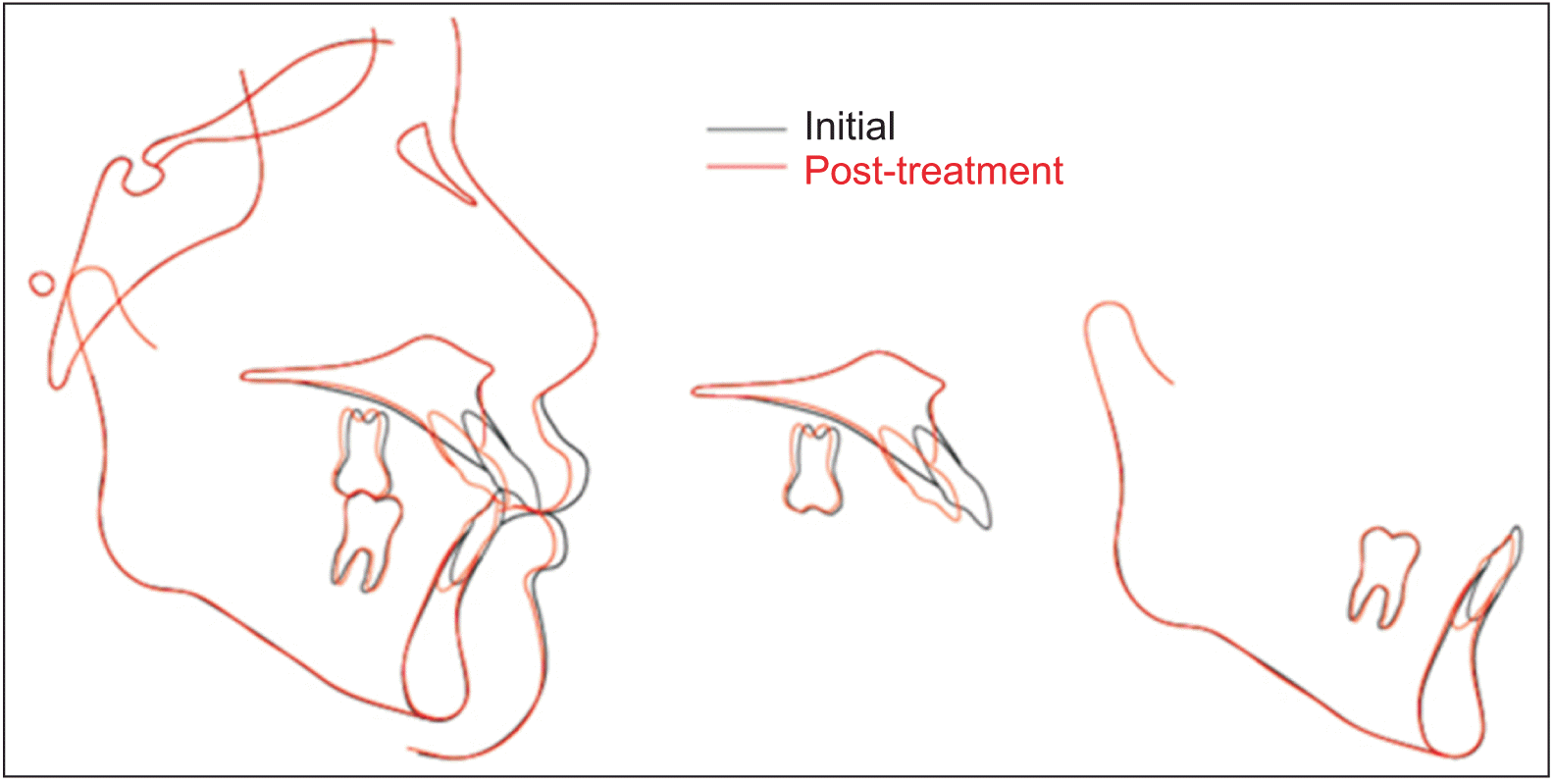
A proper and stable occlusion was established after 36 months of treatment (Figures 6–8). A combination of fixed and removable retainers was used for permanent retention. The patient was instructed to wear the retainers full time for the first month and only during night time thereafter. A new fixed prosthesis was fabricated to replace the right mandibular first molar. After 15 months of retention, the intraoral condition remained stable, and long-term follow-up was recommended (Figures 9 and 10).
The tongue is a vital structure involved in speech, mastication, and the swallowing reflex.9 Partial or total glossectomy results in tongue function disorders of varying degrees, causing uneven intraoral and extraoral pressure distribution.5 The equilibrium between the tongue and perioral muscles is essential for stabilizing occlusion.12 Total glossectomy could disrupt this equilibrium, making it a key factor in the occurrence and development of malocclusion. This patient presented with severe mandibular anterior crowding exacerbated due to the absence of the tongue, which is an extremely rare occurrence (Figures 1–3).6,7 Clinicians should be aware of this potential surgical ramification in patients undergoing total glossectomy. Moreover, radiotherapy for head and neck cancer often leads to extensive and permanent changes in the microvascular structures, mucosal structures, salivary glands, alveolar bone, enamel, and dentin.8,9,13,14 Therefore, the adaptation of alveolar bone to orthodontic forces may not be similar to that in healthy individuals. Importantly, tooth extraction is the most risk factor associated with the appearance of osteoradionecrosis, with a rate of 21.9%.15 Although cancer survivors require similar access to orthodontic care as other individuals, orthodontic care in this population could be challenging and requires special consideration during treatment planning. In this patient, the maxillary teeth were aligned first, and tooth movement was monitored through follow-up appointments. Since the maxillary teeth were aligned without alveolar bone loss and root resorption, as revealed by the periapical radiographs, the adaptation of the teeth and bone to orthodontic forces was considered to be normal in this patient (Figure 5).16 Subsequently, orthodontic treatment of the mandibular arch was initiated with light forces to minimize the risk of root resorption, increased severity of bone loss, and trauma to the mandibular incisors.17,18
The patient had a large overjet after leveling and alignment, along with the appearance of black triangles in the mandibular anterior region (Figure 4A). Extraction was selected to achieve the treatment objectives. The right and left maxillary first premolars and two periodontally hopeless mandibular incisors were extracted based on the patient’s condition (Figure 4A). The possibility of extraction was analyzed by a radiodontist due to the risk of osteoradionecrosis.18
The cumulative incidence of osteoradionecrosis is reported to be 12.4%.19 A recent study reported that osteoradionecrosis occurred after radiotherapy in 56 of 1,224 patients (4.6%), with a median time-to-event of 10.9 months (1.8–89.7), and in 90% of patients within 37.4 months (average follow-up duration of the entire cohort: 22.8 months [0.3–115.5]).20 Epstein et al.21 observed that osteoradionecrosis could occur at any time after radiotherapy; however, the risk of a second episode was low after treatment of the initial episode. Our patient had undergone radiotherapy (at a dose of < 50 Gy) 6 years and 9 months before opting for extraction and was a non-drinker and non-smoker with good oral hygiene habits; moreover, the adaptation to orthodontic force was considered normal in her case; therefore, extraction carried a low risk of osteoradionecrosis.15,22 Periodontal surgery may be attempted in advance to prevent gingival recession and improve esthetics; however, this procedure was delayed until the end of orthodontic treatment due to the severe dental crowding in this patient.23
In this study, orthodontic mini-implants were used to provide a point of force application to close the extraction space and establish a proper occlusal relationship, due to the predictability, minimal invasiveness, and low risk of osteoradionecrosis in the patient (Figure 4B). To the best of our knowledge, this is the first study to report the application of mini-implants for the orthodontic treatment of a patient after radiotherapy. In the maxillary arch, two mini-implants were placed on the mesial side of the maxillary second premolars at the mucogingival junction (Figure 4B). This position proved appropriate for the provision of intraoral bone anchorage for the magnitude and direction of force required to effect retraction and intrusion of the maxillary anterior teeth.24 However, root resorption was observed in the maxillary incisors during the last 7 months of treatment (Figure 5A). Several studies have reported that the incisors are the most susceptible teeth to external apical root resorption by virtue of their spindly apices. Furthermore, the incisors are often moved further than the other teeth during orthodontic treatment.25 Long-term orthodontic treatment could increase the possibility of root resorption, and the apical movement caused by intrusive forces is often associated with the amount of root shortening.10 Therefore, the root status of all patients should be monitored frequently through periapical radiographs.25 Slow and intermittent forces can be applied at 2- to 3-month intervals to allow for the recovery of the eroded cementum, which would retard the rapid root resorption.10,25,26
In the mandibular arch, two mini-implants were placed in the inter-radicular space between the mandibular first and second premolars. A distointrusive force was delivered directly from the mini-implants to the mandibular anterior teeth to improve the level of periodontal support (Figures 4B and 5).27,28 However, achieving pure intrusion in teeth with severe alveolar bone loss could be challenging due to the risk of uncontrolled inclination. The intrusive force must pass through a displaced center of resistance of the target segment identified according to the amount of alveolar bone loss.27-30 Additionally, the magnitude of force must be minimal and should not exceed 10 g per tooth.29 Further, trauma from occlusion and oral hygiene should be controlled strictly during treatment, and the root status should be regularly monitored.29 Alveolar bone support for the mandibular anterior teeth improved at the end of treatment. Furthermore, slight interproximal reduction of the mandibular anterior teeth to compensate for Bolton’s discrepancy resulted in the closure of the black triangles, which slightly improved the esthetic appearance.
All the treatment objectives were achieved after 36 months of treatment, despite the occurrence of root resorption in the maxillary anterior teeth (Figures 5–8). The intraoral results remained unchanged after 15 months of retention (Figures 5, 9, and 10).
A high risk of relapse was probable in this case due to the absence of the tongue and the imbalance between the labial and lingual musculature, which had originally caused severe crowding.7,12 Therefore, permanent retention and long-term follow-up were recommended for the patient, which included the lifelong use of a combination of fixed and removable retainers, with which the patient complied. The short-term favorable stability is promising; however, this does not guarantee long-term stability unless the retention protocol is properly followed. Inter-canine multi-stranded wires were used for the fixed retainer, which permitted minor physiological movement and prevented trauma from occlusion in the maxillary and mandibular incisors due to the tight incisor contact in the final occlusion. The multi-stranded wires appeared to be effective during the 15-month retention period. However, from the long-term perspective, any signs of post-treatment migration of the incisors would require replacement of the wire with a rigid stainless-steel retainer. The removable retainer would provide stability under careful monitoring for signs of irritation or inflammation.10 Further, the importance of oral hygiene should be emphasized to the patient.
Severe dental crowding with severe alveolar bone loss induced by total glossectomy and radiotherapy performed 6 years ago was successfully treated by orthodontics in a survivor of tongue SCC, using strategic extraction and intrusive forces. However, the risks of osteoradionecrosis and root resorption should be considered cautiously. Orthodontic mini-implants can be used to provide sufficient skeletal anchorage to effect closure of the extraction space and intrusion of the anterior teeth. Permanent retention and long-term follow-up are recommended for such patients.
REFERENCES
1. Chitapanarux I, Lorvidhaya V, Sittitrai P, Pattarasakulchai T, Tharavichitkul E, Sriuthaisiriwong P, et al. 2006; Oral cavity cancers at a young age: analysis of patient, tumor and treatment characteristics in Chiang Mai University Hospital. Oral Oncol. 42:83–8. DOI: 10.1016/j.oraloncology.2005.06.015. PMID: 16249113. PMID: https://www.scopus.com/inward/record.uri?partnerID=HzOxMe3b&scp=29244492025&origin=inward.
2. Patel SC, Carpenter WR, Tyree S, Couch ME, Weissler M, Hackman T, et al. 2011; Increasing incidence of oral tongue squamous cell carcinoma in young white women, age 18 to 44 years. J Clin Oncol. 29:1488–94. DOI: 10.1200/JCO.2010.31.7883. PMID: 21383286. PMID: https://www.scopus.com/inward/record.uri?partnerID=HzOxMe3b&scp=79955033400&origin=inward.
3. Santos-Silva AR, Carvalho Andrade MA, Jorge J, Almeida OP, Vargas PA, Lopes MA. 2014; Tongue squamous cell carcinoma in young nonsmoking and nondrinking patients: 3 clinical cases of orthodontic interest. Am J Orthod Dentofacial Orthop. 145:103–7. DOI: 10.1016/j.ajodo.2012.09.026. PMID: 24373660. PMID: https://www.scopus.com/inward/record.uri?partnerID=HzOxMe3b&scp=84891585784&origin=inward.
4. Mallet Y, Avalos N, Le Ridant AM, Gangloff P, Moriniere S, Rame JP, et al. 2009; Head and neck cancer in young people: a series of 52 SCCs of the oral tongue in patients aged 35 years or less. Acta Otolaryngol. 129:1503–8. DOI: 10.3109/00016480902798343. PMID: 19922105. PMID: https://www.scopus.com/inward/record.uri?partnerID=HzOxMe3b&scp=70450172632&origin=inward.
5. Costa Bandeira AK, Azevedo EH, Vartanian JG, Nishimoto IN, Kowalski LP, Carrara-de Angelis E. 2008; Quality of life related to swallowing after tongue cancer treatment. Dysphagia. 23:183–92. DOI: 10.1007/s00455-007-9124-1. PMID: 17999111. PMID: https://www.scopus.com/inward/record.uri?partnerID=HzOxMe3b&scp=44549085327&origin=inward.
6. Lin DT, Yarlagadda BB, Sethi RK, Feng AL, Shnayder Y, Ledgerwood LG, et al. 2015; Long-term functional outcomes of total glossectomy with or without total laryngectomy. JAMA Otolaryngol Head Neck Surg. 141:797–803. DOI: 10.1001/jamaoto.2015.1463. PMID: 26291031. PMID: https://www.scopus.com/inward/record.uri?partnerID=HzOxMe3b&scp=84942023954&origin=inward.
7. Yu M, Gao X. 2019; Tongue pressure distribution of individual normal occlusions and exploration of related factors. J Oral Rehabil. 46:249–56. DOI: 10.1111/joor.12741. PMID: 30375017. PMCID: PMC7379747. PMID: https://www.scopus.com/inward/record.uri?partnerID=HzOxMe3b&scp=85056469618&origin=inward.
8. Jawad H, Hodson NA, Nixon PJ. 2015; A review of dental treatment of head and neck cancer patients, before, during and after radiotherapy: part 1. Br Dent J. 218:65–8. DOI: 10.1038/sj.bdj.2015.28. PMID: 25613260. PMID: https://www.scopus.com/inward/record.uri?partnerID=HzOxMe3b&scp=84923113496&origin=inward.
9. Jawad H, Hodson NA, Nixon PJ. 2015; A review of dental treatment of head and neck cancer patients, before, during and after radiotherapy: part 2. Br Dent J. 218:69–74. Erratum in: Br Dent J 2015;218: 290. DOI: 10.1038/sj.bdj.2015.29. PMID: 25613261. PMID: https://www.scopus.com/inward/record.uri?partnerID=HzOxMe3b&scp=84923097888&origin=inward.
10. Mishra S. 2017; Orthodontic therapy for paediatric cancer survivors: a review. J Clin Diagn Res. 11:ZE01–4. DOI: 10.7860/JCDR/2017/23916.9404. PMID: 28511529. PMCID: PMC5427455. PMID: e2995bfc837a4ae49cab1677e82e1932. PMID: https://www.scopus.com/inward/record.uri?partnerID=HzOxMe3b&scp=85014366358&origin=inward.
11. Burden D, Mullally B, Sandler J. 2001; Orthodontic treatment of patients with medical disorders. Eur J Orthod. 23:363–72. DOI: 10.1093/ejo/23.4.363. PMID: 11544786. PMID: https://www.scopus.com/inward/record.uri?partnerID=HzOxMe3b&scp=0035430697&origin=inward.
12. Kurabeishi H, Tatsuo R, Makoto N, Kazunori F. 2018; Relationship between tongue pressure and maxillofacial morphology in Japanese children based on skeletal classification. J Oral Rehabil. 45:684–91. DOI: 10.1111/joor.12680. PMID: 29908035. PMID: https://www.scopus.com/inward/record.uri?partnerID=HzOxMe3b&scp=85050878287&origin=inward.
13. Santin GC, Palma-Dibb RG, Romano FL, de Oliveira HF, Nelson Filho P, de Queiroz AM. 2015; Physical and adhesive properties of dental enamel after radiotherapy and bonding of metal and ceramic brackets. Am J Orthod Dentofacial Orthop. 148:283–92. DOI: 10.1016/j.ajodo.2015.03.025. PMID: 26232837. PMID: https://www.scopus.com/inward/record.uri?partnerID=HzOxMe3b&scp=84938388818&origin=inward.
14. Martin P, Muller E, Paulus C. 2019; Alteration of facial growth after radiotherapy: orthodontic, surgical and prosthetic rehabilitation. J Stomatol Oral Maxillofac Surg. 120:369–72. DOI: 10.1016/j.jormas.2019.04.004. PMID: 30980947. PMID: https://www.scopus.com/inward/record.uri?partnerID=HzOxMe3b&scp=85065017297&origin=inward.
15. Khoo SC, Nabil S, Fauzi AA, Yunus SSM, Ngeow WC, Ramli R. 2021; Predictors of osteoradionecrosis following irradiated tooth extraction. Radiat Oncol. 16:130. DOI: 10.1186/s13014-021-01851-0. PMID: 34261515. PMCID: PMC8278595. PMID: ebac24c028d84d94b99a1309dd0b973c. PMID: https://www.scopus.com/inward/record.uri?partnerID=HzOxMe3b&scp=85109853278&origin=inward.
16. Yee JA, Türk T, Elekdağ-Türk S, Cheng LL, Darendeliler MA. 2009; Rate of tooth movement under heavy and light continuous orthodontic forces. Am J Orthod Dentofacial Orthop. 136:150.e1–9. discussion 150–1. DOI: 10.1016/j.ajodo.2008.06.027. PMID: 19651334. PMID: https://www.scopus.com/inward/record.uri?partnerID=HzOxMe3b&scp=67949094544&origin=inward.
17. Burch JG, Bagci B, Sabulski D, Landrum C. 1992; Periodontal changes in furcations resulting from orthodontic uprighting of mandibular molars. Quintessence Int. 23:509–13. DOI: 10.1016/s0889-5406(08)80083-2. PMID: 1410254.
18. Aarup-Kristensen SFM, Hansen CR, Johansen J. 2019; Osteoradionecrosis after radiotherapy for head and neck cancer: incidence, risk factors, and mandibular dose-volume effects. Int J Radiat Oncol Biol Phys. 105(Suppl 1):E400. DOI: 10.1016/j.ijrobp.2019.06.1585.
19. Kuhnt T, Stang A, Wienke A, Vordermark D, Schweyen R, Hey J. 2016; Potential risk factors for jaw osteoradionecrosis after radiotherapy for head and neck cancer. Radiat Oncol. 11:101. DOI: 10.1186/s13014-016-0679-6. PMID: 27473433. PMCID: PMC4967325. PMID: https://www.scopus.com/inward/record.uri?partnerID=HzOxMe3b&scp=84979752489&origin=inward.
20. Aarup-Kristensen S, Hansen CR, Forner L, Brink C, Eriksen JG, Johansen J. 2019; Osteoradionecrosis of the mandible after radiotherapy for head and neck cancer: risk factors and dose-volume correlations. Acta Oncol. 58:1373–7. DOI: 10.1080/0284186X.2019.1643037. PMID: 31364903. PMID: https://www.scopus.com/inward/record.uri?partnerID=HzOxMe3b&scp=85070302847&origin=inward.
21. Epstein J, van der Meij E, McKenzie M, Wong F, Lepawsky M, Stevenson-Moore P. 1997; Postradiation osteonecrosis of the mandible: a long-term follow-up study. Oral Surg Oral Med Oral Pathol Oral Radiol Endod. 83:657–62. DOI: 10.1016/S1079-2104(97)90314-0. PMID: 9195618. PMID: https://www.scopus.com/inward/record.uri?partnerID=HzOxMe3b&scp=0031157940&origin=inward.
22. Iqbal Z, Kyzas P. 2020; Analysis of the critical dose of radiation therapy in the incidence of Osteoradionecrosis in head and neck cancer patients: a case series. BDJ Open. 6:18. DOI: 10.1038/s41405-020-00044-3. PMCID: PMC7515872. PMID: 33042578. PMID: https://www.scopus.com/inward/record.uri?partnerID=HzOxMe3b&scp=85091442937&origin=inward.
23. Irie MS, Mendes EM, Borges JS, Osuna LG, Rabelo GD, Soares PB. 2018; Periodontal therapy for patients before and after radiotherapy: a review of the literature and topics of interest for clinicians. Med Oral Patol Oral Cir Bucal. 23:e524–30. DOI: 10.4317/medoral.22474. PMID: 30148466. PMCID: PMC6167093. PMID: https://www.scopus.com/inward/record.uri?partnerID=HzOxMe3b&scp=85053079904&origin=inward.
24. Upadhyay M, Yadav S, Nanda R. 2014; Biomechanics of incisor retraction with mini-implant anchorage. J Orthod. 41 Suppl 1:S15–23. DOI: 10.1179/1465313314Y.0000000114. PMID: 25138361. PMID: https://www.scopus.com/inward/record.uri?partnerID=HzOxMe3b&scp=84919455212&origin=inward.
25. Harris EF. 2000; Root resorption during orthodontic therapy. Semin Orthod. 6:183–94. DOI: 10.1053/sodo.2000.8084. PMID: https://www.scopus.com/inward/record.uri?partnerID=HzOxMe3b&scp=0042949040&origin=inward.
26. Ballard DJ, Jones AS, Petocz P, Darendeliler MA. 2009; Physical properties of root cementum: part 11. Continuous vs intermittent controlled orthodontic forces on root resorption. A microcomputed-tomography study. Am J Orthod Dentofacial Orthop. 136:8.e1–8. discussion 8–9. DOI: 10.1016/j.ajodo.2007.07.026. PMID: 19577132. PMID: https://www.scopus.com/inward/record.uri?partnerID=HzOxMe3b&scp=67649411604&origin=inward.
27. Park HK, Sung EH, Cho YS, Mo SS, Chun YS, Lee KJ. 2011; 3-D FEA on the intrusion of mandibular anterior segment using orthodontic miniscrews. Korean J Orthod. 41:384–98. DOI: 10.4041/kjod.2011.41.6.384. PMID: https://www.scopus.com/inward/record.uri?partnerID=HzOxMe3b&scp=84855297271&origin=inward.
28. González Del Castillo McGrath M, Araujo-Monsalvo VM, Murayama N, Martínez-Cruz M, Justus-Doczi R, Domínguez-Hernández VM, et al. 2018; Mandibular anterior intrusion using miniscrews for skeletal anchorage: a 3-dimensional finite element analysis. Am J Orthod Dentofacial Orthop. 154:469–76. DOI: 10.1016/j.ajodo.2018.01.009. PMID: 30268257. PMID: https://www.scopus.com/inward/record.uri?partnerID=HzOxMe3b&scp=85054341251&origin=inward.
29. Feu D. 2020; Orthodontic treatment of periodontal patients: challenges and solutions, from planning to retention. Dental Press J Orthod. 25:79–116. DOI: 10.1590/2177-6709.25.6.079-116.sar. PMID: 33503129. PMCID: PMC7869805. PMID: https://www.scopus.com/inward/record.uri?partnerID=HzOxMe3b&scp=85100261656&origin=inward.
30. Choi SH, Kim YH, Lee KJ, Hwang CJ. 2016; Effect of labiolingual inclination of a maxillary central incisor and surrounding alveolar bone loss on periodontal stress: a finite element analysis. Korean J Orthod. 46:155–62. DOI: 10.4041/kjod.2016.46.3.155. PMID: 27226961. PMCID: PMC4879318. PMID: https://www.scopus.com/inward/record.uri?partnerID=HzOxMe3b&scp=84969776657&origin=inward.
Figure 1
Serial panoramic and periapical radiographs showing the development of severe malocclusion over time. A, Before total glossectomy. B, Three years after tongue cancer treatment. C, Six years after tongue cancer treatment and pre-orthodontic treatment.
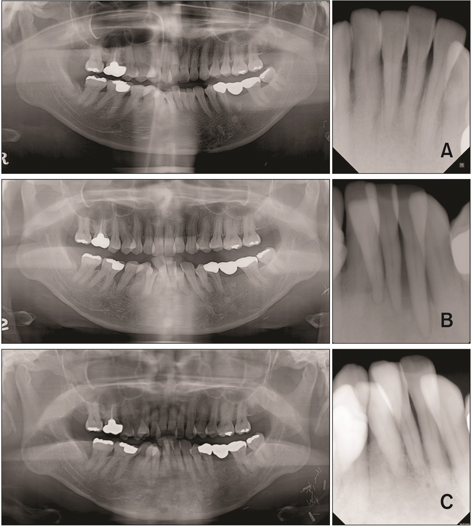
Figure 4
Intraoral photographs of treatment progress. A, Leveling and alignment. B, Closing extraction space and intruding maxillary and mandibular anterior teeth.
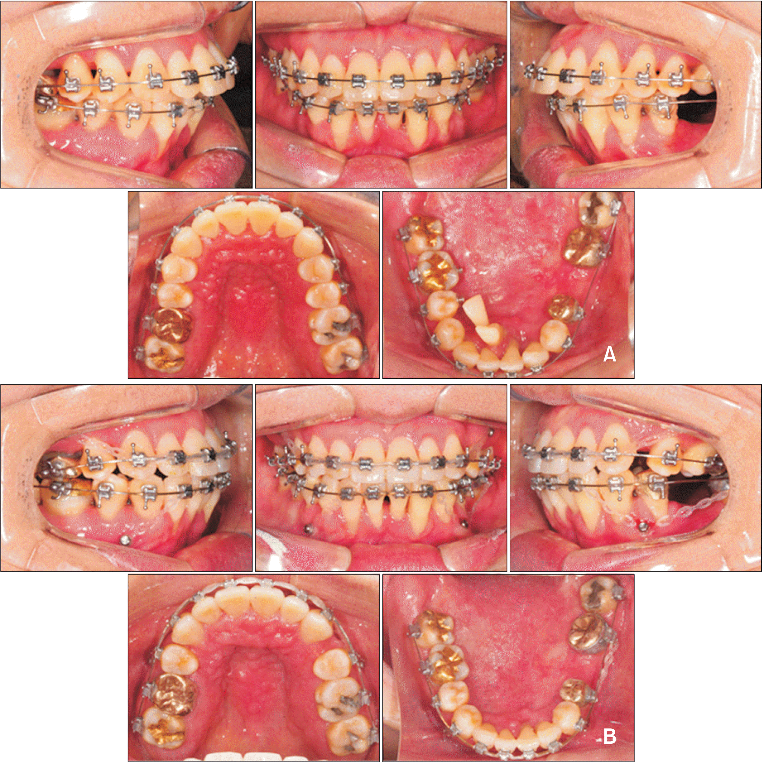
Figure 5
Periapical radiographs follow-up. A, Maxillary incisors. B, Mandibular incisors.
T0, pre-treatment; T1, re-assessment after leveling and alignment; T2, T3, treatment progress with closing extraction space and intruding maxillary and mandibular anterior teeth; T4, post-treatment; T5, one year of retention.


 PDF
PDF Citation
 Print
Print


 XML Download