METHODS
The study protocol was approved by the Institutional Review Board of Asan Medical Center (IRB No. 2017-0488), which waived the requirement for informed consent due to the retrospective nature of this study. This study was performed in accordance with the ethical guidelines of the 2013 World Medical Association Declaration of Helsinki.
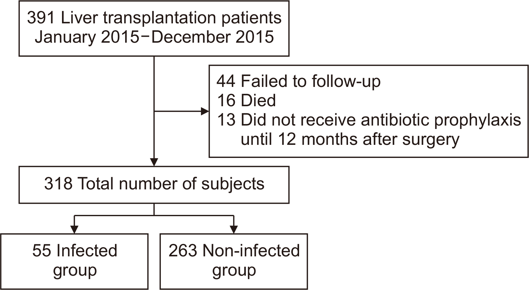
This retrospective cohort study, using electronic medical record review, identified 391 adult patients 21 years of age or older who underwent LT at a tertiary general hospital in Seoul between January 2015 and December 2015. Among these 391 LT recipients, 318 patients were included in the study; the others were lost to follow-up (n=44), died (n=16), or did not receive antibiotic prophylaxis until 12 months after LT (n=13). The remaining 318 LT recipients were reviewed for infections during hospitalization and outpatient care from January 2016 to December 2016. The selection process is presented in
Fig. 1. The case records for each patient included patient characteristics (preoperative severity-related, donor, surgery-related, postoperative, cannulation-related, and LT results). The type of infection, time of occurrence, causative microorganisms, and profiles of multi-drug-resistant bacteria were also identified.
Definition and Criteria of Infection
The criteria for identifying infections within 1 year posttransplant were either positive laboratory tests or recorded clinical signs and symptoms that met the accepted criteria for each infection. The date of occurrence was set as the date of confirmation of the test results, and when multiple infections occurred in one patient, all infections were included. Multidrug-resistant bacteria included methicillin-resistant
Staphylococcus aureus (MRSA) from nasal swab test results, and other multi-drug-resistant organisms (MDROs) isolated from clinical samples of infection or colonization. In our institution, when patients are hospitalized for LT, surveillance culture tests for the nasal cavity are performed for MRSA, but surveillance culture tests for rectal swab or stool samples are not conducted for vancomycin-resistant enterococci (VRE) or carbapenem-resistant Enterobacteriaceae (CRE). The data, therefore, came from a combination of MRSA surveillance culture results and screening culture tests performed according to the research institute’s protocol once a week from blood, urine, feces, sputum, abdominal or pigtail drains, and bile specimens before and after LT through hospital discharge. Culture tests performed in response to symptoms of suspected infection were also included. Infection was defined as microorganisms proliferating in the human body and triggering an immune response, whereas colonization was defined as microorganisms multiplying on the surface of the body without an immune response [
5].
Respiratory tract infection (RTI) criteria included a positive laboratory test [
6] or a diagnosis based on fever, cough, crackles on auscultation, or a white blood cell (WBC) count of <4,000/μL or >10,000/μL with a lesion detected by chest X-ray or computed tomography [
7].
The criteria for gastrointestinal infection varied by the location in the gastrointestinal tract, but cases of gastrointestinal infection were generally diagnosed based on one or more symptoms of dysphagia, swallowing pain, nausea, vomiting, abdominal pain, gastrointestinal bleeding, bowel perforation and diarrhea confirmed as a result of culture or image examination [
8]. Patients suffering from chronic gastrointestinal infections or diseases not related to infection were excluded [
7].
The criteria for biliary tract infection included a positive laboratory test [
9] or one or more of the following criteria: high fever (>38°C), chills, WBC count between 4,000/μL and 10,000/μL, C-reactive protein of at least 1.0 mg/dL and concurrent jaundice (total bilirubin ≥2 mg/dL), abnormal liver function tests (i.e., alkaline phosphatase, γ-glutamyl transferase, aspartate aminotransferase and alanine aminotransferase levels greater than 1.5 times the normal reference value), or confirmation by image tests [
10].
Urinary tract infection (UTI) criteria included both symptomatic UTI and asymptomatic bacteremic UTI, defined as meeting one of the healthcare-associated diagnostic criteria. Asymptomatic bacteremic UTI was defined as the absence of symptoms or signs of UTI with either ≥10
5 colony-forming units (CFU)/mL of two or fewer pathogenic microorganisms in urine culture, and at least one matching pathogenic microorganism from blood culture, or, if normal skin flora is isolated, two or more pairs of blood culture tests [
11].
The criteria for bloodstream infections were the domestic criteria used to diagnose healthcare-associated infections [
11]. The surgical site infection criteria included superficial and deep surgical site infections, for which the domestic healthcare-associated infection diagnostic criteria were applied [
11]. Skin infections were diagnosed by a dermatologist or confirmed as a result of culture tests where sufficient diagnosis was possible with characteristic clinical features [
12]. Other diagnostic criteria for infections were defined as one or more of the following [
7]: WBC count <4,000/μL or >10,000/μL, normal WBC count but immature WBC count >10%, C-reactive protein level >2 times normal, or procalcitonin level more than twice the normal reference value.
Statistical Analysis
Statistical analysis was performed using IBM SPSS ver. 21 (IBM Corp., Armonk, NY, USA). P-values of less than 0.05 were considered statistically significant. For the general characteristics of subjects, frequency, percentage, mean and standard deviation were used. The t-test and chi-square test were used as appropriate to determine the significance of differences between the infected and non-infected groups after LT. Since infection can occur multiple times in a single patient, the primary infection incidence rate was calculated as the percentage of the total number of patients who underwent LT who had a new infection at least once during the study period.
RESULTS
Infections occurred in 55 (17.3%) of 318 patients during the 1 year following LT. The patients ranged from 21 to 73 years of age, with 75.8% male, and 90.3% having undergone living-donor LT. Patients most frequently had their first infection during the first month after LT (26 patients; 47.3%). From 1 month to 3 months after LT, six patients (10.9%) had infections, and between 3 months and 1 year after LT, 23 patients (41.8%) became infected. Among these 55 patients, some experienced two concurrent RTI, UTI, or skin infections, for a total of 61 cases of infections. Including all cases, infections were most prevalent during the first month after LT (28 cases, 45.9%), followed by 3 months to 1 year (23 cases, 37.7%), with the fewest infections (10 cases, 16.4%) from 1 month to 3 months. RTI was the most common type of infection, occurring in 29 (47.5%) cases, the majority of which (14 cases) occurred between 3 months and 1 year after LT. Nine cases of RTI were recorded during the first month after LT, and six cases were identified between 1 and 3 months. The other most common types of infections were biliary tract (11 cases, 18.0%), surgical site (seven cases, 11.5%), gastrointestinal and bloodstream infections (three cases each, 6.6%) and UTI and skin infections (three cases each) (
Table 1).
The causative agents of infections were bacterial (18 cases), viral (15 cases), fungal (two cases) and unidentified (26 cases). Bacterial infections were most prevalent up to 1 month after LT, whereas viral infections occurred mainly during the following 11 months.
Pseudomonas aeruginosa and influenza virus were the most common infectious organisms (five cases each), followed by respiratory syncytial virus (four cases), and
Enterococcus faecium (three cases).
S. aureus,
Enterococcus faecalis,
Clostridium difficile, herpes simplex virus and coronavirus each caused two infections (
Table 2).
Several parameters correlated significantly with infection (
Table 3): model for end-stage liver disease (MELD) score (>17 points, P<0.001); frequency of intensive care unit (ICU) admission (18.2%, P=0.005) emergency LT operation (27.3%, P=0.007), or posttransplant renal replacement therapy (10.9%, P=0.029); ICU stay after LT (5 days, P<0.001); use of azathioprine (1.8%, P=0.029), piperacillin/tazobactam (61.8%, P<0.001), vancomycin (49.1%, P=0.002), or levofloxacin (36.4%, P<0.001); duration of placement of a central venous catheter (14 days, P=0.004), endotracheal tube (3 days, P<0.001), Foley catheter (5 days, P=0.011), Jackson-Pratt abdominal drain (18 days, P=0.002), pigtail drain (9.2±18.3 days, P=0.049), or jejunal feeding catheter (38.9±51.9 days, P=0.020; 38.2%, P=0.008); and the frequency of percutaneous transhepatic bile drainage (PTBD) (10.9%, P=0.005), endoscopic retrograde biliary drainage (ERBD) stent (12.7%, P=0.015), reoperation (21.8%, P=0.030), and multi-drug-resistant bacterial infection (25.5%, P=0.001). In the non-infected group, the frequency of endotracheal tube removal within 2 days after LT (79.1%, P<0.001), and use of metronidazole (76.8%, P=0.021) and ceftriaxone (74.1%, P=0.002) were all statistically significantly higher (
Table 3).
DISCUSSION
In the present study, the overall infection rate in patients during the year after LT was 17.3%, which is lower than the rates of 63.1% [
2] and 42.1% [
13] reported in previous studies. This may be due to differences in the length of the follow-up period, definitions of infection, and/or improvements in surgical techniques and immunosuppressive regimens since those studies were carried out. Infections occurred most frequently within the first month after LT, which is consistent with a previous study of transplant patients, in which 46 of 65 infections occurred during the first month [
2].
The most prevalent infection types were RTI (47.5% of all infections), biliary tract infections (18%), and surgical site infections (11.5%). This contrasts with the results of previous studies [
2,
14], in which intraperitoneal infection was the most common. This discrepancy may be in part due to a longer posttransplant monitoring period (3 years) in one of these previous studies [
2]. Since intraperitoneal infection typically occurs during the early period post-LT, however, the inconsistency was more likely due to differences in the study subjects. It is also possible that the frequency of surgical site infections was relatively low in the present study because patient deaths within the first 1 month after LT were excluded. In addition, and according to the RTI diagnostic criteria, this study had both cases where microorganisms were confirmed as a result of the culture test, and some cases where lesions were seen on imaging in combination with clinical symptoms. RTI was the most prevalent type of infection during the period from 3 months to 1 year after LT, and most of the causative microorganisms were respiratory viruses prevalent in the community at the time. This suggests that exposure of LT recipients to seasonal respiratory viruses should be reduced as much as possible [
4].
Pneumocystis pneumonia and cytomegalovirus infections have frequently been reported as common infectious complications after LT [
2,
14,
15], but neither of these pathogens was identified as a causal agent of posttransplant infection in the present study. In our institution, all LT patients are routinely administered sulfamethoxazole/trimethoprim prophylactically for up to 12 months after LT for prevention of pneumocystis, and prophylactic antiviral agents for 3 months after LT, which may explain these findings.
Biliary tract infection occurred less frequently than in the previous studies [
2,
16], possibly because the data collection period and subject participation criteria were different from those studies, although the diagnostic criteria were similar. Surgical wound infections were also less frequent than in a previous study [
17], in which all wound infections occurred within the first month after LT. This is likely due to improvements in surgical techniques and postoperative wound management. Bloodstream infections were also less common than in previous studies [
2], presumably because patients with bloodstream infections were excluded by the study exclusion criteria. The incidence of UTI was also lower than that of a previous study [
18], indicating that further investigation is needed to clarify these results.
The causative microorganisms of infections during the first month after LT were predominantly bacteria, but viruses became the major pathogens from 3 months to 1 year. These results are consistent with a previous long-term study (15 years from 1988) [
13], which showed that, at 3 months after LT, the frequency of bacterial infections decreased, whereas both viral (29.0%) and fungal infections (18.9%) occurred more frequently. In the present study, the causative microorganism was not identified in many cases, probably because infections were often diagnosed by imaging tests, patient symptoms, or culture tests.
During the year following LT, the number of days in the ICU, days with an endotracheal tube, and reoperation episodes were all significantly higher for infected study patients than for their uninfected counterparts. Another Korean study also showed that the period of airway intubation and ICU stay after LT were risk factors for infection [
2]. The infected group also had significantly higher MELD scores, frequency of admission to ICU before LT, and frequency of performing renal replacement therapy after LT, which is consistent with data from a previous study [
19].
For the infected group, days of central venous catheter maintenance, Foley catheter maintenance, Jackson-Pratt drainage maintenance, pigtail drainage retention, jejunal feeding catheter insertion frequency and maintenance, as well as PTBD tube and ERBD insertion frequency were higher than for their uninfected counterparts. This contrasts with a previous study that found that cause of primary liver disease, pretransplant creatinine level, intraoperative blood transfusion volume, and prothrombin time prolongation at 7 days after LT were all risk factors for early infectious complications after LT [
20]. Intravenous catheterization, biliary tract infection, intraperitoneal infection, and blood catheter indwelling for more than 22 days are recognized as causes of bacteremia after LT [
21], and drainage tubes may also pose a risk of surgical wound infection [
22], which needs to be managed carefully. In this study, pigtail drainage, PTBD, and ERBD were significantly higher in the infected group. Percutaneous pigtail drainage, PTBD, or ERBD may be performed due to infection, but it is also possible that new infections were due to the indwelling catheters. Patients with catheters, therefore, should be managed carefully.
Prednisone, tacrolimus, and mycophenolate mofetil were frequently used as immunosuppressants in both the infected and non-infected groups. This is likely the result of tacrolimus-based two- or three-drug therapies applied according to our institution’s immunosuppressive regimen protocols. Vancomycin and piperacillin/tazobactam were administered more frequently to the infected group, whereas ceftriaxone and metronidazole were given more frequently to the non-infected group, likely because, in our institution, vancomycin and piperacillin/tazobactam are routinely used when the MELD score exceeds 20 points.
In the present study, the reoperation frequency was significantly higher in the infected group, but the types of operations were different. The frequency of reoperations in the infected group was affected by bleeding control and wound repair, while in the non-infected group the frequency was higher due to tissue expander removal. Tissue expander removal is an operation to remove the implant supporting the transplanted liver graft 2 to 3 weeks after LT in patients who receiving dual liver grafts; this procedure is performed regardless of infection status.
MDRO colonization was common in the infected group in the present study; this was especially the case for MRSA and VRE especially so. As LT patients are hospitalized for a long time before LT operation and are treated with antibiotics, and since all microbiological test results confirmed within 1 year after LT were investigated, it is not known precisely whether MDRO constituted the source of infection or colonization. Follow-up studies are therefore necessary.
There are several significant strengths of this study. First, this study presented up-to-date information on infection incidence and identified risk factors following LT in the Korean setting. Second, a wide range of risk factors were included in the analysis, and distinct infection patterns were observed during different periods after LT. Third, LT recipients were retrospectively followed up for 1 year, and the incidences of infection during hospitalization and community infection after discharge were simultaneously compared and analyzed. The limitations of this study include the relatively short-term follow-up period, the use of patient data from only one tertiary general hospital, and the relatively small number of patients.
In conclusion, it is difficult to directly compare the infection incidence rates after LT in the present study to those reported in previous studies due to differences in the definitions of infection and the duration of posttransplant monitoring. The results of the present study revealed that RTI was the most common type of infection overall, especially from 3 months to 1 year after LT.


 PDF
PDF Citation
 Print
Print


 XML Download