

 PDF
PDF Citation
 Print
Print


Go to : 
INTRODUCTION
With the persistent shortage of deceased organ donations, living donor liver transplantation (LDLT) has become a legitimate alternative to deceased donor liver transplantation. LDLT is characterized by the technical complexity of transplanting a partial liver graft. Initial experiences with LDLT used the smaller left lobe and were restricted to diminutive recipients [1]. Subsequently, right-lobe LDLT was developed, which expanded the indications of LDLT to adult recipients [2,3]. The technique has been applied more liberally and the right lobe has now become the workhorse of adult LDLT [4]. Nevertheless, anatomical variations of the hepatic vasculature and biliary drainage are common in right liver grafts [5]. Recognition and management of these variations are critical for a successful transplant. In the following text, we will review the principles of designing a partial liver graft and explore the anatomical limits of safe partial liver transplantation.
Institutional Review Board is not required as no patient management was affected for writing up this manuscript. Written consent has been obtained for the operative photo shown.
Go to :
GRAFT SIZE
Graft size is the fundamental challenge in LDLT. The healthy liver has a large functional reserve, and a partial liver graft has a vast ability to regenerate. However, there is a limit to the minimal graft size to satisfy the initial metabolic demands of the recipient. When the graft size is insufficient, small-for-size syndrome (SFSS) occurs. It is characterized by the development of coagulopathy, prolonged cholestasis, and ascites, and is associated with increased mortality [6,7].
The minimal graft size for safe liver transplantation has evolved [8-16]. Graft size is expressed in terms of the graft-recipient weight ratio (GRWR) or percentage graft weight/estimated standard liver volume (GW/ESLV). In the 1990s, a GRWR of <1% and a GW/ESLV of <40% were identified as indicators of a small-for-size graft (SFSG). The pathophysiology of SFSS is related to portal hyperperfusion. A partial liver graft with half the recipient’s standard liver volume receives three to four times the portal perfusion than that before its resection from the donor [17]. Extreme portal pressure compromises the graft function and regeneration. In animal models, reperfusion injuries are inversely proportional to graft size [18]. In sizable grafts, sinusoidal congestion is minimal; whereas in SFSG, severe sinusoidal congestion, hemorrhage, endothelial damage and hepatocytic apoptosis result from increased perfusion pressure after implantation. To minimize portal hyperperfusion, the venous outflow must not be compromised [17]. The inflow can also be modulated with various surgical techniques. Unimpeded outflow and modulated inflow have allowed smaller grafts to be used safely (Table 1).
Table 1
Retrospective series reporting minimal graft sizes for living donor liver transplantation
| Study | Region | Definition | SFSG | Non-SFSG | Portal flow modulation | SFSS (%) | Short-term survival | Long-term survival |
|---|---|---|---|---|---|---|---|---|
| Kiuchi et al. (1999) [8] | Japan | GRWR <0.8% | 21 | 255 | - | - | 58% at 1 yr | - |
| Nishizaki et al. (2001) [9] | Japan | GW/ESLV ≤30% | 5 | 28 | Portocaval shunt | - | 100% at median FU of 15 mo | - |
| Ikegami et al. (2009) [10] | Japan | GV/SLV <35% | 33 | 87 | Splenic artery ligation/splenectomy | 0 | 80.7% at 1 yr | 64.2% at 5 yr |
| Moon et al. (2010) [11] | South Korea | GRWR <0.8% | 35 | 392 | - | 5.7 | 87.8% at 1 yr | 74.1% at 5 yr |
| Lee et al. (2014) [12] | South Korea | GRWR <0.7% | 23 | 294 | No | 8 | 100% at 1 yr | 91.7% at 3 yr |
| Au et al. (2015) [13] | Hong Kong | GW/ESLV ≤35% | 21 | 212 | Splenic artery ligation/portocaval shunt | - | - | 81.0% at 5 yr |
| Ikegami et al. (2016) [14] | Japan | GV/SLV <35% | 88 | 119 | Splenectomy | 11.4 | - | 91.4% at 5 yr |
| Liu et al. (2016) [15] | China | GRWR <0.7% | 26 | 220 | Splenic artery ligation/splenectomy | 11 | 85.0% at 1 yr | 73.2% at 5 yr |
| Wong et al. (2021) [16] | Hong Kong | GRWR <0.6% | 39 | 506 | Splenic artery ligation/splenectomy | 12.8 | 0% hospital mortality | 85.4% at 5 yr |
![]()
Go to :
VASCULAR OUTFLOW
Vascular outflow is essential for proper graft function. The right hepatic vein (RHV) drains the right posterior section and part of the anterior section, particularly segment 8 [19]. The middle hepatic vein (MHV) drains most of the anterior section. A right liver graft without MHV drainage suffers congestion of the right anterior section. Such congestion could lead to reduced functional hepatocytes, graft dysfunction, and even septic complications with infarct and abscess formation [20].
In a standard right-lobe liver graft, the MHV is not included. Tributaries draining the right anterior section are reconstructed using autologous or synthetic vein grafts. Options for autologous vein grafts include the great saphenous vein and inferior mesenteric vein. The patency of these interposition vein grafts is most critical in the first 2 weeks. With time, intrahepatic collaterals are expected to develop via the sinusoids, and the portal vein perfusing these segments also provides retrograde drainage [21]. Preserving the MHV to the donor reduces segment 4 congestion and minimizes donor morbidity. However, reconstruction could be complex when multiple venous tributaries are present. Variants of hepatic venous outflow are not uncommon (Fig. 1). In variants where dominant venous drainage to MHV is present for both S8 and S5, multiple reconstructions may be required, which can be performed with a Y-graft or with two vein grafts with end-to-side or end-to-end configurations.
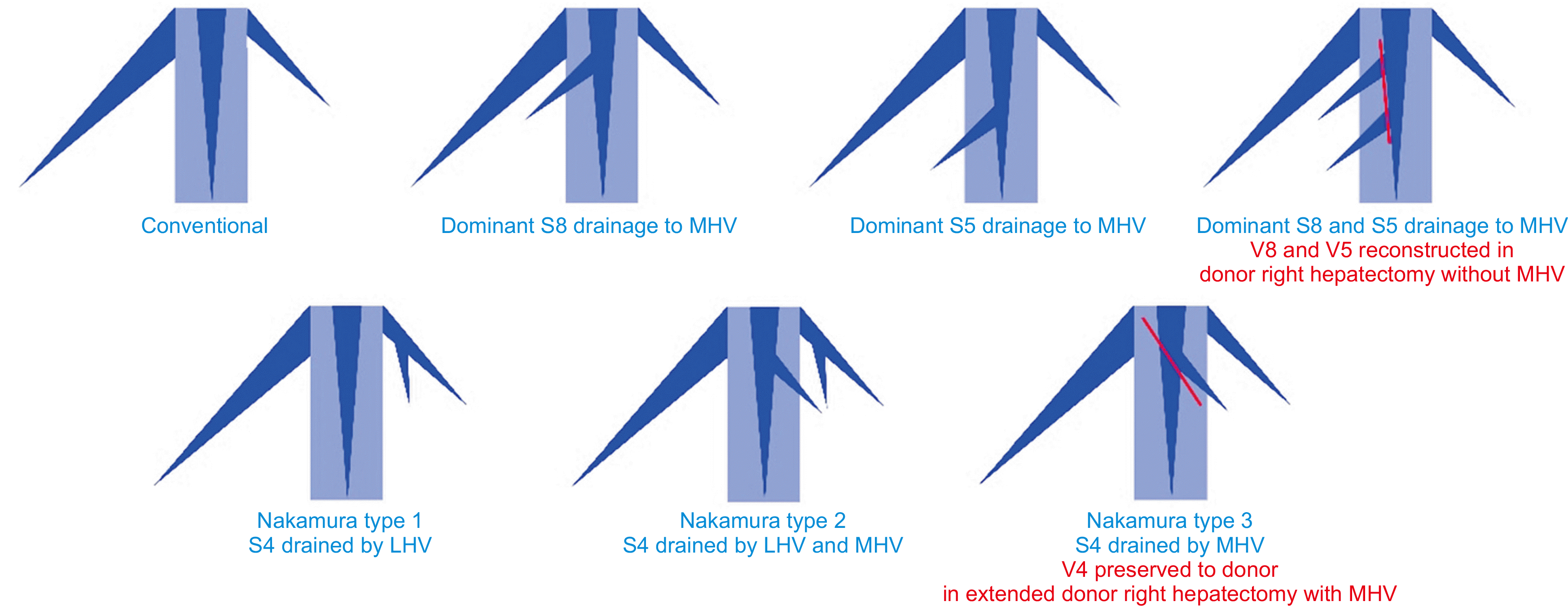
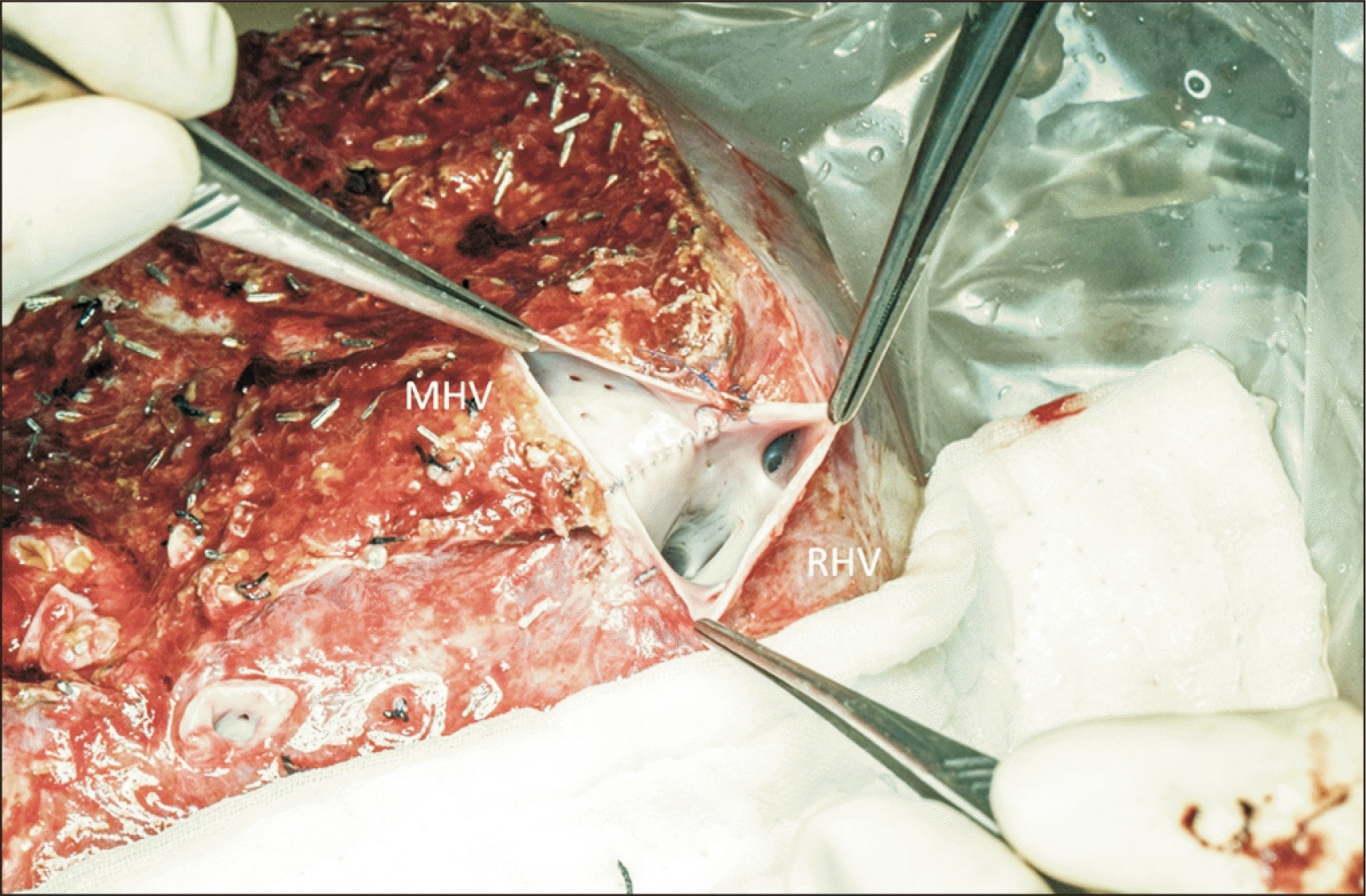
In contrast, the extended right lobe graft includes the main trunk of the MHV [2]. Several advantages are associated with the extended right lobe graft. There is no need for graft reconstruction of the venous tributaries. The RHV and MHV are joined together in the back table into a single triangular orifice [22]. Venoplasty is technically simple, and the 3-point fixation expands the venous opening and maximizes outflow capacity (Fig. 2). The resultant single venous opening offers technical ease for anastomosis in the recipient. The native outflow reconstruction is also durable and patent in the long term. The extended right lobe graft appears to be a better option in donors with hepatic venous variants. However, harvesting the MHV increases segment 4 congestion in the donor. Nakamura and Tsuzuki [23] classified drainage of the segment 4 into three types: type I, mainly via the left hepatic vein (LHV); type II, equally into the MHV and LHV; and type III, predominantly via the MHV (Fig. 1). Types II and III anatomy are less favorable for procuring extended right lobe graft. When the root of the MHV is harvested, there is increased risk of segment 4 congestion. Donor hepatectomy can be modified to preserve the segment 4 hepatic vein (V4) [24] (Fig. 1). Transection of MHV distal to V4b usually adds no technical difficulty to the venoplasty. Preservation of V4b can also be considered in patients with marginal remnant volume to avoid compromising remnant liver function. The use of the extended right lobe graft with RHV/MHV venoplasty has lowered GRWR requirement to 0.6%, with a low incidence of SFSS and excellent outcomes [16].
Go to :
INFLOW MODULATION
Liver graft with optimized outflow could tolerate portal inflow of up to 450 mL/min/100 g GW. Portal flow can be measured after implantation. When the inflow is higher than 250 mL/min/100 g, portal pressure can be obtained by cannulation of the inferior mesenteric vein. Portal modulation is considered if portal pressure exceeds 15 mmHg and can be done by splenic artery ligation or portocaval shunting [17]. Occlusion of the splenic artery reduces portal inflow from the splenic vein. It can result in reduction of the portal inflow by half [25], and the drop in portal pressure is particularly significant in patients with pre-existing portal hypertension, as evident by splenomegaly [26]. A portocaval or portomesenteric shunt can be constructed to divert mesenteric outflow [27,28]. In a porcine model, shunting reduced hepatocyte damage related to portal hyperperfusion [29].
The burden of SFSG is alleviated by experience [30]. The focus of SFSG is shifting from obtaining larger grafts to managing the use of smaller grafts with appropriate flow-modulatory measures [31]. The GRWR can be reduced to 0.6% at expert centers with careful patient selection [16]. Marginal-sized grafts are avoided for recipients with high model for end-stage liver disease scores [32] or when a difficult operation is expected, as in cases with multiple prior laparotomies and the presence of portal vein thrombosis. There is minimal occurrence of SFSS and hospital mortality [16]. A meta-analysis of pooled data indicated that SFSG use is associated with inferior medium-term, but not long-term graft survival [33]. Patients with SFSG who make it through the initial period enjoy long-term survival [13].
Go to :
ANATOMICAL VARIANTS
Portal Inflow Variant
In the conventional anatomy, the main portal vein (MPV) bifurcates at the liver hilum into the left (LPV) and right portal vein (RPV), which then divides into the right anterior (RAPV) and right posterior portal vein (RPPV). Portal vein variants are frequent and occur in 20%–30% of the population [5,34-36]. Various classifications have been proposed [5,37] (Fig. 3). The most common variant is the portal vein trifurcation, in which the MPV divides into the LPV, RAPV, and RPPV at hilum without the RPV main trunk (i.e., Covey type II). The second most common variant is the division of MPV into RPPV and a common trunk, which then divides into LPV and RAPV (i.e., Covey type III). Both variants give rise to two venous openings when a right lobe graft is harvested. The two venous openings are wide apart in type III, which poses an additional challenge for vascular reconstruction. Donors with these variations were usually rejected due to the technical difficulty in vascular reconstruction. However, the modification of surgical techniques has recently allowed the use of right liver grafts from these donors with portal vein variants [38].
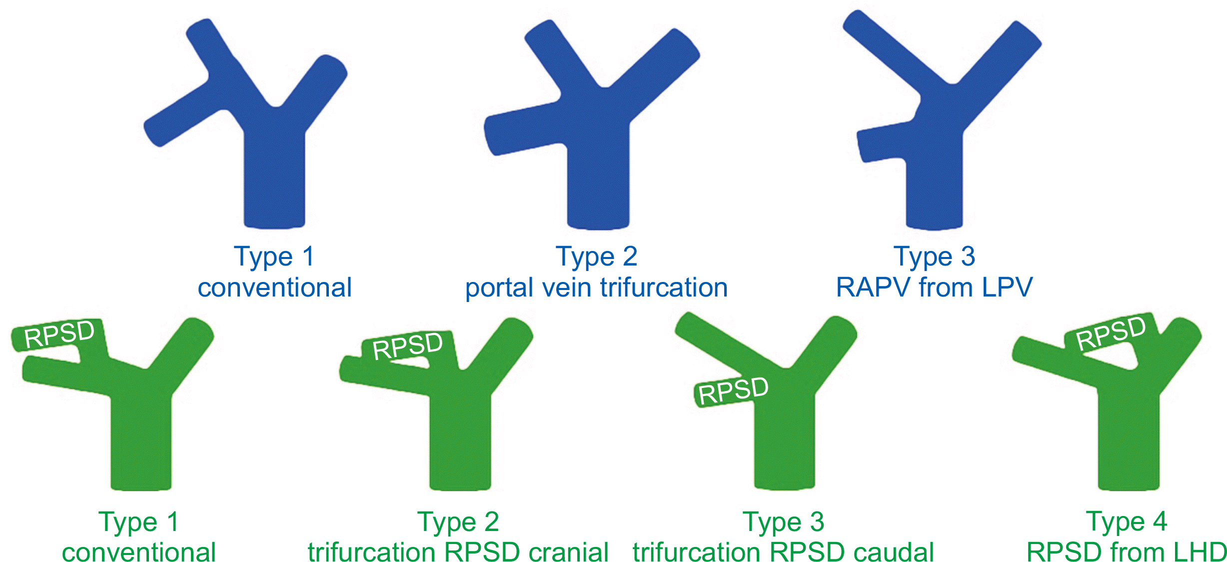
The RAPV and RPPV are usually divided separately to ensure donor safety. In type II, since the distance between the venous opening is close, they can be joined together at the back table with a venoplasty to form a single cuff. In type III, where the two portal vein openings are wide apart, venoplasty creates distortion and narrowing of both lumens. These grafts were once doubly anastomosed to the recipient LPV and RPV. Double anastomosis in the recipient is technically complex. Moreover, twisting occurs when the RAPV and RPPV, which have an antero-posterior orientation, are anastomosed to the recipient LPV and RPV. The malalignment is further exacerbated by regeneration of the liver graft, creating turbulence and predisposing to thrombosis [38]. To reduce recipient complications, a discoid patch can be harvested to include both the RAPV and RPPV openings, which are subsequently closed with a vascular patch. However, this shifts the risk to the donor. When the defect is large, a buckling deformity occurs and turbulence results. In a series of five donors with a single graft portal vein cuff harvested for the type III variant, one donor suffered portal vein thrombosis, necessitating exploration and stenting [38].
The most promising results for portal vein variant reconstruction are offered by Y-graft reconstruction [37]. The RAPV and RPPV are anastomosed to the bifurcated ends of the graft at the back table. This allows an optimal inspection of the orientation and alignment of the anastomoses. This provides a lengthy portal vein with a single opening, which is ideal for reconstruction during implantation. The explant portal vein bifurcation provides an easily available autologous Y-graft with compatible size. In patients with existing portal vein thrombus, a cadaveric iliac vein graft can be used for the same purpose. Thayer et al. [37] and Lee et al. [38] reported five right lobe grafts with Y-graft reconstruction, and no vascular complications were noted in these recipients.
Biliary Variants
Bile duct variations have long been recognized. Nakamura et al. [5] classified anatomical variants based on the take-off of the right posterior sectoral duct (RPSD). In the conventional bile duct (i.e., type I), a single right hepatic duct is present (Fig. 2). Type II and type III represent bile duct trifurcations. In type II, the RPSD branches cranial to the right anterior sectoral duct take-off, and vice versa for type III. In type IV, the RPSD branches from the left hepatic duct. In the absence of a single right hepatic duct (types II-IV), two or more bile duct openings result when the right liver is harvested. When these openings are close to each other, they could be joined to form one single orifice by ductoplasty. When these openings are wide apart, they necessitate multiple anastomoses with hepaticojejunostomy or a combination with duct-to-duct anastomosis. Multiple biliary reconstruction is technically challenging, as these openings are usually small in caliber. When combined with vascular variant anatomy, both the donor risk and recipient complications are increased. When a vascular variant is identified, preoperative magnetic resonance cholangiopancreatography can be performed to identify possible associations with biliary variants.
Go to :
SPECIAL GRAFT TYPES
Right Posterior Sectional Graft
The right posterior sectional graft is an alternative in donors with a small left lobe, which is inadequate as either the remnant or as the implant. With the conventional vascular anatomy, the short and small stump of the hepatic artery (HA) and portal vein in donors with standard anatomy make reconstruction difficult. The most favorable anatomy is RAPV arising from the LPV, which provides a long single RPPV trunk (type 3) (Fig. 2). An early bifurcation of the right anterior and right posterior HA provides a long trunk for arterial anastomosis. The anatomical relationship between the bile duct and portal vein is also important. When the RPSD is posterior to the RPPV, biliary reconstruction is extremely difficult, if not impossible. A ventrally running bile duct is most favorable.
However, the incidence of biliary stricture remains high [39,40]. Dissection around the right posterior pedicle during donor hepatectomy causes devascularization and biliary injury. A small bile duct caliber also renders anastomosis technically difficult. A thorough understanding of the anatomical variations of the portal vein, HA, and bile duct on preoperative imaging is mandatory for the use of the right posterior sectional graft, because the standard bifurcation of the portal vein and posterior bile duct on the dorsal side of the RPPV is not suitable for the right posterior section graft.
Dual Graft
Dual-graft liver transplantation is indicated when a single donation is not sufficient in size, or the remnant volume is insufficient. Options include dual left lobe transplantation and right and left lobe dual-graft transplantation. The former was first performed [41]. The larger left liver is implanted in the orthotopic position, and the smaller graft is implanted in the heterotopic right position (Fig. 4). The rotation of the heterotopic graft rotates the hilar structures into a reversed position so that the bile duct is dorsal to the HA and portal vein. Therefore, the heterotopic right graft bile duct is anastomosed to the recipient bile duct before portal vein anastomosis. After completion of portal vein and HA anastomosis, the orthotopic left graft bile duct is reconstructed with hepaticojejunostomy. A tissue expander is placed underneath the heterotopic graft to avoid graft rotation and subsequent kinking of vascular outflow.
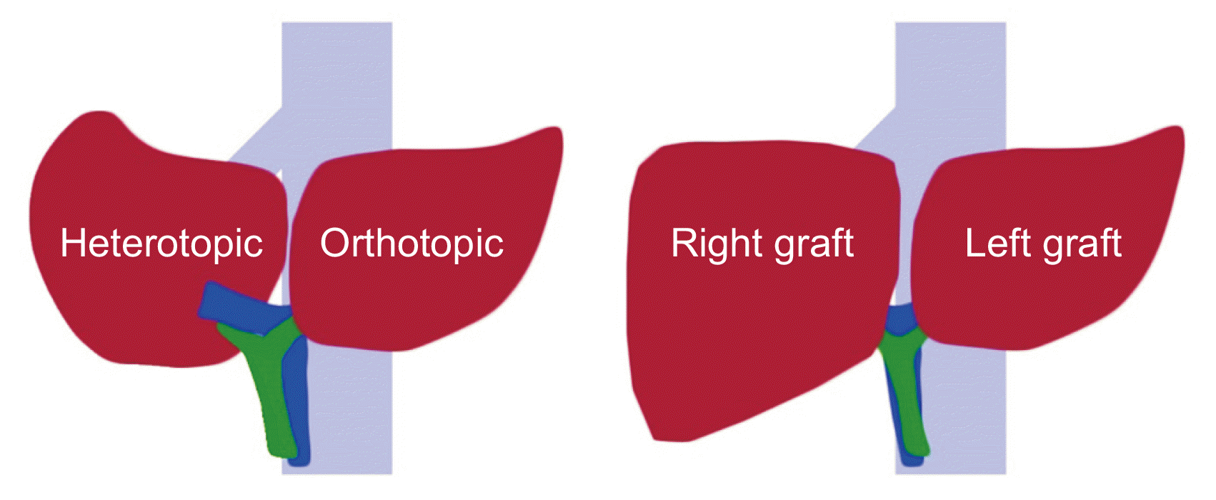
In dual right and left lobe transplantation, both grafts are implanted orthotopically (Fig. 4). The right graft is implanted first starting with the hepatic vein anastomosis. The left liver is then engrafted with the outflow reconstructed. This is followed by sequential reconstruction of the portal vein and HA anastomosis and biliary reconstruction. Dual-graft liver transplantation is technically complex, and there are ethical concerns regarding the involvement of two living donors [42]. Dual-graft liver transplantation should be prudently performed when single donation is proven unacceptable and elaborate surgical planning is crucial. At expert centers, the recipient outcomes of dual-graft transplantation are comparable to those of single-donor LDLT [43].
Go to :
CONCLUSIONS
Graft size is the fundamental challenge in partial liver transplantation. Insufficient graft size leads to SFSS, graft failure, and graft loss. However, smaller grafts can be used safely with surgical techniques to optimize outflow and modulate inflow, thereby minimizing portal hyperperfusion. Meanwhile, anatomical variations are common in the vascular and biliary systems. These variants pose additional challenges for vascular and biliary reconstruction. Recognition and appropriate management of these variants ensure donor safety and reduce recipient morbidity. The ultimate principle of partial liver transplantation is to ensure sufficient graft volume with unimpeded outflow and re-constructable vascular and biliary systems. On this basis, the anatomical limits of LDLT can be safely expanded.
Go to :
 XML Download