

 PDF
PDF Citation
 Print
Print


INTRODUCTION
Liver diseases remain a significant public health problem worldwide, especially in Asian nations, where liver cancer is one of the top 10 causes of death from malignancies [1]. In Vietnam, viral hepatitis plays a key role in causing liver disease. Hepatitis B virus (HBV) is the most common cause of viral hepatitis in the population, with 8.4% of Vietnamese infected with chronic HBV, but only 1.34% receive suitable treatments for the infection [2]. Nguyen et al. [3] further revealed 15,000 cases of hepatitis C virus (HCV) related to decompensated cirrhosis. An estimated 9,000 cases of hepatocellular carcinoma (HCC) cases were also due to HCV. Alcoholic liver cirrhosis has also attracted more attention with rising alcohol consumption, which exacerbates liver disease in patients with viral hepatitis or HCC [4].
Due to all these factors, the number of patients on the waiting list for liver transplantation (LT) has increased over time, even though some have dropped out because the deceased donor pool was insufficient. Some other patients, unable to locate a suitable deceased donor, turned to living donor LT (LDLT) as a last resort. The goal was to shorten waiting time for surgery—hopefully preventing disease progression—over the time needed to wait for a deceased donor [5,6]. Given the positive outcomes published all over the world, LDLT has become an effective and popular therapy in Vietnam [7,8].
Despite advancements in LDLT surgical techniques and post-LT management, complications after LT still affect the overall survival rates of patients and especially living donors. For this reason, initial LTs at our center were supported by staff from Asan Medical Center, Seoul, Korea. Following initial successful outcomes, we went on to perform LT by ourselves. This study discusses not only the LT outcomes and associated risk factors in the transplanted patient, but also the challenges faced on the way to successful operations.
METHODS
This study was approved by the Institutional Review Board of the University Medical Center Ho Chi Minh City (IRB No. 823/QD-BVDHYD). The patients provided written informed consent for the publication of clinical details and images.
Study Design and Data
This study used data from the liver cohort of University Medical Center, Ho Chi Minh City, from August 2018 to December 2021. The center had 18 LT cases during that time. The study data included the recipients’ and living donors’ demographics and comorbidities, as well as immunological assessments and viral markers collected at admission. The profiles of the deceased donors were collected at registration and included the cause of brain death and cold ischemic time. For the LT recipients, laboratory assessments, immunosuppressants used, surgical complications, posttransplant outcomes, and complications were collected at admission and at 1, 6, 12, 24, and 36 months after transplant. All patients and donors provided informed consent before their registration in the Vietnamese National Coordination Center for Organ Transplantation (VNCCOT).
Subject Selection
The study population consisted of patients and donors in the center's registry who had operations between August 1, 2018, and December 31, 2021.
Study Measures
Study measures included recipients’ demographics (age, sex, and body mass index), comorbidities, underlying liver disease, Child-Turcotte-Pugh and model for end-stage liver disease (MELD) scores for chronic liver diseases or cirrhosis, urgent status in VNCCOT, donor type (living or deceased donor), donor demographics (age, sex, and body mass index), graft related variables (graft type, graft-recipient weight ratio, and type of donor operation), ABO blood group compatibilities, laboratory parameters, and the types of immunosuppressant used. If HCC was the underlying reason for LT, the pretransplant HCC status, including Milan criteria and tumor markers, was also collected. All study measures were used to identify risk factors for post-LT outcomes.
Liver Transplant Procedure Protocol
Intraoperative issues
Portal vein thrombosis
Portal vein thrombosis (PVT) has long been considered a contraindication for graft survival following LT because of decreased portal flow. Emerging portal flow restoration techniques, however, have improved the outcomes of such patients (Supplementary Fig. 1) [9,10]. Thrombectomy was used in cases of PVT to counter the high risks after portal vein replacement and jump grafts. For increased safety during thrombectomy, temporary ligations of the splenic vein (SV) and superior mesenteric vein (SMV) were performed before the portal veins were cut. In addition to decreasing blood lost during the thrombectomy, dissection of the SV revealed the apparent confluence of SV and SMV to avoid their damage during thrombotic dissection (Fig. 1).
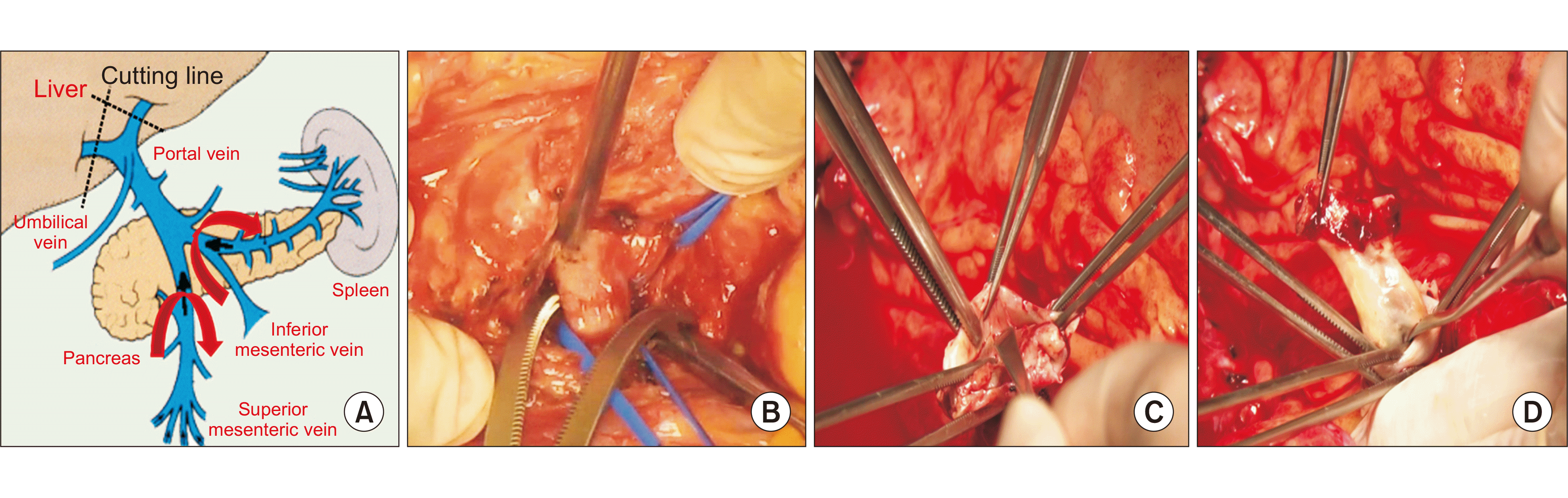
After portal vein cutting, a spatula tool was used to remove the thrombosis out of the portal lumen. To decrease the risk of re-thrombosis, care was taken not to injure the inner layer of the lumen (Fig. 1). After portal anastomosis, the patent was evaluated by an intraoperative angiogram and post-LT computed tomography (CT) images (Supplementary Fig. 2).
Venous outflow reconstruction
At our center, all LDLT cases were right liver grafts (RLGs) with reconstructed middle hepatic vein (MHV). RLG outflow reconstruction helped to decrease congestion of the left liver remnant and keep the minimum liver remnant volume of a living donor above 30%, according to the graft steatosis and donor age (Supplementary Table 1). This increases the living donor pool at the center.
Preoperative planning is critical to identify how many orifices of the RLG need to be reconstructed. (1) Anterior section hepatic vein (HV) reconstructions represent the ultimate outflow design work required at the back-table. HVs are reconstructed with the recipient’s saphenous vein (Fig. 2). (2) All drainage veins of segments 5 and 8 larger than 5 mm in diameter should be reconstructed. In multiple V5 or V8 cases, adjacent tributaries can be united into a single orifice (Fig. 2). (3) Prosthetic interposition grafts were used for HV reconstruction and final all-in-one anastomosis (right HV, MHV of graft, and inferior vena cava [IVC] of recipient) (Fig. 2). (4) The inferior right hepatic vein (IRHV) from the posterior section requires unification venoplasty of multiple short HVs for a single-wide orifice anastomosis to a corresponding orifice on IVC instead of individual anastomosis of these veins. After reconstruction, the anastomosis patent was evaluated by Doppler ultrasonography and postoperative CT (Supplementary Fig. 3).
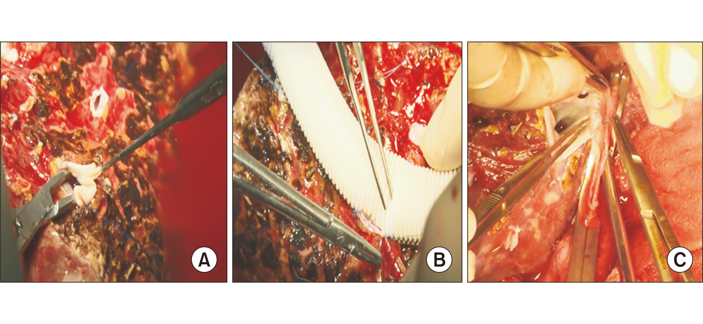
Fig. 2
Venous outflow reconstruction by artificial interposition and the saphenous vein. (A) V5–V8 revascularization using the saphenous vein (diameter after suture ≥1 cm). (B) Inner suture between V5 or V8 with interposition. (C) Artificial middle hepatic vein in alignment with the right hepatic vein. One outflow (≥4 cm) of the right liver graft.
![]()
Arterial anastomosis
For the hepatic arterial anastomosis, Prolene 9-0 or 10-0 sutures were used in an end-to-end manner, with a continuous suture technique supported by 4× loupes (instead of a digital microscope). The patency and flow of the artery were regularly evaluated by intraoperative Doppler ultrasonography and CT angiography for 1 week (Supplementary Fig. 4).
Biliary anastomosis
End-to-end anastomosis following the septoplasty technique was used where possible for anastomoses with a diameter <3 mm. Interrupted sutures were used to decrease the risk of anastomosis stenosis. The patency of biliary anastomoses was regularly evaluated using intraoperative cholangiography for the early detection of biliary leakage. All cases had an external biliary stent to allow assessment of liver function using biliary amount and application of cholangiogram if biliary complications were suspected. The stent was then ligated before the patients were discharged (Supplementary Fig. 5).
Treatments after LT
In all cases, steroids and Simulect (Novartis, Basel, Switzerland) were used to induce immunosuppression. To maintain immunosuppression, all recipients were given calcineurin inhibitors such as tacrolimus, then antimetabolite agents such as mycophenolate mofetil and mammalian target of rapamycin inhibitors such as everolimus (11.1%) in cases of renal dysfunction and de novo or recurrent malignant diseases. Steroids were stopped as soon as possible—at 1 month after LT for patients (16.7%) with stable liver function test (LFT) results.
For post-LT HBV prophylaxis, all HBV-positive recipients were prescribed anti-HBV-specific immunoglobulins with antiviral therapies. Thus the trough level of HBsAb was generally maintained above 500 IU/L during the first year and 200 IU/L in the following years.
Statistical Analysis
Continuous variables were expressed as mean±standard deviation or median (range), and categorical variables as counts and percentages. Calculations to estimate the survival chances of recipients according to post-LT outcomes at 1 year were affected by the small sample size. The censoring time, therefore, was defined as the final documented date.
To assess the potential risk factors for post-LT outcomes, chi-square analysis was conducted with each variable as an independent predictor for each outcome. Variables found to be significant in the univariate analysis (P<0.05) were analyzed to determine which factors independently predicted post-LT outcomes and patient survival. All analyses were performed using IBM SPSS ver. 26.0 (IBM Corp., Armonk, NY, USA).
RESULTS
Baseline Characteristics
Eighteen adult recipients were included in the analysis, with a mean follow-up duration of 17.2±2.7 months, depending on the LT date. Among these patients, 16 cases had deceased donor LT (DDLT). The mean ages of adult LDLT and DDLT recipients were 57.6±2.3 and 36.5 years, respectively. Recipients’ demographics and clinical characteristics by donor type are summarized in Table 1. HCC and alcoholic liver disease were the two most common underlying liver diseases among the LDLT (55.6% and 33.3%) and DDLT (100% and 0%) recipients. All HCC cases met the Milan criteria, and 44% of cases needed bridging therapies during the waiting period (Table 1). RLGs were used for all LDLT procedures, while whole-liver grafts were used in DDLT.
Table 1
Demographics and clinical characteristics of adult liver transplant recipients overall and by donor types
LDLT, living donor liver transplantation; DDLT, deceased donor liver transplantation; BMI, body mass index; HCC, hepatocellular carcinoma; TACE, trans-arterial chemoembolization; RFA, radiofrequency ablation; MELD, model for end-stage liver disease; CTP, Child-Turcotte-Pugh; mTOR, mammalian target of rapamycin.
![]()
Donor Characteristics
The mean age of living donors was 36.1±6.5 years, and 50% were male. A small proportion of donors (18.7%) were overweight (body mass index, 25–29 kg/m2), and five donors (31.3%) with latent HBV infection were used for patients with HBV. Donors were relatives of recipients in eight cases (50%). Conventional open surgery was performed in all cases (Table 2). The selected donor criteria were graft steatosis by biopsy, liver remnant volume, and LFTs (Supplementary Table 1) [11]. The indocyanine green (ICG) test was used to evaluate liver function, and standard tests like aspartate aminotransferase, alanine aminotransferase, gamma-glutamyl transferase, bilirubinemia, and platelets were also considered. If the ICG test yielded a result of less than 10%, right hepatectomy could be performed according to Makuuchi’s algorithm (Supplementary Fig. 6) [12].
In deceased donors (n=2), the principal cause of death was the progression of underlying disease, followed by trauma. The mean cold ischemic time was 2.5 hours. Stable LFTs were used instead of biopsy before surgery to estimate graft steatosis. The donors did not have a history of disease.
Outcomes after LT
Complications
The surgical post-LT complications included middle hepatic venous stenosis (22.2%), portal stenosis below 50% (11.1%), biliary leakage (5.6%), splenic abscess after splenic arterial embolization (5.6%), intestinal perforation (5.6%), and hepatic arterial stenosis (5.6%). All complications occurred in LDLT patients. Among them, two cases with a Clavien-Dindo classification of grade 3B had to be re-operated because of splenic abscess and small intestinal perforation, while one patient needed endoscopic retrograde cholangiopancreatography (ERCP) stenting for biliary leakage. Others with a Clavien-Dindo classification of grade I-II were treated without intervention or operation.
The non-surgical complications were rejection (22.2%), pneumonitis (11.1%), and renal dysfunction (5.6%) (Supplementary Table 2). For graft rejection, high doses of steroids (25 mg) were administered three times per day for the first 3–5 days, followed by tapering doses for the next 2–3 days; in all cases the response to treatment was positive. No deaths during operations or hospital stays were recorded, and no recurrent HCCs have yet been detected.
Overall survival rate
With the first LT occurring in 2018, patients' follow-up time was 17.2±2.7 months, with the longest follow up time 40 months. Two LDLT cases (11.1%) died from bacterial pneumonitis after 4 months. The rest were monitored through the study period without additional complications or deaths. The survival rates at 3 months, 6 months, and 1 year were 100%, 88.9%, and 88.9%, respectively. The median length of hospital stay was 28 days, with 25th and 75th percentiles of 25 days and 41 days, respectively. There were no deaths during the hospital stays. Most patients went back to daily life after 1 month and returned to work after 3 months.
Living donor outcomes
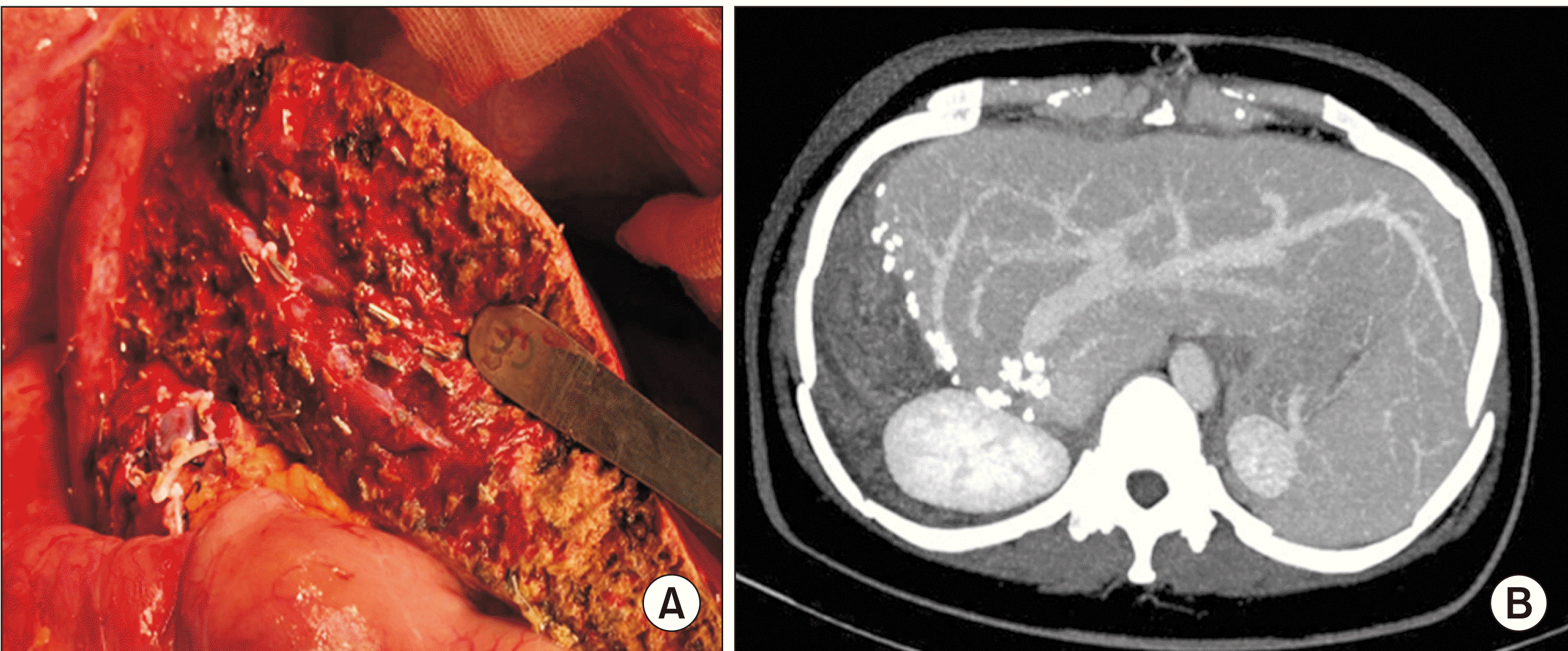
For the living donors, right livers without the MHV were harvested and conventional open procedures were performed. The operating time was 4±1 hours, and the median length of hospital stay was 7 days, with 25th and 75th percentiles of 6 days and 7.75 days, respectively. There were no deaths during the hospital stays. All donors went back to daily life and work after 2–3 weeks. CT scans were used to evaluate their abdominal conditions after a month (Fig. 3). There was one case (6.25%) of biliary leakage detected on postoperative day 6 and treated with ERCP stenting. The donor was rechecked and the stent was removed after 6 months.
DISCUSSION
This study showed the outcomes and risk factors in patients who underwent LT and the potential risks and recovery of living donors after the operation. Among surgical complications, vascular stenosis after suturing always receives considerable attention. As shown here, 35% of complications were related to vascular anastomosis.
Arterial Hepatic Stenosis
One case (5.6%) had hepatic arterial stenosis suspected due to thrombosis. On postoperative day 7, intrahepatic arterial flow was not detected by CT angiography (Supplementary Fig. 7). As the hepatic artery plays a significant physiological role in providing blood for the liver parenchyma and the biliary tree, after arterial reconstruction, the interruption or the reduction of arterial flow during liver transplant is frequently associated with biliary tree complications due to ischemic processes with the absence of collaterals in a liver transplant recipient [13]. In one case, peripheral vascular perfusion was detected using Doppler ultrasonography (Supplementary Fig. 7) following stable LFTs. Antithrombic medications were prescribed, and the patient closely monitored using ultrasound and LFTs. At postoperative day 14, the intrahepatic arterial flow was evaluated using CT. The patient was discharged on postoperative day 21, and graft rejection has not been seen since then.
Middle Hepatic Venous Stenosis
Patency of the MHV was essential in reducing the risk of suboptimal graft function and graft failure [14]. In right-lobe LDLT, the focus was on donor safety and MHV reconstruction to avoid congestion injury of the anterior segment. All right lobe grafts were harvested without their MHV to decrease the risk of donor liver failure. The MHV was reconstructed by prosthetic interposition graft, and all-in-one anastomosis was performed. The patency rates of the all-in-one anastomosis were 87.5% at 1 month, 87.5% at 3 months, 81.3% at 6 months, and 68.8% at 12 months. This compared favorably to the proportions of separate outflow reported by Kirchner et al. [15] of 90%, 65%, and 37% at 3, 6, and 12 months, respectively. The graft rejection rate of LDLT recipients was not statistically different in the group with MHV obstruction (P=0.07) (Table 3).
Graft Rejection
Beyond the non-surgical complications, rejection was the most common problem. There were four cases of graft rejection during the first month after LT (22.2%). Graft rejection was treated with high doses of steroids. Unfortunately, these high doses—although only short courses—increased susceptibility to infections such as hospital pneumonitis, impaired wound healing, and raised the risk of metabolic disorders [16]. These unexpected side-effects of high steroid doses resulted in a switch to a lower-dose protocol. Among all cases of graft rejection, the mean time was 6±2.1 days, with 11 improving without steroid resistance, and no chronic rejection so far recorded. No statistically significant relationship between graft rejection and donor status (living or deceased) (P=1.004), pretransplant viral hepatitis (P=0.161), graft steatosis below or above 5% (P=0.303), and ABO blood type (P=2.296) were found (Supplementary Table 3).
Overall Survival Rate
In this study, the overall survival rates at 3 months, 6 months, and 1 year were 100%, 88.9%, and 88.9%, respectively. The longest follow-up time was 40 months. These outcomes are comparable to those in other studies, which showed survival rates of 80%–90% at 1 year [17-20]. After discharge, most patients were checked at regular intervals in the outpatient department.
Living Donor Outcomes
The rate of living donor complications was 6.25%, with biliary leakage the only complication reported. These outcomes were similar to other studies showing rates of complications of 9%–15%, with biliary leakage one of the most common complications after hepatectomy [21,22]. After their operations, no donors suffered psychological disorders or difficulties with social integration. Most went back to work after a few weeks without physical assistance.
Limitations
The small sample size limited tests of statistical reliability, so more time would be needed to confirm the results. Although LT requires painstaking preparation and many precise, well-ordered steps, the positive outcomes reported herein suggest that LT is a valuable therapy for patients with end-stage liver disease and early HCCs.
Supplementary Materials
Supplementary materials can be found via https://doi.org/10.4285/kjt.22.0010.
 XML Download