

 PDF
PDF Citation
 Print
Print


INTRODUCTION
Endovascular treatment with coil embolization is considered a primary management strategy for ruptured intracranial aneurysms. Despite its effectiveness and safeness compared to surgical management, the question of long-term durability remains controversial [5,7,10,14,20]. Coil embolization has some drawbacks, one of which is not being able to completely block the blood flow into the aneurysm with the first treatment. Therefore, the recanalization of a coiled aneurysm can present a great risk to the treated patients [10,22,31].
Since the International Subarachnoid Aneurysm Trial report in 2002, questions have been raised about the durability of endovascular coiling, and reports on this have continued [19-21]. However, as in the following report, the focus has mainly been on long-term clinical results, and it seems that long-term radiologic results and imaging follow-up plans after endovascular treatment, especially in ruptured cases, have been insufficiently dealt with.
To the best of our knowledge, there exist no clear protocols for when to follow up and how to do it [28,29,32]. Also, the imaging modalities and intervals of follow-up can vary according to each institution [15,32]. The present study aimed to evaluate the long-term follow-up catheter-selected angiography protocol by considering the balance between safety and efficacy after endovascular coiling for small ruptured intracranial aneurysms.
Go to : 
MATERIALS AND METHODS
This study was approved by the Institutional Review Board (IRB) of Dankook University Hospital (2019-06-014). Due to the retrospective design of the study, consent was neither required by the IRB nor by the study team.
Study population
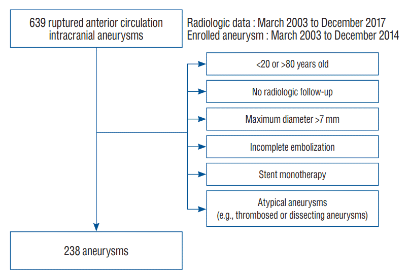
This was a retrospective review of patients treated with coil embolization at a single institution. From March 2003 to December 2014, we performed endovascular coil embolization for 639 patients with intracranial ruptured aneurysms of the anterior circulation. The radiological records between March 2003 and December 2017 were retrospectively reviewed. Patients 20 to 80 years old who received endovascular treatment only for ruptured saccular aneurysms were included in the study if they had follow-up imaging using catheter-selected angiography at least once. To reduce the factors known to greatly influence recurrences, such as aneurysm size and the completeness of embolization, the study subjects were limited to a ruptured aneurysm of 7 mm or less [13,31]. Patients were excluded if they underwent incomplete embolization including stent monotherapy or had atypical aneurysms (e.g., thrombosed or dissecting aneurysms) or aneurysms with a maximum diameter of greater than 7 mm. Of 639 patients, we enrolled 238 ruptured cerebral aneurysms treated with coil embolization as a result of Raymond-Roy Occlusion Classification (RROC; also known as the Montreal Scale, or the Raymond Montreal Scale) [18] class I or II and followed up with at least one transfemoral cerebral angiography (TFCA) (Fig. 1). Patient data including age, sex, aneurysm size, stent assistance, location of the ruptured aneurysm, follow-up period, and clinical and radiologic results were collected. Postembolization angiographic appearance was evaluated using the RROC, a widely accepted system for evaluating aneurysm occlusion class [18,25,26]. In this scheme, class I is defined as complete obliteration without a residual neck, class II as contrast filling of the residual neck, and class III as contrast filling of the residual aneurysm. It has been shown that class III aneurysms have a higher propensity to remain incompletely occluded [25,26].
Coil and stent devices
Bare platinum coils including Guglielmi detachable coil (Boston Scientific, Marlborough, MA, USA), Target coil (Stryker, Cork, Ireland), and MicroPlex coil (MicroVention, Aliso Viejo, CA, USA) were used in all cases. Neuroform 2 or Neuroform 3 stent (Boston Scientific), Neuroform EZ (Stryker), and Enterprise (Codman, West Chester, PA, USA) were used when stent assistance was required.
Follow-up protocol
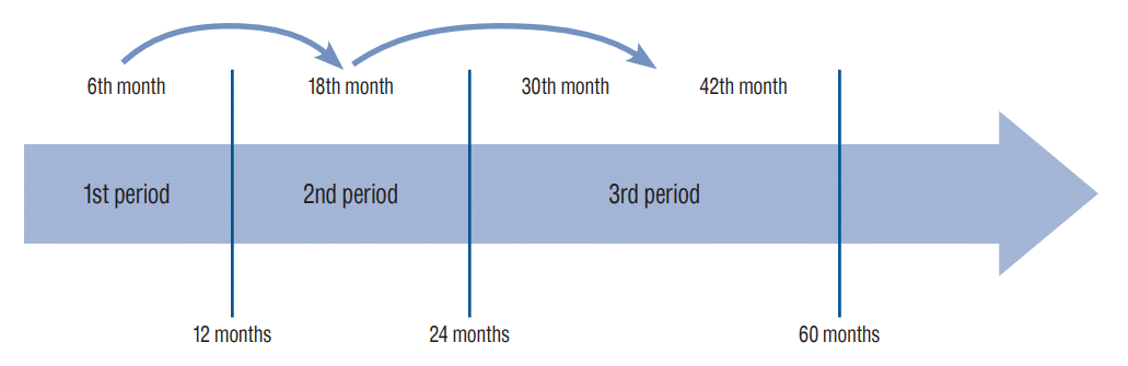
Our protocol for angiographic follow-up using only catheter-selected angiography was as follows (Fig. 2). The first angiographic follow-up was conducted 3 to 6 months postoperatively according to clinical outcomes and the immediate angiographic results. The next one was performed 12 months later depending upon the angiographic results of the first follow-up. The third one was performed 12 to 24 months later, depending on the results of the second follow-up. As above, our institution, in principle, reported at least three follow-up observations and performed digital subtraction angiography (DSA) to detect relevant aneurysm recurrences. According to our protocol, we classified four periods of angiographic follow-up as follows: follow-up within post-treatment 1 year (defined as the first period), from 1 to 2 years (the second period), 2 to 5 years (the third period), and over 5 years (long-term, the fourth period). During the follow-up test, if the appearance of a radiolucent band indicating isolation by thick neointimal tissue formation at the aneurysm neck (known as the white-collar sign) was confirmed, the follow-up test was terminated. On the other hand, aneurysms were treated with confirmed recurrence or recanalization due to coil mesh compaction or aneurysm growth [11]. Two neurointerventionalists independently interpreted the radiological images.
Go to :
RESULTS
Details of follow-up data
Of the total 238 patients, 218 were followed up in the first period, 143 in the second period, and 97 in the third period. We identified 14 cases (6.4%) of recurrence that required retreatment from 218 aneurysms in follow-up angiography in the first period. Among 143 aneurysms in the second period, five cases (3.5%) of recurrence were identified. There were no findings suspicious of recanalization in 97 patients who received angiography in the third period. Of the total 238 cases, there were 19 recurrences, for a recurrence rate of 8.0%. Seventy-six patients underwent follow-up only once during three periods (64 patients only in the first period, eight patients only in the second period, and four patients only in the third period). One hundred fifty-eight patients completed the test after two or more follow-up tests. Of the 158 patients, 92 had two follow-up examinations during three periods. Specifically, 65 patients had them in the first and second periods, four patients had them in the second and third periods, and 23 patients had them in the first and third periods. Sixty-six patients underwent follow-up during all three periods. In this study, all recurrences were confirmed within 2 years (the first or second period), and patients without recurrences up to 2 years had no recurrences at follow-ups within 5 years. The details of the follow-up data are shown in Table 1.
Recurred aneurysms
Additional treatment was performed for aneurysms with marked recurrence or recanalization due to coil mesh compaction. Among 19 cases of recurrent aneurysms that underwent additional treatment, 10 cases (52.6%) were located at the anterior communicating artery, six (31.6%) at the bifurcation of the middle cerebral artery, and three (15.8%) at the posterior communicating artery. Fourteen cases (73.7%) of recurrence were revealed in the first period of follow-up angiography, and five cases (26.3%) in the second period. Of the 14 patients who showed recurrence in the first period follow-up, seven (50.0%) had RROC class I aneurysms on the immediate post TFCA, and seven (50.0%) had RROC class II [18]. Of the five patients who showed recurrence in the second-period test, three (60.0%) had RROC class I aneurysms in the immediate post-treatment TFCA, and two (40.0%) of them had aneurysms that had changed to class II in the first period follow up [18]. Six (31.6%) out of 19 recurrences showed a tendency toward repeat recurrences even after additional treatment. Five (83.3%) out of six repeated recurrences were located at the anterior communicating artery and the other one (16.7%) was at the posterior communicating artery. The details of the recurred aneurysms data are shown in Table 2.
Table 2.
Details of recurred aneurysms
![]()
Long-term follow-up data
Twenty-eight patients received long-term follow-up angiography over 5 years (the fourth period) after the initial coil embolization. In our study, all 28 aneurysms had good outcomes without recurrence. In 21 (75.0%) out of 28 long-term follow-ups, favorable radiologic results were confirmed in more than two follow-ups within 5 years. In the remaining seven cases (25.0%), less than one follow-up test was performed during three periods, and in four cases (14.3%), no recurrence was observed in the first long-term follow-up test of more than 7 years after the immediate post-treatment TFCA. Moreover, in six cases of follow-up over 10 years, there were no recurrences. The details of the long-term follow-up data are shown in Table 3.
Table 3.
Details of long-term follow-up data
Post-op : post-operative, F : female, MCAbif : the bifurcation of the middle cerebral artery, class I : Raymond-Roy Occlusion Classification (RROC) class I, M : male, AcomA : the anterior communicating artery, class II : RROC class II, PcomA : the posterior communicating artery, Lt. : left, M1 : the M1 segment of the middle cerebral artery, Rt. : right, A1 : the A1 segment of the anterior cerebral artery, AChoA : the anterior choroidal artery, DACA : the distal anterior cerebral artery
![]()
Go to :
DISCUSSION
To the best of our knowledge, there exists no guidelines or scientific data defining the optimal protocol for when and how long to follow-up ruptured intracranial aneurysms after endovascular treatment [28,29]. It is often challenging for clinicians to determine when or how many times follow-up images should be acquired, which recurrent aneurysms need to be treated again, and which kinds of imaging modality should be applied for follow-up.
In our study, of the 238 patients with ruptured cerebral aneurysms treated with coil embolization and who underwent at least one follow-up TFCA, 19 patients (8.0%) showed recurrence requiring additional treatment. Fourteen cases (73.7%) of recurrence were revealed in the first period of follow-up, and five cases (26.3%) in the second period. There were no patients who did not show recurrence in the previous examination but showed recurrence in the 3rd period or more. Based on these data, we could infer that most recurrences have a high probability of occurring within 2 years of the first coil embolization. As far as we know, few prior studies or protocols have clearly specified the follow-up period for ruptured cerebral aneurysms that have undergone coil embolization [14,25,27,28,33]. In our institution, follow-up was performed on all patients with ruptured cerebral aneurysms according to a strict and identical schedule, and most recurrence findings were reported to be found during the first and second years after the procedures. A strict follow-up schedule is required, and our institution recommends at least two follow-ups within 2 years. Furthermore, if there is a possibility of recurrence, it is considered appropriate to perform an early follow-up within 6 months regardless of the imaging modality [28]. Objective interpretation of the results and accuracy not to ignore even small changes are required. When these conditions are observed, satisfactory long-term results of more than 5 years can be expected.
In a previous report, Rezek et al. [27] stated that unfavorable angiographic appearance was noted almost twice as frequently by an independent core laboratory compared to the operators at the treating centers treating aneurysms with coil embolization. This may emphasize that clinicians should not underestimate a minor difference in follow-up images and that the objective interpretation of surgical outcomes could achieve favorable long-term results. In our study, 19 out of 238 cases with completely occluded aneurysms confirmed in the initial treatment had a recanalization rate of 8.0%. There were no rebleeding or reruptured events during the follow-up period. We assume that the study subjects were limited to small-sized aneurysm which underwent complete embolization with good compliance in follow-up resulted in early detection in recanalization which prevented rebleeding or reruptured events. However, there were a few cases of rebleeding or reruptured events from patients who were excluded from current study which includes large aneurysm, inadequate packing density and follow up losses. In our institution, small cerebral aneurysm recurrences with sizes of 7 mm or less that require additional treatment, which was included in the study criteria, were defined as cases where contrast filling of more than 2 mm of the residual aneurysm was observed. It was judged that 2 mm was the minimum size that could be filled using one minimum size coil [12,17,30]. Of the 19 recurrence cases, six cases (31.6%) showed repeated recurrence due to coil mesh compaction even after additional treatment and for this, additional coil embolization was repeatedly performed. As in other previous studies, we found that retreated aneurysms were predisposed to recanalization compared to initially treated aneurysms [6,23,31]. Of the 238 cases in this study, a total of 58 cases (24.3%) were treated with stent assisted coil embolization. To add an additional explanation, among the 19 recurrences, there was one case (5.2%) in which the stent was assisted during the first treatment (Fig. 3). As previously demonstrated in other studies, it was confirmed that the use of stents was associated with lower rates of aneurysm recurrence [1,3,4,9,16,34]. Lawson et al. [16] reported that stent-assisted coiling caused the progression of occlusion and the complete thrombosis of incompletely coiled aneurysms, possibly by a flow remodeling effect.
 | Fig. 3.A 45-year-old male treated with stent assisted coil embolization for right middle cerebral artery ruptured aneurysm (case 12 in Table 2). A : Transfemoral angiographic finding of the right middle cerebral artery aneurysm. B : Immediate post-embolization angiography shows nearly completely occluded aneurysm with small amount of contrast filling of the residual neck (Raymond-Roy Occlusion classification class II, white arrowhead). C : On post-embolization 10-month angiography, the aneurysm was filled with contrast, which showed recurrence which required retreatment.
|
Recently, the techniques of magnetic resonance angiography (MRA) have advanced remarkably and are used in many institutions [8,15,28,29,33]. Due to concerns that DSA is an invasive procedure that can cause complications such as cerebral thromboembolism (from silent microemboli to transient or permanent neurological deficit in 0.5% to 3% of procedures), nephrotoxicity, or anaphylactic reactions due to contrast and puncture site hematoma, many institutions use MRA for follow-up examinations as the noninvasive imaging of choice [8,15,28,29,33]. There are no clear recommendations in clinical practice. However, we think that DSA is still the gold standard for evaluating aneurysmal occlusion after coiling and is an essential tool for follow-up imaging. Three T high-field time-of-flight MRA is a better method for detecting recurrences compared to 1.5 T field MRA, but still signal voids and metallic materials creates artifacts, especially when using stents. Although other studies have reported that these artifacts did not hamper recurrence identification, even if recurrence is eventually detected by MRA, it should be confirmed through DSA [2,8,15,24,33]. Since the presence or absence of recurrence has a clinically important impact, the most accurate diagnosis is considered necessary. Based on these consensuses, even if follow-up study using MRA is performed after the initial treatment, we recommend that DSA be performed at least once 2 years after the treatment to confirm the recurrence.
The present study conducted by our institution had several limitations. First, this was a retrospective chart review at a single institution. It is possible that these situations might have increased the risk of selection bias. Second, the sample size enrolled in this study was too small to reach statistically significant conclusions. In addition, there was another limitation in that it is difficult to apply the study results to cases of cerebral aneurysms with sizes exceeding 7 mm or atypical aneurysms (e.g., thrombosed or dissecting aneurysm). A future study with a larger number of patients is needed to establish a protocol that clearly specifies the follow-up period for ruptured cerebral aneurysms that underwent coil embolization. Despite these limitations, our study might provide guidance for planning treatment strategies.
Go to :
CONCLUSION
In this study, most of the recurrence findings were found during the first and the second year after the treatments. As a result of these findings, we suggest that at least one DSA examination may be necessary around post-treatment 2 years, especially in ruptured cases. A strict radiologic follow-up schedule is required, and if the angiographic results are favorable at 2 years post-treatment, long-term angiographic results should be favorable.
Go to :
 XML Download