

 PDF
PDF Citation
 Print
Print


INTRODUCTION
Transvenous embolization (TVE) is the mainstream endovascular therapy for treating the cavernous sinus dural arteriovenous fistula (CSDAVF) [11,24]. Transfemoral venous routes to access the cavernous sinus (CS) are the ipsilateral or contralateral inferior petrosal sinus (IPS), superior petrosal sinus, and superior ophthalmic vein via the facial or superficial temporal vein [1,2,4,15,21,22]. The most favorite route is the ipsilateral IPS since it is relatively direct and short from the internal jugular vein (IJV) [11,24]. Sometimes, however, the IPS is found to be occluded due to thrombosis or fibrosis [2,5,10,12,16,17,20,25].
Some studies have reported successful access to the CS in TVE via the occluded IPS [5,12]. Cho et al. [5] reported that the microguidewire looping technique was a safe and effective technique to enter the CS via the occluded ipsilateral IPS during transvenous coil embolization of CSDAVF. Jia et al. [12] previously identified an accessible and successful approach—the frontier-wire technique—using a regular 0.035-inch guidewire. These techniques emphasized the anatomic knowledge of the IPS and technical safety due to the blind approach. Furthermore, these studies considered reinforcing support necessary when the occluded IPS was cannulated, and the microcatheter was advanced into the CS. The use of these reopening techniques provided a high technical success rate of 70–80%.
However, even successful probing most courses of the occluded IPS using the reopening techniques, a fibrotic stricture between the end of the occluded IPS and dilated ipsilateral CS made final entry into the CS challenge. Furthermore, not infrequently, even after a successful approach into the CS, further access to the fistula or shunted pouch (SP), which is the target of the coil embolization, was challenging [13,16,17,19,25]. It would be attributed to the neurointerventional microguidewires unable to overcome the fibrotic membranous barriers or septum. All these obstacles may make us choose more prolonged and more difficult venous routes or increase the chance of unsuccessful results.
We find that using a rigid-tipped microguidewire, such as the chronic total occlusion (CTO) wire, can be a method to enter the SP in the situations mentioned above. It can facilitate to advance of the microcatheter into the SP by puncturing the fibrotic stricture and membranous barrier. CTO wire originally has more penetrability and trackability to revascularize the CTO lesions in coronary intervention [18]. This report aims to share our initial experience with the rigid-tipped microguidewire to overcome the challenges of access to the CS or SP through the occluded IPS in the TVE of the CSDAVF.
Go to : 
MATERIALS AND METHODS
The Institutional Review Board (IRB) of Asan Medical Center, University of Ulsan College of Medicine approved this study (IRB No. 2020-1839), and the requirement for patient consent was waived due to its retrospective and anonymized design.
Study population
We conducted a retrospective review of prospectively collected data for all neurointerventional procedures in our institution from January 2015 to July 2020. We searched the patients with the CSDAVF who underwent transvenous coil embolization via the occluded IPS and whose access to the CS or SP was achieved by the reopening technique followed by using the rigid-tipped microguidewire. A total of four patients met the searching criteria. The patients underwent a total of five procedures using the rigid-tipped microguidewire. All were females with a mean age of 52 years old. One patient underwent a second procedure due to CSDAVF recurrence 5 days post-procedure. We obtained symptoms and neurological signs of the patients from the medical records. Characteristics of CSDAVFs were evaluated according to findings of a preoperative 6-vessel cerebral angiography. The clinical and angiographic characteristics of the study cohort are summarized in Table 1.
Table 1.
Summary of clinical and angiographic characteristics
![]()
Endovascular procedures using the frontier-wire technique
Under general anesthesia, arterial and venous approaches are gained through the left femoral artery and right femoral vein. And then systemic heparinization is performed using intravenous unfractionated heparin. A 4-F diagnostic catheter (Jungsung Medical, Seoul, Korea) with a continuous heparinized flush is introduced through the left femoral artery and then placed in the proximal portion of the internal carotid artery (ICA) or external carotid artery for better observation of the SP in control arterial angiogram. A 6-F guide catheter (GC) (Envoy, Codman Neurovascular; or Fubuki, Asahi Intecc, Aichi, Japan) is introduced through the right femoral vein and initially advanced toward the IJV ipsilateral to the SP and placed inferior to the jugular bulb. Approaching the contralateral IJV and jugular bulb is considered if the ipsilateral IJV cannot be accessed anatomically. In our institution, the 0.035-in polymer-jacketed guidewires (Radifocus; Terumo; or Crescendo; Sungwon Medical, Cheongju, Korea) are used to cannulate the invisible course of the IPS as previously described, known as the frontier-wire technique [12]. Briefly, under the guidance of the jugular venographic roadmap, the tip of the GC and the 0.035-in hydrophilic guidewire are turned anteromedially to locate the ostium of the occluded IPS. Following the selection of the ostium of the occluded IPS, the guidewire is rotated gently and advanced along an imaginary anatomic course to the CS. Once the guidewire tip is advanced as much as possible, the jugular venographic roadmap is acquired. By removing the guidewire, we can obtain the jugular venographic roadmap with a white line as a footprint of the guidewire indicating the course of the IPS. The microcatheter system is then advanced into the CS along the white line in the roadmap. After the microcatheter is further moved into the SP, subsequent coil embolization is performed. We usually use the neurointerventional microguidewires such as Traxcess 14 (Microvention), Transend 14 (Stryker), or Synchro 14 (Stryker).
Application of the rigid-tipped guidewire
Even the reopening technique enables us to cannulate nearly the entire course of the IPS, the fibrotic stricture or membranous barrier sometimes prevents entrance into the CS or SP, respectively [9,15,16]. It cannot be penetrated with 0.014-in neurointerventional microguidewires or 0.035-in guidewire. To penetrate these fibrotic and short-segment structures, we apply the rigid-tipped microguidewire such as the CTO wire as the second-line method following the reopening technique such as the frontier-wire technique. When the microcatheter tip is placed just before the target, such as the CS or SP, the microcatheter is then meticulously controlled to direct towards the target using the neurointerventional microguidewire, under the guidance of the biplane arteriographic roadmaps showing the CS or SP and synchronized three-dimensional (3D) rotational angiography. Removing the neurointerventional microguidewire, the rigid-tipped microguidewire is prepared without any tip shaping, which prevents the unexpected navigation of the microguidewire tip. The rigid-tipped microguidewire is gently introduced and inserted an additional 1-mm in length from the microcatheter tip under biplane fluoroscopes. Then, firmly fixing the position of the CTO wire, the microcatheter is advanced carefully over the wire until both ends are overlapped. Removing the rigid-tipped microguidewire, a control angiogram is performed to confirm the entrance into the target. After the microcatheter is further advanced into the SP using the neurointerventional microguidewires, subsequent coil embolization is performed (Fig. 1).
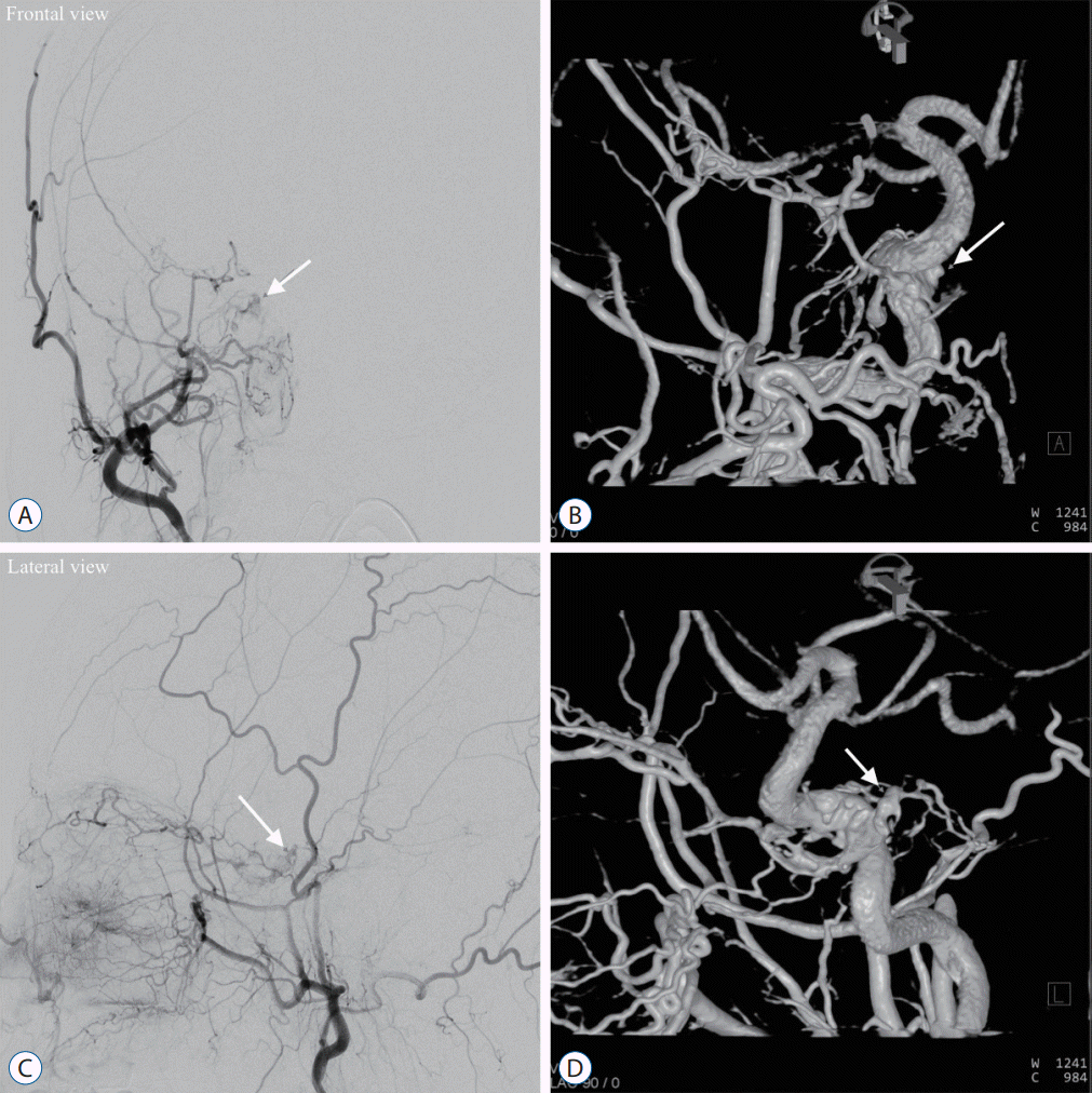 | Fig. 1.Showing illustration of the case (case 4). The patient complained of the right conjunctival injection and sixth cranial nerve palsy. Right cavernous sinus dural arteriovenous fistula (CSDAVF) was demonstrated in both frontal (A and B) and lateral views (C and D) of the right external carotid artery (ECA) angiography and 3D rotational angiography. The 3D rotational angiography of the right ECA was contaminated by the ipsilateral ICA opacification. Ipsilateral superior petrosal sinus, inferior petrosal sinus, and intercavernous sinus were occluded. Venous drainage via a mainly ipsilateral superior ophthalmic vein and partly inferior ophthalmic vein were noted. The white solid arrow in each panel (A-D) indicated the shunted pouch (SP). Multiple dural feeders fed the SP from the bilateral internal carotid artery and ECA, such as a middle meningeal artery, accessory meningeal artery, ascending pharyngeal artery, artery of foramen Rotundum, and meningohypophyseal trunk.
|
Evaluations and analyses
Puncture success, shunt occlusion, and complications are evaluated. Puncture success is defined as the opacification of the space in the control angiography through the microcatheter. If the entrance of the microcatheter tip into the CS is not achieved, even the reopening technique cannulate most course of the IPS, that space should be the CS; If the entrance of the microcatheter tip into the SP is not achieved after the microcatheter tip is verified inside the CS, that space should be the SP. Shunt occlusion is defined based on the results of the immediate final angiography as complete occlusion (no residual shunt flow), near-complete occlusion (minimal residual shunt flow), and partial occlusion (apparent residual shunt flow). Lastly, the complication is defined as the composite of bleeding and cranial nerve palsy.
Go to :
RESULTS
The results are summarized in Table 2. Contralateral IPS access was sought due to the failure of the ipsilateral IJV access (case 1). Of the four patients, all procedures were performed in one session with an immediate, complete angiographic obliteration and a subsequent clinical improvement. One patient (patient 2) developed CSDAVF recurrence and required another session of TVE. This recurrence (case 3) was also treated with TVE using the rigid-tipped microguidewire in the same manner as its first procedure (case 2). Therefore, a total of five transvenous coil embolizations in four patients with CSDAVFs were performed with the rigid-tipped microguidewire. The rigid-tipped microguidewire was used five times in our study cohort. The rigid-tipped microguidewire was used at the following sites : before the CS in three cases (cases 1, 2, and 3) and before the SP in two cases (cases 4 and 5; case 4 described in Figs. 1 and 2). The rigid-tipped microguidewire used in this study was Conquest pro 12 (Asahi Intecc).
 | Fig. 2.Showing intraoperative procedures (case 4). A : A 4-F diagnostic catheter was placed at the right proximal external carotid artery (ECA) for the arteriographic roadmap. A 6-F guide catheter was introduced through the right internal jugular vein. The 6-F guide catheter and another 4-F diagnostic catheter were used by means of the coaxial technique. To reinforce the 0.035-in guidewire, the 6-F guide catheter was inserted as close as possible to the right inferior petrosal sinus (IPS) orifice. The 4-F diagnostic catheter was also inserted into the distal part of the right IPS. Performing the frontier-wire technique, arteriographic and venographic roadmaps were acquired simultaneously with the presence of the 0.035-in guidewire. The microcatheter was successfully advanced with the guidance of the white footprint of the guidewire (white arrows). The microcatheter tip seemed to be in the shunted pouch (SP) (white arrowheads). B : Control angiogram, the microcatheter tip was in the isolated posterior aspect of the right cavernous sinus (CS) (white arrow). The membranous barrier could not be overcome by the neurointerventional microguidewires or 0.035-in guidewire. C : Under the biplane right ECA roadmaps, the direction of the microcatheter tip was controlled precisely using the neurointerventional microguidewire. With the removal of the neurointerventional microguidewire, the rigid-tipped microguidewire was introduced gently and then inserted only 1 mm further than the microcatheter tip (white arrows). Holding firmly the rigid-tipped microguidewire, the microcatheter could be advanced beyond the obstacle under the guidance of the rigid-tipped microguidewire. D : After removing the rigid-tipped microguidewire, control angiogram showed that the microcatheter tip was in the SP (white arrowhead). E : The microcatheter was able to navigate the CS using the microguidewire. Subsequent coil embolization of both the entry of the venous drainage and SP was done. The white arrow indicated the coil mass packed in the SP. In the final right ECA arteriography, the cavernous sinus dural arteriovenous fistula was obliterated and no residual flow.
|
Table 2.
Summary of the procedural results
![]()
Complete coil embolization of the SP and the superior or inferior ophthalmic veins’ origin was achieved in all cases. On the immediate final angiography, arteriovenous shunt flow disappeared in all cases. There was no evidence of intracranial hemorrhage. Neurological deterioration was not detected at the follow-up neurological examination.
Go to :
DISCUSSION
TVE for CSDAVF
Endovascular treatment of the CSDAVF is revolutionized with the development of innovative endovascular devices [7]. Transfemoral TVE through the ipsilateral or contralateral IPS has been considered the safest and most effective option for treating the CSDAVF [2,7,9,11,24]. Once access to the CS is achieved, subsequent coil embolization is not that difficult, leading to successful treatment in most cases. Therefore, the reopening techniques with the knowledge of the IPS anatomy are introduced to take a shortcut to the CS even though the IPS is occluded and the unrevealed individual IPS anatomy can be unusual [2,5,12,28]. There are alternative transfemoral venous routes such as the superior petrosal sinus and superior ophthalmic vein. More invasive approaches such as direct transorbital puncture or surgery can also be performed in rare circumstances, but these may increase patient comorbidity [3,6,11,24,26,27].
The authors experienced that the resistive stricture and membranous barrier hindered the entrance into the next space just before the CS and SP, respectively. The neurointerventional devices did not overcome these obstacles due to insufficient penetrability and rigidity against the rigid structures. The mechanism of venous sinus occlusion in the dural arteriovenous fistula was considered thrombogenesis with activated coagulopathy or hemodynamic hypertrophy of the sinus wall [20]. The neurointerventional microguidewires or the 0.035-in guidewire could overcome the thrombogenesis-induced occlusion. However, these devices could not penetrate the fibrotic occlusive portion despite the short-segment lesion.
The creative usage of CTO wire
Thus, another device with greater penetrability and rigidity was sought. CTO wires generally have higher tip load and lateral support than the neurointerventional microguidewires, originally designed for crossing chronic occlusive lesions in coronary intervention [18]. Higher tip load indicates greater penetrability. Higher lateral support provided by the wire positively affects the wire’s penetrability and the microcatheter’s trackability. Conquest Pro 12 used in this study has a 0.014-in diameter, a 20-cm, tapered, hydrophilic-coating tip portion with a radiopaque spring coil, and a 12.0-gf (gram force) tip load. Its distal end is not coated to allow transmission of tactile sensation from the tip [18].
The mode of the transvenous coil embolization needed to be discussed [24]. The entire sinus packing is the standard method. But there are some risks, including cranial nerve palsy due to the mass effect. When the fistula is found by thoroughly reviewing the biplane digital subtraction angiography and 3D rotational angiography, obliterating the fistula is ideal. In terms of the fistula in the CSDAVF, a small and restricted space in which numerous feeders converge is called the SP [14,23]. To perform the obliteration of the SP is called selective embolization or super selective shunt occlusion. Some studies reported that the frequent locations of the SP were posteromedial and posterosuperior to the CS [23]. They also reported a good clinical outcome, including a low rate of cranial nerve palsy. In our cases, all cases were treated by selective embolization without neurological complications. Therefore, access to the SP and performing selective embolization would be necessary for a better clinical outcome, even though there are some obstacles on the way to the SP.
The literature and our experience with CTO wires
There were no reports for transvenous coil embolization of the CSDAVF using the CTO wires in the literature review. The usage of the CTO wire in neurointerventional procedures may be avoided considering its rigid property. But, regarding the result of our cases, the use of the rigid-tipped microguidewire seems feasible as a second-line technique. The technical concepts of the CTO wire puncturing are to use its high tip load and high lateral support for penetrability and microcatheter advancement. Once the CTO wire is gradually pushed only 1 mm further from the microcatheter tip, the microcatheter should be advanced. But advancing the microcatheter could be difficult due to the difference in an external diameter between the microcatheter and microguidewire, and relatively insufficient support of the guiding system [5].
The reopening techniques also have similar challenges. Fortifying guiding system support and using the rigid portion of the neurointerventional microguidewire are suggested as technical tips [5,12]. To use the rigid portion of the neurointerventional microguidewire, the microguidewire should be advanced far enough into a draining vein or further looped within the CS [12]. As the high lateral support of the CTO wire will provide enough support and rigidity, the use of the CTO wire can facilitate the microcatheter advancement into the CS or SP even though the wire is advanced only 1-mm more from the microcatheter tip.
There are some important technical points to avoid any risk of extravascular protrusion. The direction of the wire should be meticulously selected. Effective targeting of the CS or SP is crucial to prevent mispositioning of the microcatheter. Therefore, a thorough review of the 3D rotational angiography and the guidance of the biplane arteriographic roadmaps showing the target are required. It is also crucial that the rigid-tipped microguidewire should be prepared without any tip shaping. And the rigid-tipped microguidewire should be pushed only 1 mm to puncture the short-segment lesion without any tip rotation. However, extravasation from the obliterated sinus or even the intact sinus can result in minor clinical consequences. It is said that the venous origin subarachnoid hemorrhage is usually self-limiting [8].
Limitations and recommendations
Prospective studies or studies on a larger scale would be required to validate the feasibility and safety of this technique due to the limited number of cases in this study. Till a larger number of cases of an occluded IPS in a CSDAVF approached with this novel technique, our future recommendation is that the use of a rigid-tipped CTO microguidewire should be carried out in experienced high-volume centers by highly experienced interventionists.
Go to :
CONCLUSION
While performing the TVE of the CSDAVF with the occluded IPS, the short-segment stricture or membranous barrier can be encountered right proximal to the CS or the SP even after successful navigation through the occluded IPS. With this novel technique, the success rate of access to the CS and SP via the occluded IPS could be improved, leading to better outcomes of the TVE of the CSDAVF. The rigid-tipped microguidewire originally developed for the coronary intervention may help overcome those resistive obstacles effectively and safely.
Go to :
 XML Download