This article has been
cited by other articles in ScienceCentral.
Abstract
Primary hyperparathyroidism (PHPT) is a common endocrine disorder characterized by elevated parathyroid hormone levels. In most cases, the disease involves a single parathyroid adenoma, followed by parathyroid hyperplasia, but the incidence of ectopic parathyroid adenoma is rare. However, in cases where the parathyroid hormone level remains high even after parathyroid surgery, an ectopic parathyroid gland should be considered. Here in a case of a 60-year-old female who presented PHPT is reported. She had undergone a surgical removal of the parathyroid gland of suspected hyperplasia, but still represented persistent PHPT, postoperatively. Ectopic mediastinal parathyroid adenoma was identified by 99mTc-sestamibi parathyroid scan and surgical excision via a median sternotomy approach was performed. Thirty-eight months postoperatively, there was no evidence of recurrence. Preoperative localization assessment is critical for minimizing surgical failure in cases of PHPT.
Go to :

Keywords: Hyperparathyroidism, Parathyroid adenomas, Parathyroid hormone
Introduction
Primary hyperparathyroidism (PHPT) is an endocrine disorder that may lead to hypercalcemia and hypophosphatemia and consequent neurologic, musculoskeletal, renal and cardiovascular symptoms. Most cases of hyperparathyroidism are associated with certain general symptoms resulting from metabolic abnormalities. As chronic hypercalcemia is also associated with non-specific symptoms such as general weakness, osteoporosis, gastric ulcer, nephrolith and loss of consciousness, the diagnosis of PHPT is often delayed [
1,
2]. Recent developments in biochemical evaluations have led to the standardization of serum calcium level measurements and the rate of PHPT diagnosis has increased following the implementation of a parathyroid hormone radioimmunoassay. Among diseases associated with PHPT, 85% of cases involve a single parathyroid adenoma, followed by parathyroid hyperplasia (12%-15%) and multiple parathyroid adenomas (1%-2%). In contrast, parathyroid carcinoma, supernumerary parathyroid, and ectopic parathyroid are extremely rare [
2,
3]. This report presents a literature review and discusses a case of supernumerary ectopic parathyroid combined with parathyroid hyperplasia.
Go to :

Case
A 60-year-old female was hospitalized with the chief complaint of the sensation of a foreign body in her throat. She had no prior history of medical abnormalities except for hypertension. Laryngoscopic findings indicated laryngeal edema, injection, and pachydermia. A nodule measuring 2×6 mm, suspected to be a thyroid adenoma, was detected in the right thyroid in neck CT. A thyroid function test and immune antibody test showed normal results. The patient was diagnosed with laryngopharyngeal reflux disease and thyroid nodule.
Ultrasound-guided fine-needle aspiration of the thyroid mass revealed a benign follicular lesion with cystic changes leading to regular tracing inspections for one year. The patient requested precautionary surgery when the suspected right thyroid nodule exhibited signs of growth. A serum analysis showed the following values: creatinine, 1.5 mg/dL (reference range, 0.5-1.4); calcium, 11.4 mg/dL (reference range, 8.4-10.2); and phosphate, 2.2 mg/dL (reference range, 2.6-4.5), indicating creatinemia, hypercalcemia and hypophosphatemia, respectively. A parathyroid hormone test verified an increase in levels to 395.0 pg/mL (reference range, 15-55). Neck CT and ultrasonography did not identify a mass in the parathyroid. Therefore, PHPT consequent to parathyroid hyperplasia was suspected and a hemithyroidectomy with partial parathyroidectomy were scheduled. During the surgery, three of the four parathyroid glands with the suspected hyperplasia were removed. Postoperatively, thyroid nodular hyperplasia was identified via biopsy and parathyroid chief cell hyperplasia was observed in one of the three removed parathyroid glands.
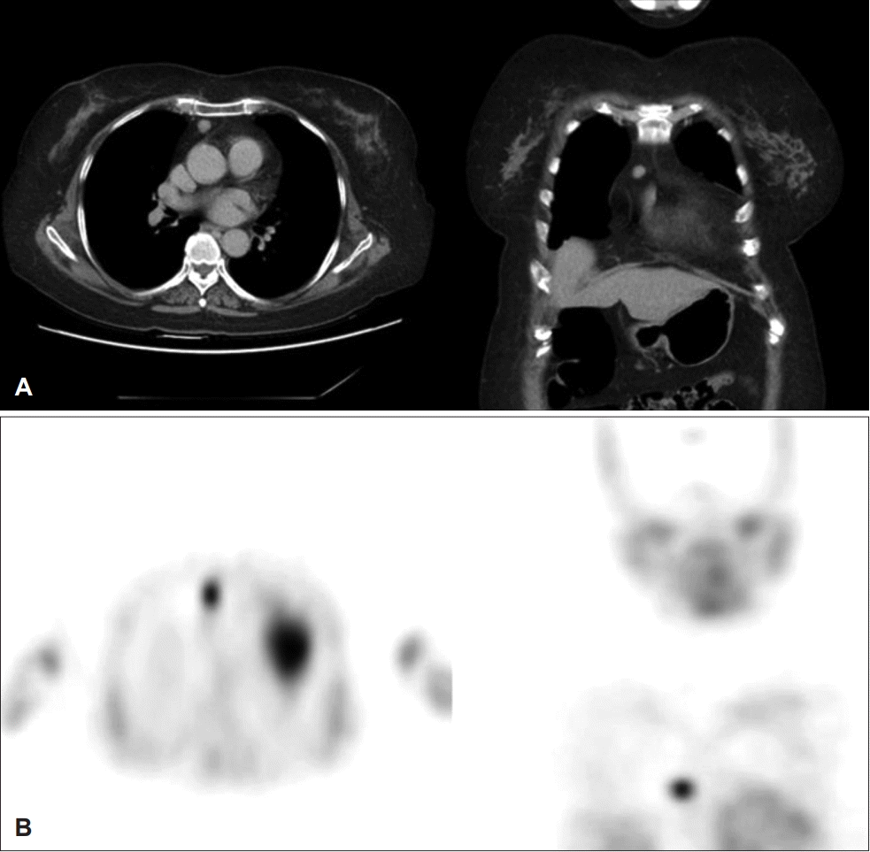
However, the patient’s parathyroid hormone level remained high (509 pg/mL). Blood analysis continued to reveal hypercalcemia (blood calcium level, 11.6 mg/dL) and hypophosphatemia (blood phosphoric acid level, 2.4 mg/dL) even after surgery. A 99mTc-sestamibi parathyroid scan which included the chest area and chest CT (
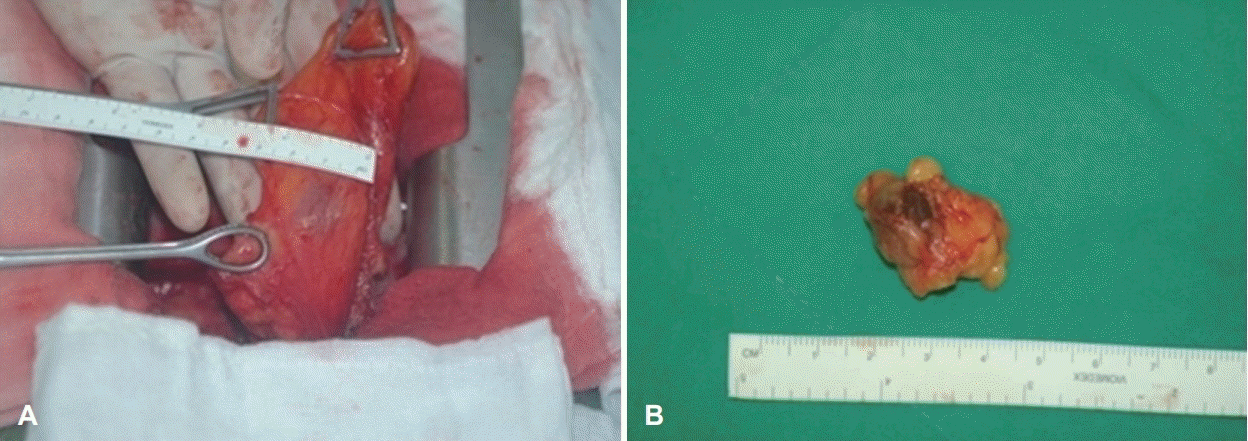
Fig. 1) revealed a suspected ectopic parathyroid adenoma in the chest, which was removed via a median sternotomy approach (
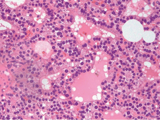
Fig. 2). Biopsy confirmed the presence of an ectopic parathyroid adenoma (
Fig. 3). Postoperatively, the patient’s parathyroid hormone level returned to the normal range (26.4 pg/mL). Her blood calcium level decreased to 9.3 mg/dL and her phosphoric acid level increased to 4.2 mg/dL. No complication due to surgery was observed and the patient’s disease progression is under observation at the outpatient clinic.
 | Fig. 1.Pre-operative image study. A: Contrast-enhanced CT of the chest shows a small round mass in the right anterior mediastinum. B: 99mTc sestamibi scan of the parathyroid after the first operation shows high uptake in a nodular lesion in the right anterior mediastinum. 
|
 | Fig. 2.Intra-operative findings. A: Total thymectomy was performed via a median sternotomy approach, with ectopic parathyroid excision. B: A 1.5×1.3 cm yellow–brown round mass was excised. 
|
 | Fig. 3.The surgical specimen contains hypercellular parathyroid tissue with follicular structures lined by cuboidal cells that lack cytologic atypia (hematoxylin and eosin stain; magnification ×400). 
|
Go to :

Discussion
Ectopic parathyroid adenoma is reported to be the etiologic agent in 1%-3% of the cases of persistent, relapsing pseudohyperparathyroidism [
4]. These lesions are usually found in the thymus, mediastinum, thyroid, thyroid-thymus ligament and lower mandible. Ectopic lesions are a critical cause of postoperative relapse. Most normal adults usually have both upper and lower pairs of parathyroid glands; however, 6%-13% of adults have more than four parathyroid glands [
5]. Gilmour reported that 6.7% of evaluated adults had a supernumerary parathyroid; 5.8% had five parathyroid glands and cases involving six, eight, and even 12 glands were also reported [
5]. The patient in the present case had a single supernumerary parathyroid gland in the mediastinum, which was confirmed to be parathyroid adenoma. Parathyroid hyperplasia was also observed in the remaining parathyroid glands. Therefore, this is a unique case, since it involved both parathyroid hyperplasia and supernumerary ectopic parathyroid.
Among the various causes of surgical failure for hyperparathyroidism, the primary reason is the inability to differentiate the abnormal parathyroid gland from the ectopic parathyroids, supernumerary parathyroids, multiple parathyroid adenomas, thyroid adenoma and multiple parathyroid hyperplasia [
6-
9]. Therefore, preoperative localization assessments are critical to minimizing surgical failure. Various opinions exist regarding the localization of parathyroid tumors. The 99mTc sestamibi scan has been the most frequently used method since its introduction. A 99mTc sestamibi scan and neck ultrasonography are performed initially with CT and MRI recommended in cases involving poor localization. The 99mTc sestamibi scan is an effective diagnostic modality in cases involving revision surgery for persistent hyperparathyroidism [
10]. If localization remains inadequate, invasive methods such as angiography and selective venous sampling are incorporated.
An incidental finding of hypercalcemia in the present case led to the detection of hyperparathyroidism. Despite neck CT and two rounds of ultrasonography, the parathyroid lesion could not be localized. Accordingly, multiple parathyroid hyperplasia was suspected, and selective parathyroidectomy was accordingly performed. During this operation, three enlarged parathyroid glands were identified, and a biopsy revealed an increased number of parathyroid chief cells in one of them. As a result, the patient was diagnosed with parathyroid hyperplasia. However, her postoperative parathyroid hormone level remained relatively high and a supernumerary ectopic parathyroid in the mediastinum was identified using localization methods, such as chest CT and 99mTc sestamibi parathyroid scan, leading to the precautionary surgery. Supernumerary ectopic parathyroid adenoma is extremely rare; no cases have been reported so far. To the best of our knowledge, the present case is the first such documented case. We note that in cases when the parathyroid level remains high even after parathyroid surgery, an ectopic parathyroid gland should be considered. Localization tests, such as a 99mTc sestamibi or CT scans, will be helpful in such cases.
Go to :


 PDF
PDF Citation
 Print
Print


 XML Download