Abstract
Maffucci syndrome is a non-hereditary disease, in which benign cartilage tumor called enchondroma occurs throughout the body, causing various symptoms. In particular, when chondrosarcoma occurs in the skull base, various neurologic symptoms can appear. Many of these symptoms have been diagnosed and reported by neurosurgeons. This paper presents a rare case of Maffuci syndrome. The patient is a 34-year-old female who first visited the otolaryngology outpatient department for ear fullness without any other neurological symptoms. The initial diagnosis was adult onset otitis media with effusion (OME), but further examination and biopsy revealed skull base chondrosarcoma. Moreover, the mass was extensively invading the skull base, so surgical treatment would have been dangerous; thus, careful follow-up has been conducted for the patient in the outpatient clinic. This report highlights the importance of nasopharyngoscopy as well as other imaging tests to observe nasopharyngeal masses in OME patients with congenital or acquired diseases, which are known to sporadically develop tumor.
Enchondromas are benign cartilaginous tumors. Maffucci syndrome is a non-hereditary disorder of multiple enchondromas with three types of vascular lesions: cavernous hemangiomas, phlebectasias, and lymphangiectasia-lymphangiomas [1,2]. Malignancy, such as chondrosarcoma, glioma, and ovarian juvenile granulosa cell tumors, develops in approximately 50% of Ollier disease or Maffucci syndrome cases [3].
Enchondromas usually appear around the growth plate cartilage and may result from the deregulated proliferation and differentiation of chondrocytes during natural endochondral ossification [4]. Skull base lesions have been reported in 27 of 209 (13%) Maffucci syndrome cases [5]. In addition, a higher risk of sarcomatous degeneration and secondary chondrosarcoma of enchondromas have been reported in Maffucci syndrome [6-8].
In patients with Maffucci syndrome, early resection is recommended to facilitate prompt diagnosis and prevent sarcomatous degeneration. However, these lesions can be large and multicentric, making resection dangerous. Although the treatment of choice is surgical resection, resection of a skull base chondrosarcoma should be considered carefully because of the risk of surgery. In particular, patients with non-fatal/no symptoms should be monitored closely with conservative treatment prior to considering surgery [9].
A 34-year-old insulin-dependent diabetic female with Maffucci syndrome presented to the Department of Otorhinolaryngology-Head and Neck Surgery, Medical University of Hallym, with complaints of right ear fullness.
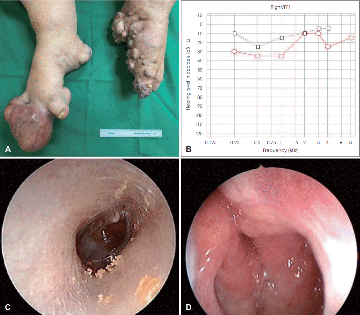
The patient experienced relapsing hemangiomas in the vagina and enchondromas in both upper and lower extremities (Fig. 1A).
On the first outpatient day, tympanometry showed right B-type and left A-type. Audiometry revealed an air-bone gap at the right hearing level of 22 (13) dB, and normal left hearing level (Fig. 1B). These tests and an otoscope evaluation led to the diagnosis of otitis media with effusion (OME) of the right ear (Fig. 1C).
With antibiotic treatment (Clarithromycin 500 mg/dayx1 week initially followed by Cefitoren 300 mg/day after aspiration of the effusion) OME resolved after one month and tympanometry showed bilateral A-type. After 3 months, the right OME recurred. Aspiration was performed again, and a nasopharyngeal mass was confirmed with a nasopharyngoscope (Fig. 1D).
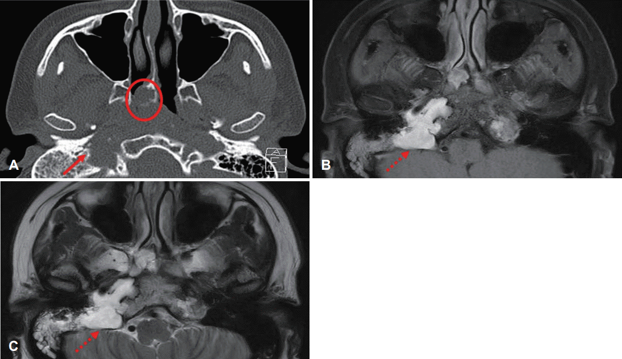
Paranasal sinus (PNS) CT confirmed a bulging mass in the right nasopharynx, destructive changes of the right skull base, clivus, right mastoid, and petrous bone, and widening of the petroclival fissure and right jugular foramen, extending to the right choana (Fig. 2A). PNS MRI was performed for further evaluation (Fig. 2B and C).
On MRI, the differential was nasopharyngeal cancer with skull base and intracranial invasion, chondrosarcoma (originating from the right petroclival joint), or chondral myxoid fibroma. Furthermore, the mass appeared to be vascularly abundant. However, transfemoral cerebral angiography revealed that no blood flow to tumor from external carotid artery and no internal carotid artery invasion by tumor.
As the site was high risk for surgery, endoscopic biopsy was performed under general anesthesia to establish a treatment plan. Incision is performed on the mucosa above the tumor, the tumor is resected while bleeding control is achieved. The resected tumor was taken out through the nasal cavity, and the tumor was myxoid with what appeared to be a cartilaginous component.
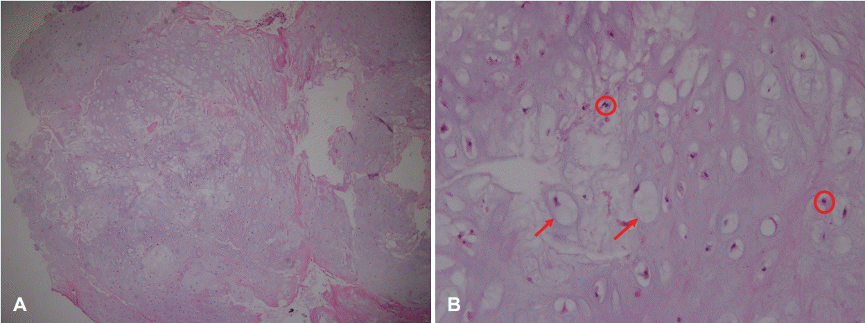
Histological examination revealed a mesenchymal tumor in a hyaline cartilaginous matrix with a few enlarged chondrocytes with hypochromic nuclei and bland histological appearance. In addition, characteristic physaliferous cells with multiple cytoplasmic vacuoles within the myxoid stroma as in chordoma were noted (Fig. 3). Based on immunohistochemistry, S-100 positive, Cytokeratine negative and EMA negative were confirmed. Therefore, it was possible to observe more chondrosarcoma and enchondroma rather than chordoma. Although it was difficult to clearly distinguish low-grade chondrosarcoma from pathological enchondroma, it enabled the observation of binucleated chondrocytes and the bone destruction clinically caused by the tumor [9]. The final diagnosis was low-grade chondrosarcoma.
Chondrosarcoma requires extensive surgical resection. However, for sites considered high risk for surgery, close observation should be performed prior to considering surgery as a treatment option [9]. The mass in this case was extensively invading the skull base; therefore, surgical treatment would be dangerous and careful follow-up is being conducted in outpatient clinics. Follow-up would be performed monthly for 6 months, and then once every 6months thereafter. PNS CT would be taken every 2 years.
The occurrence of skull base chondrosarcoma in Maffucci syndrome is very low (5%), and they account for only approximately 0.1% of all head and neck tumors [6]. In most such cases, the main presenting symptom is headache or other neurological symptoms. Therefore, the otolaryngologist may not be consulted during the initial diagnostic process. To our knowledge, this patient was the first case of skull base chondrosarcoma in which the patient presented with the main symptom of ear fullness.
There is a case report by an otolaryngologist of nasal cavity chondrosarcoma with Maffucci syndrome. The main symptom was nasal obstruction at the first visit as the nasal cavity mass could be easily identified through endoscope. The nasal cavity chondrosarcoma could be completely resected through endoscopic sinus surgery [10].
The patient was diagnosed with adult-onset OME when she first visited the hospital. Although OME is a common disease in children, it is not common in adults; therefore, it is necessary to promptly identify the cause. The main causes of adult-onset OME include infiltration of the eustachian tube by nasopharyngeal carcinoma and other malignancies in the vicinity of the Eustachian tube, changes in the middle ear and eustachian tube induced by radiotherapy, and systemic disease [11].
The association between OME in adults and nasopharyngeal tumor is well established. There are two potential mechanisms for effusion formation by nasopharyngeal tumor: tubal dysfunction due to muscle involvement and direct infiltration of the eustachian tube, with the latter being the most important [12].
The most common cause of adult-onset OME is sinonasal disease [11]. Furthermore, since adult-onset OME may be associated with acid reflux disease, eustachian tube dysfunction, and smoking, intensive efforts are needed to identify the cause.
In our case, at the time of the first visit, OME was assumed to be caused by rhinosinusitis as the patient complained of nasal obstruction and rhinorrhea as comorbid symptoms. Since it is the most common causes of adult-onset OME, no further evaluation was performed. Faster diagnosis would have been possible if PNS CT and nasopharyngoscopy had been performed earlier.
In particular, in adult-onset OME patients with congenital or acquired diseases that are known to sporadically develop tumors, nasopharyngoscopy as well as other imaging tests should be used to discriminate nasopharyngeal masses, nasal cavity masses, and skull base masses.
ACKNOWLEDGMENTS
This study was approved by the Institutional Review Board of Chuncheon Sacred Heart Hospital, Hallym University College of Medicine (Approval Number: CHUNCHEON2021-07-005).
Notes
Author Contribution
Conceptualization: Chan Hum Park. Data curation: Gil Myeong Son. Formal analysis: Gil Myeong Son. Investigation: Gil Myeong Son. Methodology: Chan Hum Park. Project administration: Gil Myeong Son. Resources: Ki Joon Park, Seung Su Ha. Supervision: Chan Hum Park. Visualization: Gil Myeong Son. Writing—original draft: Gil Myeong Son. Writing—review & editing: Chan Hum Park.
REFERENCES
1. Verdegaal SH, Bovée JV, Pansuriya TC, Grimer RJ, Ozger H, Jutte PC, et al. Incidence, predictive factors, and prognosis of chondrosarcoma in patients with Ollier disease and Maffucci syndrome: An international multicenter study of 161 patients. Oncologist. 2011; 16(12):1771–9.
2. Jermann M, Eid K, Pfammatter T, Stahel R. Maffucci’s syndrome. Circulation. 2001; 104(14):1693.
3. El Abiad JM, Robbins SM, Cohen B, Levin AS, Valle DL, Morris CD, et al. Natural history of Ollier disease and Maffucci syndrome: Patient survey and review of clinical literature. Am J Med Genet A. 2020; 182(5):1093–103.
4. Amary MF, Damato S, Halai D, Eskandarpour M, Berisha F, Bonar F, et al. Ollier disease and Maffucci syndrome are caused by somatic mosaic mutations of IDH1 and IDH2. Nat Genet. 2011; 43(12):1262–5.
5. Dini LI, Isolan GR, Saraiva GA, Dini SA, Gallo P. Maffucci’s syndrome complicated by intracranial chondrosarcoma: Two new illustrative cases. Arq Neuropsiquiatr. 2007; 65(3B):816–21.
6. Ranger A, Szymczak A. Do intracranial neoplasms differ in Ollier disease and Maffucci syndrome? An in-depth analysis of the literature. Neurosurgery. 2018; 65(6):1106–13. discussion 1113-5.
7. Herget GW, Strohm P, Rottenburger C, Kontny U, Krauß T, Bohm J, et al. Insights into enchondroma, enchondromatosis and the risk of secondary chondrosarcoma. Review of the literature with an emphasis on the clinical behaviour, radiology, malignant transformation and the follow up. Neoplasma. 2014; 61(4):365–78.
8. Altay M, Bayrakci K, Yildiz Y, Erekul S, Saglik Y. Secondary chondrosarcoma in cartilage bone tumors: Report of 32 patients. J Orthop Sci. 2007; 12(5):415–23.
9. Oushy S, Peris-Celda M, Van Gompel JJ. Skull base enchondroma and chondrosarcoma in Ollier disease and Maffucci syndrome. World Neurosurg. 2019; 130:e356–61.
10. Steinbichler TB, Kral F, Reinold S, Riechelmann H. Chondrosarcoma of the nasal cavity in a patient with Maffucci syndrome: Case report and review of the literature. World J Surg Oncol. 2014; 12:387.
11. Finkelstein Y, Ophir D, Talmi YP, Shabtai A, Strauss M, Zohar Y. Adult-onset otitis media with effusion. Arch Otolaryngol Head Neck Surg. 1994; 120(5):517–27.
12. Mills R, Hathorn I. Aetiology and pathology of otitis media with effusion in adult life. J Laryngol Otol. 2016; 130(5):418–24.
Fig. 1.
Skin lesions gross examination and audiometry, otoscopy, and nasopharyngoscopy findings. A: Photographs of both feet. Multiple enchondromatosis with vascular malformations are seen. In the left foot, multiple cutaneous lesions are evident. B: Audiometry. Right hearing level 22 (13) dB; air-bone gap is seen. C: Otoscope revealed otitis media with effusion of the right ear. D: Nasopharyngoscope revealed a nasopharyngeal mass. A bulging mass was seen in rostrum sphenoidale and torus tubarius. Obliteration of rosenmuller fossa and mucosal irregularity were also observed.

Fig. 2.
CT and MRI of paranasal sinuses (PNS). A: PNS CT. Asymmetric bulging mass in right nasopharynx compressing the eustachian tube orifice (circle): destructive changes of the right skull base, erosion and destructive changes of the clivus, right mastoid, and petrous bone, and widening of the petroclival fissure and right jugular foramen, extending to the right choana (arrow). B: PNS MRI T1-weighted image. C: PNS MRI T2-weighted image. Low signal intensity on T1-weighted image, mainly high signal intensity on T2-weighted image with partial isosignal intensity. Huge tumor in right skull base, located in petrous apex, petroclival joint, clivus, petroclival fissure (dotted arrows).

Fig. 3.
Histopathology photomicrographs. A: Low-power microscopy shows abundant cartilaginous matrix with chondrocytes embedded in lacunae (H&E, ×40). B: High-power microscopy shows permeation of intertrabecular spaces and myxoid changes chondroid matrix liquefaction (H&E, ×200). physaliferous cells with multiple cytoplasmic vacuoles (arrows), binucleated chondrocyte (circles). H&E, hematoxylin and eosin.


 PDF
PDF Citation
 Print
Print


 XML Download