Journal List > J Liver Cancer > v.22(2) > 1516079696

Jo, Rhu, Kim, Choi, and Joh: Indications for open hepatectomy in the era of laparoscopic liver resection: a high volume single institutional study

Abstract

Background/Aim

Since the introduction of laparoscopy for liver resection in the 1990s, the performance of laparoscopic liver resection (LLR) has been steadily increasing. However, there is currently no data on the extent to which laparoscopy is used for liver resection. Herein, we investigated the extent to which laparoscopy is performed in liver resection and sought to determine whether surgeons prefer laparoscopy or laparotomy in the posterosuperior (PS) segment.

Methods

For this retrospective observational study, we enrolled patients who had undergone liver resection at the Samsung Medical Center between January 2020 and December 2021. The proportion of LLR in liver resection was calculated, and the incidence and causes of open conversion were investigated.

Results

A total of 1,095 patients were included in this study. LLR accounted for 79% of the total liver resections. The percentage of previous hepatectomy (16.2% vs. 5.9%, P<0.001) and maximum tumor size (median 4.8 vs. 2.8, P<0.001) were higher in the open liver resection (OLR) group. Subgroup analysis revealed that tumor size (median 6.3 vs. 2.9, P<0.001) and surgical extent (P<0.001) in the OLR group were larger than those in the LLR group. The most common cause of open conversion (OC) was adhesion (57%), and all OC patients had tumors in the PS.

Conclusions

We investigated the recent preference of practical surgeons in liver resection, and found that surgeons preferred OLR to LLR when treating a large tumor located in the PS.

INTRODUCTION

Since the introduction of laparoscopy for liver resection in the 1990s, the number of laparoscopic liver resections (LLR) has steadily increased.1-4 The use of this surgical technique has recently been expanded, with further applications to living-donor liver resection for transplantation.5 Despite this wide application, there is controversy regarding the use of LLR for tumors in areas (segments 1, 4a, 7, and 8) which are difficult to resect laparoscopically.6
For tumors located in the posterosuperior (PS) segment of the liver, the laparoscopic approach is challenging and risky owing to limited visualization and difficulty in controlling bleeding.7 Nevertheless, in recent years, LLR of the PS segment has been increasingly performed owing to the development of 3-D scope and surgical instruments.8,9 Furthermore, several studies have indicated that LLR has superior outcomes than does open liver resection (OLR).10
However, to the best of our knowledge, there are no data on the actual use of laparoscopy in liver resection. This study therefore aimed to investigate the extent to which laparoscopy is performed in liver resection, and to determine whether surgeons prefer laparoscopy and laparotomy when operating on tumors in the PS segment. In addition, the rate and causes of open conversion (OC) in LLR were investigated.

METHODS

We conducted a retrospective observational study of patients who underwent liver resection at the Samsung Medical Center between January 2020 and December 2021. The exclusion criteria were as follows: 1) age ≤18 years, 2) living donor liver resection, and 3) non-mass-forming liver resection. This study was conducted in adherence with Strengthening the Reporting of Observational Studies in Epidemiology (STROBE) guidelines (Supplementary Table 1).
Data on sex, body mass index, American Society of Anesthesiologists (ASA) score, liver function test results, previous surgical history, and postoperative course were collected from medical records. Tumor data regarding histologic subtype, surgical margin, maximal size, multifocality, and location were investigated through pathology records. The resection extent, cause of OC, time of OC decision, estimated blood loss, operative time, and transfusion rate were investigated through operation records. Operative techniques for LLR have been described previously.11 Data on hospital stay was collected including admission days before the actual operation. Usually, the patient is admitted to the hospital 2 days before the operation.
Proximity to the major vessels was evaluated using preoperative computed tomography, and the distance was calculated from the margin of the main tumor lesion to the major vessel (main or second branches of Glisson’s tree, major hepatic vein, and inferior vena cava).
Liver function was evaluated using the Child-Pugh classification.12 The prior surgical history, including the number and type of surgery, was reviewed and classified into four types (hepatectomy, upper gastrointestinal surgery, lower gastrointestinal surgery, genitourinary surgery). Patients who underwent various types of surgery were prioritized and classified in the following order: hepatectomy, upper gastrointestinal surgery, lower gastrointestinal surgery, and genitourinary surgery.
The tumor locations were classified into two groups (PS and anterolateral [AL]). The PS segment was defined as segments 1, 4a, 7, and 8, and the AL segment was defined as the remaining segments (2, 3, 4b, 5, 6).8 Patients with multiple tumor locations were classified as having PS segments when at least one tumor was located in the PS segment.
The extent of resection was classified into four groups: subsegmentectomy, segmentectomy, bisegmentectomy, and hemihepatectomy. Multiple wedge resections were classified as subsegmentectomy.
The selection criteria for the laparoscopic approach were surgeon-dependent. When considering LLR, the tumor location, history of portal vein embolization, and trisectionectomy were not considered. The indications for OLR included a tumor size >10 cm, except when the tumor was a pedunculated type, reconstruction of a vascular or biliary conduit was required, the tumor was close to an important vital structure making difficult to dissect laparoscopically, the tumor had invaded adjacent organs necessitating concomitant resection and reconstruction, future remnant liver was <25%, and Child–Pugh classification was class B. One surgeon performed laparoscopic surgery, while the remaining three surgeons used both approaches. The selection criteria for LLR were described in our previous article.13

Statistical analysis

Normally distributed continuous variables are shown as mean±standard deviation, and non-normal continuous variables are expressed as median (range). Fisher’s exact test or Pearson’s chi-square test were used to compare proportions between groups, as deemed appropriate. For comparison of continuous variables, normality test was performed with Shapiro–Wilk test. The Student’s t-test was used when the normal distribution was followed, and the Mann–Whitney U test was used for variables that were not normally distributed. P-value <0.05 was considered statistically significant. All analyses were performed using the R 4.0.4 software (The R Core Team, Vienna, Austria).

RESULT

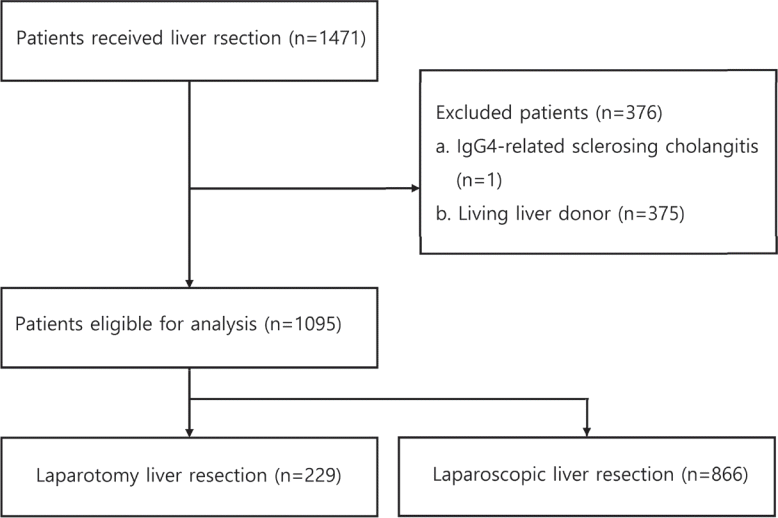
Among 1,471 patients, 1,095 were eligible for inclusion after excluding living liver donors (n=375) and non-massforming liver lesions (immunoglobulin G4-related sclerosing cholangitis, n=1). There were 229 patients with OLR and 866 patients with LLR, and LLR accounted for 79% of total liver resections. A flow diagram showing the selection process of patients included in this study is given in Fig. 1.

1. Surgeon type and preference

Four surgeons participated in this study. One was a specialized liver surgeon with >20 years of experience, two were surgeons with >10 years of experience, and the other was a surgeon with >5 years of experience. The surgeon with more than 20 years of experience had a higher OLR rate (97.8%) compared to other surgeons (9.7%, 27%, and 15%, respectively).

2. Comparison of characteristics between OLR and LLR

A comparison of the characteristics of the OLR and LLR groups is summarized in Table 1. There were no significant differences in sex (male 73.8% vs. 71.8%, P=0.61), liver function (Child–Pugh score A 99.1% vs. 99.7%, P=0.1), ASA score (two 79% vs. 78.5%, P=0.34), previous number of abdominal operations (P=0.079), or PS location (PS 50.7% vs. 43.4%, P=0.06) between the two groups. However, there were significant differences in age (62.0±11.3 vs. 59.9±11.5, P=0.012), previous type of surgery (hepatectomy 16.2% vs. 5.9%, P<0.001), and maximum tumor size (median 4.8 vs. 2.8, P<0.001).
In terms of operation-related details, the OLR group demonstrated a larger surgical extent (P<0.001), longer operation time (P<0.001), higher transfusion rate (P<0.001), and longer hospital stay (P<0.001) than the LLR group.

3. Comparison between OLR and LLR in PS location

In the analysis of patients in the PS location, there were no significant differences in baseline characteristics, such as age, sex, ASA, liver function, or previous surgical history. However, in terms of tumor-related characteristics, the OLR group demonstrated a larger tumor size (median 6.3 cm vs. 2.9 cm, P<0.001) and a higher proportion of hepatocellular carcinoma (80.2% vs. 70.5%, P=0.007) than the LLR group.
Similar to the whole group analysis, in the operation-related details, the OLR group demonstrated a larger surgical extent (69.8% vs. 39.4% for hemihepatectomy or more, P<0.001), longer operation time (190.6±63.4 minutes vs. 176.2±59.0 minutes, P=0.031), and higher transfusion rate (8.6% vs. 1.6%, P=0.001) than the LLR group. The details of this comparison are summarized in Table 2.

4. Laparoscopic liver resection and open conversion

Among the 866 patients who underwent LLR, 14 had OC, and the incidence of OC was 1.6%. Comparison between the non-OC and OC groups revealed no significant differences, except for in liver function (Child–Pugh score B, 7.1% vs. 0.1%, P=0.032). However, in terms of previous surgical history and tumor characteristics, the OC group demonstrated a trend towards a higher number of previous abdominal operations (P<0.001), a higher proportion of previous hepatectomy (P=0.009), and a higher proportion of PS location (P=0.003). In addition, in the operation-related details, the OC group also showed a longer operative time (243.9±52.4 minutes vs. 154.3±60.1 minutes, P<0.001), larger amount of blood loss (median 500 cc with interquartile range [IQR] 250.0-887.5 vs. median 150 cc with IQR 100-300, P<0.001), and higher transfusion rate (14.3% vs. 0.8%, P=0.008). A comparison of the characteristics of the OC and non-OC groups is shown in Table 3.

5. Cause of OC

Descriptive data showing the reasons for OC are summarized in Table 4. The most common cause of OC was adhesion (n=8), followed by bleeding (n=3). The time determined for OC was less than 30 minutes in more than half of the patients with adhesions (5/8). All the patients in the OC group had at least one tumor located in the PS segment. Two patients (cases 2 and 9) underwent surgery for tumors larger than 10 cm, while 10 patients had tumors smaller than 5 cm.

Discussion

With the advancement of surgical techniques, liver resection has become a safe procedure for liver tumors when performed by experienced surgeons in patients with adequate indications.14 In particular, LLR is considered a standard method because of the various advantages of postoperative outcomes in patients with tumors in areas such as the anterolateral segment, including S2, 3, 4b, 5, and 6, which are easily accessible by laparoscopy.6
In contrast to the AL location of most liver tumors, PS tumors are located deep below the right diaphragm and are surrounded by the rib cage. This induces several disadvantages for surgery, including poor visibility and difficult bleeding control during laparoscopic surgery. Thus, over the past decade, LLR of PS location tumors has been considered a challenging and dangerous procedure, and a scoring system has been developed to evaluate it.7,15 However, in recent years, the number of LLRs performed has rapidly increased, and with the development of flexible scopes and various surgical instruments, laparoscopy for tumors located in the PS is being actively applied.16-18 The aim of this study was to investigate the extent to which laparoscopy is actually performed in liver resection, and to determine whether surgeons prefer laparoscopy and laparotomy in the PS segment.
Laparoscopic liver resection was performed in 79% of patients. In the comparison between the OLR and LLR groups, previous hepatectomy, maximum tumor size, and wide surgical extent were significantly different. This suggests that surgeons prefer OLR if patients have a previous history of surgery in the liver, large tumors, or wide resection areas.
PS location was not an important factor in determining surgical procedures in the whole-group analysis. However, in the subgroup analysis of patients with a PS location, differences in the preference of surgeons according to the surgical method were identified. For example, surgeons preferred OLR over LLR in patients who needed to undergo hemihepatectomy or more extended resection with a large tumor in the PS location.
In our study, the incidence rate of OC during LLR (1.6%) was lower than that reported in previous studies (4.4-21.9%).3,19,20 The reason for this difference is that previous studies included patients with early LLR, and the development of technology also influenced the results. In the details of OC, the most common cause was adhesion. In contrast, previous studies have reported that bleeding is the major cause of OC.20 Although the incidence was low, adhesions have also been reported as a minor cause of OC.21-23 In addition, another feature of our results was that all the OC cases had a tumor located in the PS. This indicates that PS location may be a risk factor for OC, although this needs to be analyzed in future studies. The location is also important in cases with adhesions. The anterolateral location is covered by the anterior and right lateral abdominal walls, and gastrointestinal organs, including the greater omentum. These are covered with the peritoneum, and adhesiolysis can be performed relatively easily. However, the posterolateral location was covered with the diaphragm. Adhesiolysis of the posterolateral part can be complicated by bleeding from both the diaphragm and the liver, and opening of the diaphragm can occur.
This study has several limitations which should be discussed. Firstly, this was a single-center retrospective study, which limits the generalizability of the results. Additionally, the lack of details regarding tumor characteristics, such as proximity to major vessels or exact segment tumor location, made it impossible to evaluate the difficulty score. In future studies, research including the difficulty score index should be conducted. The OLR and OC rates may be higher in data from other centers; because of the low number of OC in our study, we did not perform multivariate analysis to analyze the risk factors for OLR or OC. However, these data could be a good guide not only for surgeons, but also for oncologists, when deciding on the treatment plan for patients with liver malignancy.
To the best of our knowledge, this is the first study to investigate the actual preference of surgeons for surgical techniques when performing liver resection. Although this study was limited by complications inherent to the single-center retrospective design, the results may nevertheless be helpful to surgeons who are inexperienced with few people to turn to for advice when deciding to perform liver resection.

Notes

Conflicts of Interest

The authors have no conflicts to disclose.

Ethics Statement

The study protocol conformed to the ethical guidelines of the Declaration of Helsinki and was approved by the Institutional Review Board (IRB) of Samsung Medical Center (IRB No. SMC 2022-05-076). The need for informed consent was waived by the IRB of Samsung Medical Center due to the retrospective nature of the study

Funding Statement

This study was supported by the Scientific Research Fund of the Korean Liver Cancer Association (2021).

Data Availability

The data presented in this study are available upon request from the corresponding authors.

Author Contribution

Conceptualization: SJJ, JR

Data curation: SJJ

Formal analysis: SJJ, JR

Investigation: SJJ

Methodology: SJJ, JR

Project administration: JWJ

Resources: JR, JMK, G-SC, JWJ

Supervision: JMK, G-SC, JWJ

Validation: JMK, G-SC, JWJ

Visualization: SJJ

Writing–original draft: SJJ

Writing–review & editing: JR, JMK, G-SC, JWJ

Approval of final manuscript: all authors

Supplementary Material

Supplementary data can be found with this article online https://doi.org/10.17998/jlc.2022.08.22.

REFERENCES

1. Buell JF, Cherqui D, Geller DA, O'Rourke N, Iannitti D, Dagher I, et al. The international position on laparoscopic liver surgery: the Louisville Statement, 2008. Ann Surg. 2009; 250:825–830.
2. Troisi RI, Montalti R, Van Limmen JG, Cavaniglia D, Reyntjens K, Rogiers X, et al. Risk factors and management of conversions to an open approach in laparoscopic liver resection: analysis of 265 consecutive cases. HPB (Oxford). 2014; 16:75–82.
3. Nguyen KT, Gamblin TC, Geller DA. World review of laparoscopic liver resection-2,804 patients. Ann Surg. 2009; 250:831–841.
4. Reich H, McGlynn F, DeCaprio J, Budin R. Laparoscopic excision of benign liver lesions. Obstet Gynecol. 1991; 78:956–958.
5. Kwon CHD, Choi GS, Joh JW. Laparoscopic right hepatectomy for living donor. Curr Opin Organ Transplant. 2019; 24:167–174.
6. Wakabayashi G, Cherqui D, Geller DA, Buell JF, Kaneko H, Han HS, et al. Recommendations for laparoscopic liver resection: a report from the second international consensus conference held in Morioka. Ann Surg. 2015; 261:619–629.
7. Ban D, Tanabe M, Ito H, Otsuka Y, Nitta H, Abe Y, et al. A novel difficulty scoring system for laparoscopic liver resection. J Hepatobiliary Pancreat Sci. 2014; 21:745–753.
8. Teo JY, Kam JH, Chan CY, Goh BK, Wong JS, Lee VT, et al. Laparoscopic liver resection for posterosuperior and anterolateral lesionsa comparison experience in an Asian centre. Hepatobiliary Surg Nutr. 2015; 4:379–390.
9. Zheng B, Zhao R, Li X, Li B. Comparison of laparoscopic liver resection for lesions located in anterolateral and posterosuperior segments: a meta-analysis. Surg Endosc. 2017; 31:4641–4648.
10. Liu F, Li Q, Wei Y, Li B. Laparoscopic versus open liver resection for difficult lesions: a meta-analysis. J Laparoendosc Adv Surg Tech A. 2018; 28:1428–1436.
11. Cho W, Kwon CHD, Choi JY, Lee SH, Kim JM, Choi GS, et al. Impact of technical innovation on surgical outcome of laparoscopic major liver resection: 10 years' experience at a large-volume center. Ann Surg Treat Res. 2019; 96:14–18.
12. Pugh RN, Murray-Lyon IM, Dawson JL, Pietroni MC, Williams R. Transection of the oesophagus for bleeding oesophageal varices. Br J Surg. 1973; 60:646–649.
13. Kim JM, Kim S, Rhu J, Choi GS, Kwon CHD, Joh JW. Elderly hepatocellular carcinoma patients: open or laparoscopic approach? Cancers (Basel). 2020; 12:2281.
14. Orcutt ST, Anaya DA. Liver resection and surgical strategies for management of primary liver cancer. Cancer Control. 2018; 25:1073274817744621.
15. Hasegawa Y, Wakabayashi G, Nitta H, Takahara T, Katagiri H, Umemura A, et al. A novel model for prediction of pure laparoscopic liver resection surgical difficulty. Surg Endosc. 2017; 31:5356–5363.
16. Ciria R, Cherqui D, Geller DA, Briceno J, Wakabayashi G. Comparative short-term benefits of laparoscopic liver resection: 9000 cases and climbing. Ann Surg. 2016; 263:761–777.
17. Scuderi V, Barkhatov L, Montalti R, Ratti F, Cipriani F, Pardo F, et al. Outcome after laparoscopic and open resections of posterosuperior segments of the liver. Br J Surg. 2017; 104:751–759.
18. Guro H, Cho JY, Han HS, Yoon YS, Choi Y, Jang JS, et al. Laparoscopic liver resection of hepatocellular carcinoma located in segments 7 or 8. Surg Endosc. 2018; 32:872–878.
19. Tzanis D, Shivathirthan N, Laurent A, Abu Hilal M, Soubrane O, Kazaryan AM, et al. European experience of laparoscopic major hepatectomy. J Hepatobiliary Pancreat Sci. 2013; 20:120–124.
20. Li L, Xu L, Wang P, Zhang M, Li B. The risk factors of intraoperative conversion during laparoscopic hepatectomy: a systematic review and meta-analysis. Langenbecks Arch Surg. 2022; 407:469–478.
21. Goh BK, Chan CY, Wong JS, Lee SY, Lee VT, Cheow PC, et al. Factors associated with and outcomes of open conversion after laparoscopic minor hepatectomy: initial experience at a single institution. Surg Endosc. 2015; 29:2636–2642.
22. Cauchy F, Fuks D, Nomi T, Schwarz L, Barbier L, Dokmak S, et al. Risk factors and consequences of conversion in laparoscopic major liver resection. Br J Surg. 2015; 102:785–795.
23. Levi Sandri GB, Ettorre GM, Aldrighetti L, Cillo U, Dalla Valle R, Guglielmi A, et al. Laparoscopic liver resection of hepatocellular carcinoma located in unfavorable segments: a propensity score-matched analysis from the I Go MILS (Italian Group of Minimally Invasive Liver Surgery) Registry. Surg Endosc. 2019; 33:1451–1458.

Figure 1.
Flow diagram showing the selection criteria. IgG4, immunoglobulin G4.
jlc-2022-08-22f1.tif
Table 1.
Comparison of characteristics between OLR and LLR
OLR (n=229) LLR (n=866) P-value
Age (years) 62.0±11.3 59.9±11.5 0.012
Sex
 Male 169 (73.8) 622 (71.8)
 Female 60 (26.2) 244 (28.2) 0.610
BMI 24.1±3.3 24.6±3.1 0.028
ASA
 1 12 (5.2) 69 (8.0) 0.340
 2 181 (79.0) 680 (78.5)
 3 36 (15.7) 113 (13.0)
 4 0 (0.0) 4 (0.5)
Child-Pugh score
 A 227 (99.1) 864 (99.8) 0.195
 B 2 (0.9) 2 (0.2)
Previous surgical history
 Number of abdominal operation
  None 153 (66.8) 624 (72.1) 0.079
  1 61 (26.6) 196 (22.6)
  2 9 (3.9) 39 (4.5)
  3 6 (2.6) 7 (0.8)
 Type of previous surgery
  None 153 (66.8) 624 (72.1)
  Hepatectomy 37 (16.2) 51 (5.9) <0.001
  Upper gastrointestinal surgery 18 (7.9) 37 (4.3)
  Lower gastrointestinal surgery 10 (4.4) 94 (10.9)
  Genito-urinary surgery 11 (4.8) 60 (6.9)
Tumor characteristics
 Tumor location
  Posterior superior 116 (50.7) 376 (43.4) 0.060
  Anterolateral 113 (49.3) 490 (56.6)
 Histologic subtype
  Hepatocellular carcinoma 176 (76.9) 596 (68.8) <0.001
  Cholangiocarcinoma 19 (8.3) 29 (3.3)
  Metastatic tumor 16 (7.0) 104 (12.0)
  Benign tumor 15 (6.6) 137 (15.8)
  Other malignancy 3 (1.3) 0 (0.0)
 Maximum tumor size (cm) 4.8 (2.7-9.5) 2.8 (2.0-4.5) <0.001
 Number of tumor
  1 175 (76.4) 725 (83.7) 0.034
  2 30 (13.1) 87 (10.0)
  3 13 (5.7) 31 (3.6)
  >3 11 (4.8) 23 (2.7)
Operation-related characteristics
 Resection extent
  Subsegmentectomy 20 (8.7) 170 (19.6) <0.001
  Segmentectomy 29 (12.7) 166 (19.2)
  Bisegmentectomy 30 (13.1) 233 (26.9)
  Hemihepatectomy or more 150 (65.5) 297 (34.3)
 Operative time 187.0 ± 56.6 155.8 ± 61.0 <0.001
 Estimated blood loss 300.0 (200.0-450.0) 165.0 (100.0, 300.0) <0.001
 Transfusion (RBC)
  Yes 18 (7.9) 9 (1.0)
  No 211 (92.1) 857 (99.0) <0.001
 Postoperative outcome
  ICU stay (days) 0.5±1.6 0.1±1.1 0.003
  Hospital stay (days) 15.3±8.6 9.6±8.6 <0.001

Values are presented as number (%), median (interquartile range), or mean±standard deviation.

OLR, open liver resection; LLR, laparoscopic liver resection; BMI, body mass index; ASA, American Society of Anesthesiologists; RBC, red blood cell; ICU, intensive care unit.

Table 2.
Comparison between OLR and LLR in posterosuperior location patients
OLR (n=116) LLR (n=376) P-value
Age (years) 61.2±10.5 59.9±11.5 0.235
Sex
 Male 89 (76.7) 269 (71.5)
 Female 27 (23.3) 107 (28.5) 0.329
BMI 24.3±3.3 24.6±3.1 0.303
ASA
 1 6 (5.2) 33 (8.8) 0.585
 2 93 (80.2) 292 (77.7)
 3 17 (14.7) 50 (13.3)
 4 0 (0.0) 1 (0.3)
Child-Pugh score
 A 114 (98.3) 375 (99.7) 0.140
 B 2 (1.7) 1 (0.3)
Previous surgical history
 Number of abdominal operation
  None 86 (74.1) 279 (74.2) 0.266
  1 23 (19.8) 76 (20.2)
  2 4 (3.4) 19 (5.1)
  3 3 (2.6) 2 (0.5)
 Type of previous surgery
  None 86 (74.1) 279 (74.2)
  Hepatectomy 12 (10.3) 21 (5.6) 0.083
  Upper gastrointestinal surgery 6 (5.2) 13 (3.5)
  Lower gastrointestinal surgery 4 (3.4) 36 (9.6)
  Genitourinary surgery 8 (6.9) 27 (7.2)
Tumor characteristics
 Histologic subtype
  Hepatocellular carcinoma 93 (80.2) 265 (70.5) 0.007
  Cholangiocarcinoma 5 (4.3) 8 (2.1)
  Metastatic tumor 10 (8.6) 47 (12.5)
  Benign tumor 7 (6.0) 56 (14.9)
  Other malignancy 1 (0.9) 0 (0.0)
 Maximum tumor size (cm) 6.3 (3.3-10.2) 2.9 (2.0-4.7) <0.001
 Number of tumor
  1 84 (72.4) 298 (79.3) 0.367
  2 18 (15.5) 44 (11.7)
  3 6 (5.2) 19 (5.1)
  >3 8 (6.9) 15 (4.0)
Operation-related characteristics
 Resection extent
  Subsegmentectomy 6 (5.2) 71 (18.9) <0.001
  Segmentectomy 15 (12.9) 65 (17.3)
  Bisegmentectomy 14 (12.1) 92 (24.5)
  Hemihepatectomy or more 81 (69.8) 148 (39.4)
 Operative time 190.6±63.4 176.2±59.0 0.031
 Estimated blood loss 300.0 (150.0, 505.0) 200.0 (100.0, 300.0) <0.001
 Transfusion (RBC)
  Yes 10 (8.6) 6 (1.6)
  No 106 (91.4) 370 (98.4) 0.001
 Postoperative outcome
  ICU stay (days) 0.4±1.3 0.2±1.6 0.096
  Hospital stay (days) 13.0 (11.0-18.0) 9.0 (8.0-10.0) <0.001

Values are presented as number (%), median (interquartile range), or mean±standard deviation.

OLR, open liver resection; LLR, laparoscopic liver resection; BMI, body mass index; ASA, American Society of Anesthesiologists; RBC, red blood cell; ICU, intensive care unit.

Table 3.
Characteristics of open conversion patients in laparoscopic liver resection
Open conversion (n=14) Non-open conversion (n=852) P-value
Age (years) 59.2±12.5 59.9±11.4 0.836
Sex
 Male 11 (78.6) 611 (71.7)
 Female 3 (21.4) 241 (28.3) 0.768
BMI 24.4±4.4 24.6±3.1 0.873
ASA
 1 0 (0.0) 69 (8.1) 0.232
 2 10 (71.4) 670 (78.6)
 3 4 (28.6) 109 (12.8)
 4 0 (0.0) 4 (0.5)
Child-Pugh score
 A 13 (92.9) 851 (99.9) 0.032
 B 1 (7.1) 1 (0.1)
Previous surgical history
 Number of abdominal operation
  None 5 (35.7) 619 (72.7) <0.001
  1 3 (21.4) 193 (22.7)
  2 6 (42.9) 33 (3.9)
  3 0 (0.0) 7 (0.8)
 Type of previous surgery
  None 5 (35.7) 619 (72.7)
  Hepatectomy 1 (7.1) 50 (5.9) 0.011
  Upper gastrointestinal surgery 1 (7.1) 36 (4.2)
  Lower gastrointestinal surgery 4 (28.6) 90 (10.6)
  Genitourinary surgery 3 (21.4) 57 (6.7)
Tumor characteristics
 Tumor location
  Posterior superior 12 (85.7) 364 (42.7) 0.003
  Anterolateral 2 (14.3) 488 (57.3)
 Histologic subtype
  Hepatocellular carcinoma 8 (57.1) 588 (69.0) 0.317
  Cholangiocarcinoma 0 (0.0) 29 (3.4)
  Metastatic tumor 4 (28.6) 100 (11.7)
  Benign tumor 2 (14.3) 135 (15.8)
 Maximum tumor size (cm) 6.8±8.8 3.9±3.3 0.241
 Number of tumor
  1 8 (57.1) 717 (84.2) 0.015
  2 3 (21.4) 84 (9.9)
  3 1 (7.1) 30 (3.5)
  >3 2 (14.3) 21 (2.5)
Operation-related characteristics
 Resection extent
  Subsegmentectomy 1 (7.1) 169 (19.8) 0.629
  Segmentectomy 2 (14.3) 164 (19.2)
  Bisegmentectomy 5 (35.7) 228 (26.8)
  Hemihepatectomy or more 6 (42.9) 291 (34.2)
 Operative time 243.9±52.4 154.3±60.1 <0.001
 Estimated blood loss 500.0 (250.0-887.5) 150.0 (100.0-300.0) <0.001
 Transfusion (RBC)
  Yes 2 (14.3) 7 (0.8)
  No 12 (85.7) 845 (99.2) 0.008
 Postoperative outcome
  ICU stay (day) 0.5±0.9 0.1±1.1 0.114
  Hospital stay (day) 12.0 (10.0-19.0) 9.0 (7.0-10.0) <0.001

Values are presented as number (%), median (interquartile range), or mean±standard deviation.

LLR, laparoscopic liver resection; BMI, body mass index; ASA, American Society of Anesthesiologists; RBC, red blood cell; ICU, intensive care unit.

Table 4.
Cause of open conversion and details
Case Sex Age BMI ASA Diagnosis Cause of open conversion Number of previous operation Details of previous operation Tumor location Maximum tumor size (cm) Tumor number Surgical margin (mm) Open time (min) Proximity to major vessel (vessel, mm)
1 M 56 28.7 2 HCC 1. Unfavorable anatomy for laparoscopy 0 S4, 8 1.6 1 5 139 MHV, 18
2. Bleeding
2 F 47 25.7 2 Hemangioma Remnant tumor on margin 2 1. C-sec S5, 6, 7, 8 34 1 N/A 11 abutting MHV, RHV
2. C-sec
3 F 55 19.8 2 HCC 1. Tumor rupture status 0 S5, 8 6.7 2 15 54 MHV, 14
2. Diaphragm invasion
4 M 30 31.2 2 CRLM Poor visualization 1 1. Laparoscopic right hemicolectomy S4, 6, 7, 8 3.2 >3 10 98 RHV, 7
5 M 78 23.8 2 HCC Severe adhesion 2 1. Bowel obstruction S7 3.1 1 35 5 RHV, 7
2. Prostate cancer
6 F 54 29.1 2 CRLM Severe adhesion 2 1. Hysterectomy S6, 7, 8 2.3 >3 10 19 RHV, 7
2. Right hemicolectomy with duodenum wedge resection
7 M 52 16.0 2 Hemangioma Severe adhesion 1 Anterior resection S6, 7, 8 8.2 2 1 8 Abutting RHV, MHV
8 M 69 22.3 2 CRLM Severe adhesion 1 Laparoscopic extended right hemicolectomy S7 2.5 1 13 6 MHV 16
9 M 71 24.0 3 HCC Tumor adhesion with stomach 1 Laparoscopic cholecystectomy S1 17.1 1 N/A 41 Abutting IVC
10 M 71 24.3 3 HCC Severe adhesion 1 Total gastrectomy S6, 7 2.6 2 15 25 Abutting RHV
11 M 60 20.7 2 HCC Severe adhesion 0 S7 2.6 1 1 57 Abutting RHV, MHV
12 M 69 25.2 2 CRLM Severe adhesion 2 1. Laparoscopic low anterior resection S1 2.5 3 2 34 Portal bifurcation, 29
2. Laparoscopic extended left hemihepatectomy
13 M 51 20.1 3 HCC Bleeding 2 1. Kidney transplantation S6, 7 4.1 1 15 103 RHV, 9
2. Graftectomy
14 M 66 30.2 3 HCC 1. Bleeding 0 S6, 7 4.2 1 19 137 RHV, 22
2. HCMP patient

Values are presented as number (%), median (interquartile range), or mean±standard deviation.

M, male; F, female; BMI, body mass index; ASA, American Society of Anesthesiologists; HCC, hepatocellular carcinoma; CRLM, colorectal cancer liver metastasis; C-sec, cesarean section; N/A, not applicable; MHV, middle hepatic vein; RHV, right hepatic vein; IVC, inferior vena cava; HCMP, hypertrophic cardiomyopathy.

TOOLS
Similar articles