Journal List > Cancer Res Treat > v.54(4) > 1516079664

Kim, Kim, Jung, Shin, Im, Kim, Kim, Chang, Kim, Choi, Park, Kim, Kim, Choi, Lee, Kim, Kwon, Kang, Chung, Kim, Nam, Yoon, Kim, Cha, Oh, and Kim: The Pattern of Care for Brain Metastasis from Breast Cancer over the Past 10 Years in Korea: A Multicenter Retrospective Study (KROG 16-12)

Abstract

Purpose

We aimed to investigate manifestations and patterns of care for patients with brain metastasis (BM) from breast cancer (BC) and compared their overall survival (OS) from 2005 through 2014 in Korea.

Materials and Methods

We retrospectively reviewed 600 BC patients with BM diagnosed between 2005 and 2014. The median follow-up duration was 12.5 months. We categorized the patients into three groups according to the year when BM was initially diagnosed (group I [2005–2008], 98 patients; group II [2009–2011], 200 patients; and group III [2012–2014], 302 patients).

Results

Over time, the median age at BM diagnosis increased by 2.2 years (group I, 49.0 years; group II, 48.3 years; and group III, 51.2 years; p=0.008). The percentage of patients with extracranial metastasis was 73.5%, 83.5%, and 86.4% for group I, II, and III, respectively (p=0.011). The time interval between BC and BM was prolonged in patients with stage III primary BC (median, 2.4 to 3 years; p=0.029). As an initial brain-directed treatment, whole-brain radiotherapy alone decreased from 80.0% in 2005 to 41.1% in 2014. Meanwhile, stereotactic radiosurgery or fractionated stereotactic radiotherapy alone increased from 13.3% to 34.7% during the same period (p=0.005). The median OS for group I, II, and III was 15.6, 17.9, and 15.0 months, respectively, with no statistical significance.

Conclusion

The manifestations of BM from BC and the pattern of care have changed from 2005 to 2014 in Korea. However, the OS has remained relatively unchanged over the 10 years.

Introduction

Up to 40% of cancer patients with systemic disease experience brain metastasis (BM) [1]. The incidence of BM has been steadily increasing [2]. Some tumors have a high propensity to BM and the reported incidence are as follows: melanoma, 28.2%; lung, 26.8%; renal, 10.8%; and breast, 7.6% [3]. Given the worldwide high incidence of breast cancer [4], BM management of breast cancer (BC) patients is a crucial issue.
The treatment options for BM from BC consist of surgical resection and brain-directed radiotherapy (RT) [5]. Historically, whole-brain radiotherapy (WBRT) has been the first choice of treatment if BM is unresectable [5]. Concerning WBRT-induced neurocognitive toxicity, WBRT with memantine or hippocampal-sparing WBRT has been introduced [6,7]. And finally, as the results of several randomized trials comparing stereotactic radiosurgery (SRS) with or without WBRT, there has been a paradigm shift from WBRT to SRS, especially in limited BM [811].
Accompanying changes in RT, there has also been a breakthrough in systemic treatment. Several innovative cancer treatments such as molecular targeted therapy and immunotherapy have shown satisfactory results in metastatic BC patients [1114]. With a higher control rate of extracranial disease, more patients are now presented with BM. Due to the low penetration efficacy of drugs into the blood-brain barrier [15], however, it is still an unmet clinical need to find effective systemic drugs for BM management. Although currently, there are a few systemic treatment options for BM [16], substantial progress would be expected and the treatment patterns for BM could be changed accordingly.
This is the first pattern-of-care study of patients with BM from BC in Korea past decade. In this study, we tried to find changes in the manifestations of BM, the evolution of treatment modalities, and improvement of overall survival (OS) during the decade of the study period.

Materials and Methods

1. Patients

Among a total of 730 patients with BM from BC who were enrolled in the Korean Radiation Oncology Group (KROG) 16-12 study from 17 high-volume institutions in Korea, 600 patients were identified with available initial BC stage from 2005 to 2014. The inclusion and exclusion criteria of the KROG 16-12 study were previously described [17].
The median follow-up duration was 12.5 months (interquartile range [IQR], 5.1–23.3). Based on the year of initial BM diagnosis, patients were classified arbitrarily into three groups: group I, from 2005 to 2008, n=98; group II, from 2009 to 2011, n=200; and group III, from 2012 to 2014, n=302, respectively.
We categorized tumor subtypes into three by the results of immunohistochemical staining of primary BC: hormone receptor (estrogen receptor and/or progesterone receptor)–positive/human epidermal growth factor receptor 2–negative (HR+/HER2−), HER2+, and triple-negative BC (TNBC). The initial stage of BC was described according to the seventh edition of the American Joint Committee on Cancer staging criteria. The BC-specific graded prognostic assessment (breast-GPA) score was calculated using three factors, Karnofsky performance status, tumor subtype, and age [18]. According to the breast-GPA score, we divided patients into four groups: GPA 0–1.0, 1.5–2.0, 2.5–3.0, and 3.5–4.0, respectively.

2. Statistical analysis

Comparisons of continuous variables were done using a one-way anlaysis of variance or the Kruskal-Wallis test. Tukey’s multiple comparison test was used for post-hoc analysis. For categorical data, chi-square or Fisher exact test was used. The Cochran-Armitage trend test was performed to calculate p-values for trend. OS was calculated from the date of BM diagnosis to that of any death, with the Kaplan-Meier method. And its difference between groups was compared using the log-rank test. A two-sided p-value less than 0.05 was considered statistically significant. Figures without a p-value mean no statistical significance. All analyses were carried out using the R statistical software ver. 4.1.0 (https://www.r-project.org/). Graphics except for the Kaplan-Meier curve were made by GraphPad-Prism Analysis software ver. 8.3.0 (San Diego, CA) or Microsoft Excel 2019 (Redmond, WA).

Results

Baseline characteristics among the three groups are compared in Table 1. During the study period, the median age at diagnosis of primary BC and BM has been increased from 45.1 to 48.1 years (p=0.019) and from 49.0 to 51.2 years (p=0.008), respectively. Without statistical significance, BM tended to develop latest in the group III. Regarding tumor subtypes, tumors with HR+/HER2− marginally increased their portion, meanwhile, those of other subtypes decreased (p=0.082). However, HER2+ occupied the largest portion during the study period. Patients with extracranial metastasis in group I, II, and III accounted for 73.5%, 83.5%, and 86.4%, respectively (p=0.011). Over 80% of each group had neurologic symptoms at BM diagnosis. There were no significant differences in intracranial tumor burden but, the largest number of patients in group III had BM in both tentorial regions, compared to that of group I and II (p=0.068). Overall, the distribution of breast-GPA showed no difference.
In terms of the change of the number of BM according to tumor subtypes, patients with HER2+ BM of 4 or less showed a tendency to increase more recent, nevertheless with no statistical significance (group I, n=21, 45.7%; group II, n=45, 51.7%; and group III, n=73, 57.9%, p for trend=0.134). In HR+/HER2−, the opposite trend was observed (group I, n=11, 64.7%; group II, n=31, 57.4%; and group III, n=50, 52.1%, p for trend=0.296), and no specific trend in TNBC.
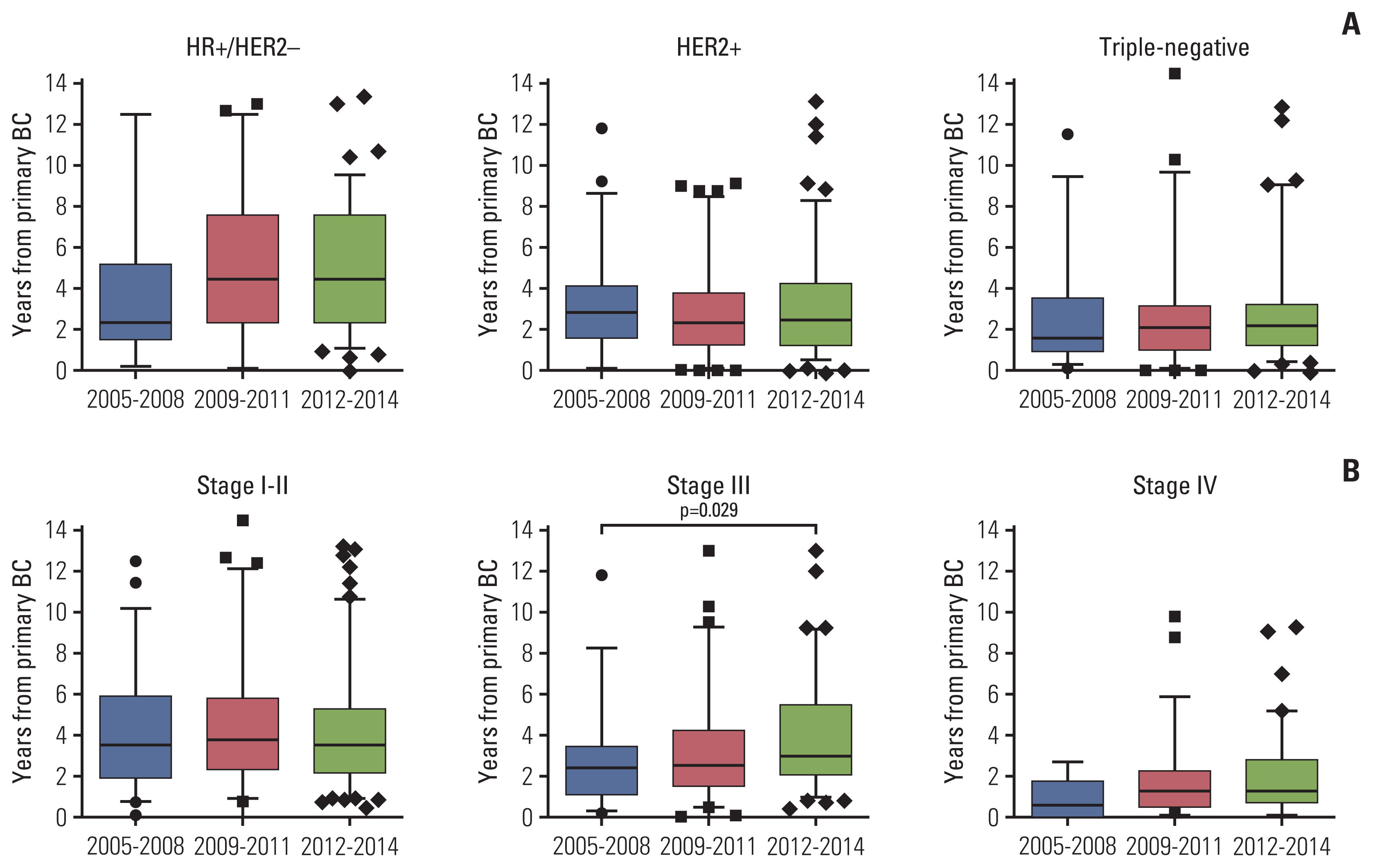
Fig. 1 shows the interval between BC and BM based on tumor subtypes or the initial stage of BC. The changes of this interval according to the times were not found except for that of stage III patients. In these patients, the median time interval has steadily protracted from 2.4 to 3 years (p=0.029).
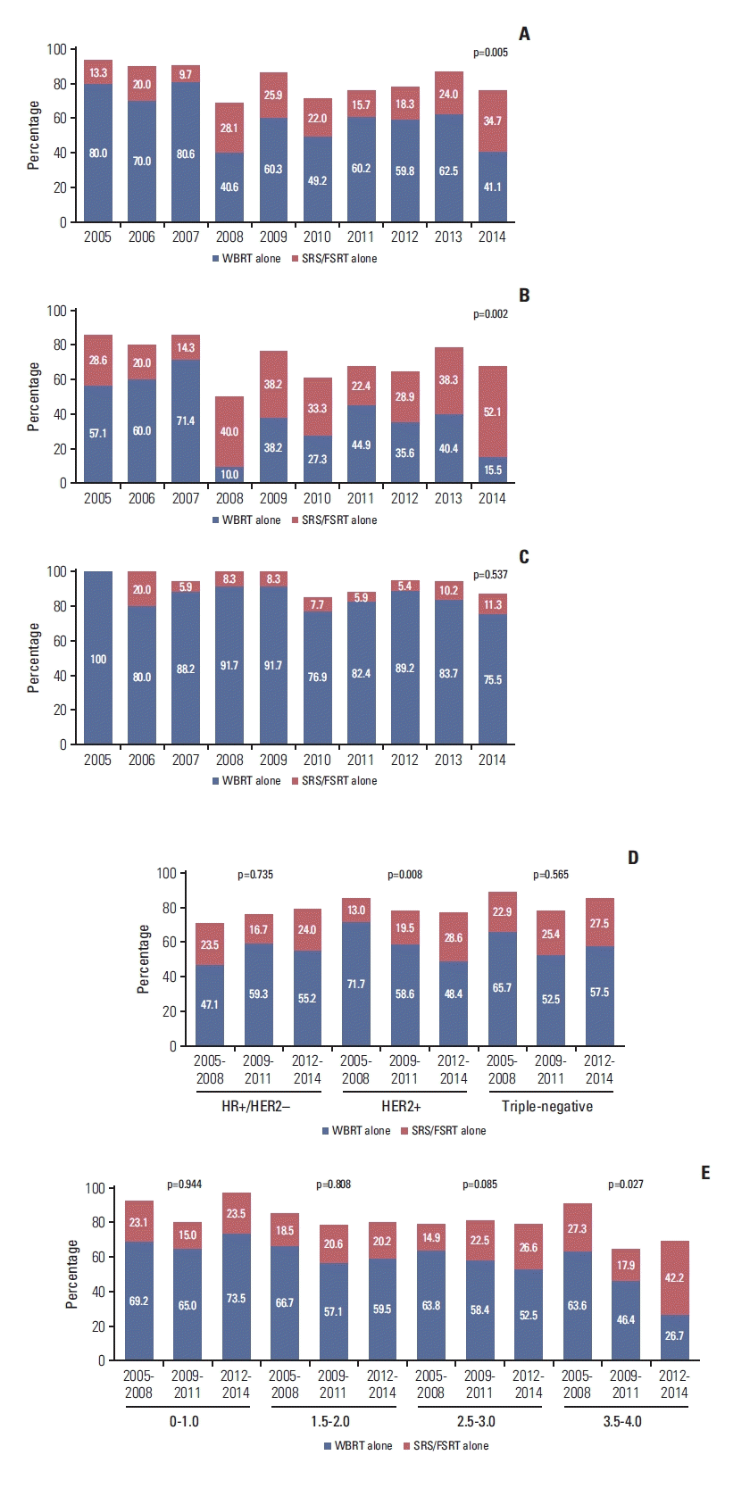
Brain-directed local treatment was immediately administered approximately 5 days after the initial diagnosis of BM (S1 Fig.). The largest portion of the initial brain-directed treatment was WBRT alone, followed by SRS or fractionated stereotactic radiotherapy (FSRT) alone (Table 2). However, WBRT decreased from 80.0% to 41.1% and SRS/FSRT alone increased from 13.3% to 34.7% over the past 10 years (p for trend=0.005) (Fig. 2A). These changes were prominent in patients with limited BM (1–4 BM, p for trend=0.002) (Fig. 2B), but not in those with BM > 4 (p for trend=0.537) (Fig. 2C). This paradigm shift in treatment strategy into SRS/FSRT alone was observed for patients with HER2+ BM (p for trend=0.008, Fig. 2D) and with breast-GPA scores of 3.5–4.0 (p for trend=0.027, Fig. 2E). About 80% of patients were treated with systemic treatment following brain-directed treatment. Especially, for patients with TNBC, the use of systemic therapy after initial brain-directed treatment did not increase (77.1% in group I, 78.0% in group II, and 68.8% in group III, respectively; p for trend=0.255). With respect to HER2+ patients, more than half of patients received anti-HER2 therapy. Among 417 patients with WBRT, only 21 patients received WBRT using 3-dimensional conformal RT or intensity-modulated RT (one patient was treated with hippocampal avoidance WBRT). Additional boost ranging from 3 to 25 Gy was administered after WBRT in 25 patients (19 patients after conventional WBRT) and a simultaneous-integrated boost was done in another patient.
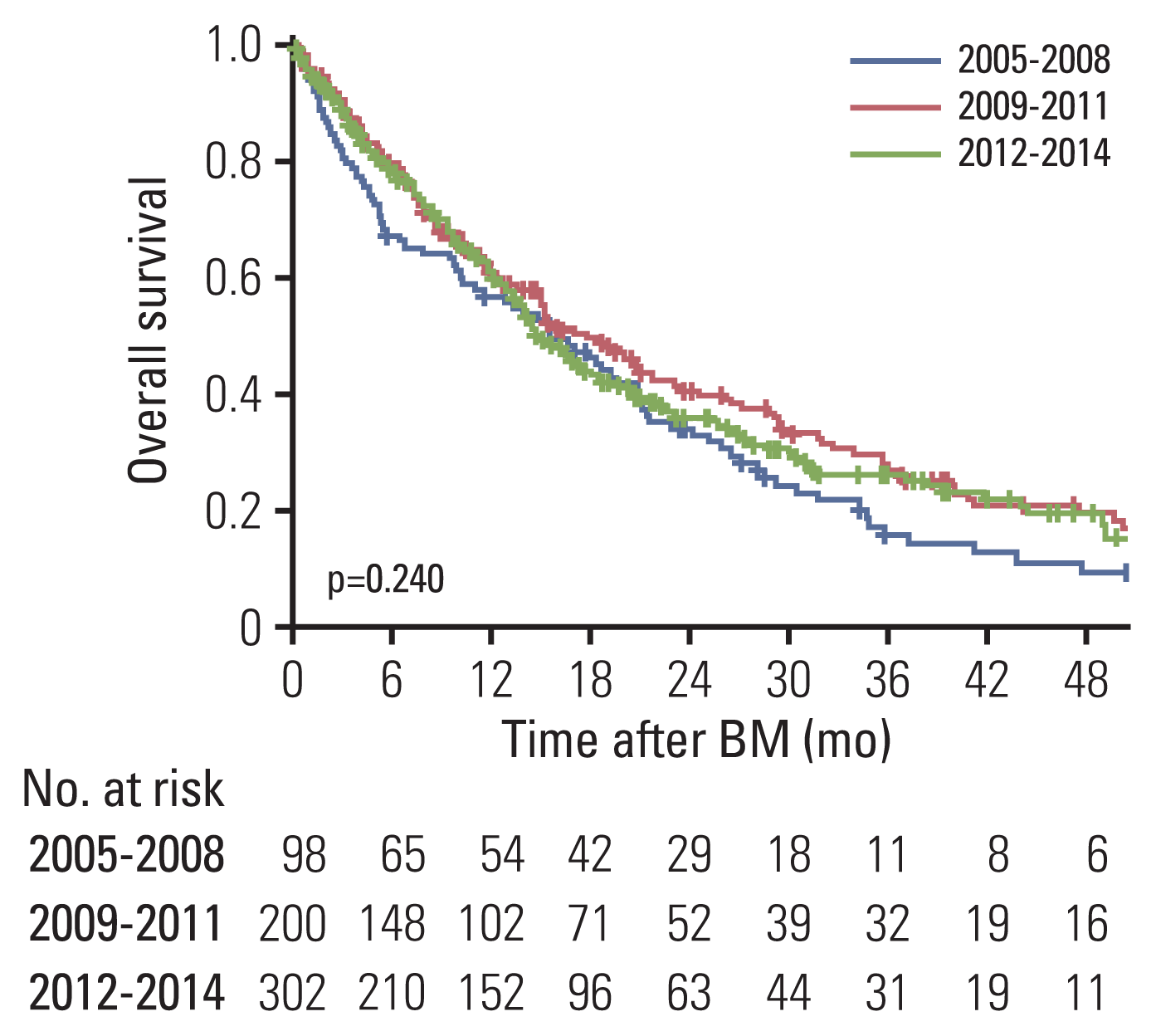
For the entire cohort, OS did not change significantly from 2005 to 2014 (median, 15.6 months in group I, 17.9 in group II, and 15.0 in group III, respectively; p=0.240) (Fig. 3, S2 Table). The 1-year OS rate of group I, II, and III was 57.0%, 61.0%, and 61.0%, respectively. In subgroup analysis, shown in S2 Table, only patients with the highest breast-GPA scores improved their median OS by a factor of two from 15.5 to 30.0 months (p=0.03). According to tumor subtype, the initial stage of primary BC, number of BM, brain-directed treatment for initial BM as well as other breast-GPA groups, we did not find any improvement in OS.

Discussion

In our study, the proportion of older BM patients with extracranial metastasis significantly increased over the past 10 years. Regardless of tumor subtypes, the time of BM diagnosis has been prolonged after primary BC with stage III disease. The first choice of brain-directed treatment for BM was primarily WBRT alone, but the use of SRS/FSRT alone has been increased during the period, especially in limited BM, which had 1–4 BM. Also, subsequent systemic treatment was frequently given and emphasized that multidisciplinary approaches based on the individualized situation were important in these patients. Unfortunately, there has been no such dramatic improvement in OS over 10 years.
Median age at initial BM diagnosis increased by 2.2 years in the current study. However, due to the increased median age at primary BC, there was no statistically significant increment in the time interval from primary BC to BM. This result was contrary to the report by Nieder et al. [19] which found the significantly lengthened time to development of BM. They explained this result by the increased use of systemic treatment. While the study by Nieder et al. [19] had a time interval of more than 25 years, this conflicting result might also be related to the fact that our study described a change over a short period of 10 years.
However, in terms of the initial stage of BC, patients with stage III, high-risk localized disease, showed a longer period until the brain failure was experienced. This interval seemed to be increasing recently in stage IV patients as well. It has been known that the stage of BC, as well as subtypes, is a prognostic factor for the time from BC to BM [20]. Concerning that advanced stage is associated with an earlier BM development [20], it is important to note that the time to BM was prolonged in stage III–IV patients in this study. In addition, a greater portion of extracranial metastasis could reflect the effectiveness of systemic treatment.
We would readily expect early identification of asymptomatic and tiny BM in recent years on account of the progress of brain imaging modalities. However, over 80% of the included patients had neurologic symptoms in the present study. Furthermore, no changes in the number of BM at diagnosis and slightly more patients with BM in both supra- and infra-tentorial regions were found. This might result from the timing of brain imaging after patients have symptoms. Currently, controversies exist on the role of brain magnetic resonance imaging (MRI) as a screening tool for BM [21]. However, recent studies emphasize and favor the use of MRI because early detection of BM could be managed by SRS with less invasiveness and toxicities [21,22]. In view of cost-effectiveness, it is necessary to select the optimal candidates for BM screening.
Our significant observation was that WBRT accounted for the largest proportion among the brain-directed local therapies. However, its use was decreasing while the use of SRS increased. This was observed especially in patients with 1–4 BM and over half of these patients in 2014 were treated with SRS/FSRT alone. These findings coincide with previous reports [19,23,24]. Several factors have affected this paradigm shift in initial approaches for BM. Increased awareness of late toxicities after WBRT, including neurocognitive dysfunction, has made physicians avoid choosing WBRT in selected patients [10,25]. Recently amended guidelines recommending SRS for patients with limited BM have changed the choice of RT as well [11]. Besides, as the distinctive situation in Korea, the relaxed reimbursement guidelines for SRS of National Health Insurance Service might play a part since April 2007.
We performed analyses on the relation of the shift to SRS not only with the number of BM but also with tumor subtypes and breast-GPA. Among three subtypes, the first course of RT for HER2+ BM solely has preferred SRS/FSRT alone over WBRT. Although we could not determine the obvious reasons for this alteration, it might be affected by the synergism of the increasing number of patients with limited BM and SRS utilization in these patients. The reasons for the low number of BM at initial, especially in HER2+ patients, were not clear. Other possible causes of this propensity beyond our data should also be perceived. Although SRS/FSRT is being widely used in HER2+ BM, the risk of distant intracranial failure should be considered. The risk of new BM without initial WBRT was higher in HR+/HER2− followed by HER2+ subtypes according to our previous report for the new BM development after the initial brain-directed local treatment according to the tumor subtypes [26]. Contemporary patients with breast-GPA scores of 3.5–4.0 were largely treated with SRS/FSRT alone. In the next high breast-GPA group, WBRT was still mainly used, but the use of SRS/FSRT alone increased marginally. These indicated that SRS/FSRT was favored in patients with a better prognosis.
Overall, survival has not altered during the study period. This was disappointing but, from another point of view, might be an encouraging result. As Nieder et al. [19] described, recently treated patients had more extracranial metastasis and few options of systemic treatments since several systemic agents were heavily administered to these patients before BM diagnosis. Even though in this situation, 77.8% of patients in group III received systemic treatment after brain-directed treatment, and there was no decrease in OS rate at 1 year.
The current study has clear limitations; the retrospective design had inherent flaws such as selection bias and the cohort was relatively small compared to population-based studies. We did not look at the socioeconomic status of enrolled patients, which could influence decisions making of the treatment modality. A lack of detailed information on systemic treatment, especially chemotherapeutic agents or novel molecular targeted therapy, limited the interpretation of our analysis. In spite of these shortcomings, this study was currently the best way possible to show the evolving strategies of BM treatment in BC patients over the past 10 years in Korea, as it analyzed much more detailed data not covered in large-scale population-based studies.
In conclusion, presentations of BM from BC have profoundly changed from 2005 to 2014 in Korea. In accordance with these changes, management for BM has also been evolved. Still, WBRT had a large portion of the brain-directed treatment however, it has been reserved for salvage option after initial use of SRS/FSRT. Although patients with unfavorable features have been increasing, there has been no significant change in OS over the past decade. Patients with good prognostic factors showed an improvement in OS.

Electronic Supplementary Material

Supplementary materials are available at Cancer Research and Treatment website (https://www.e-crt.org).

Notes

Ethical Statement

This study was approved by the institutional review board of each institution. All procedures performed in studies involving human participants were in accordance with the ethical standards of the institutional and/or national research committee and with the 1964 Helsinki declaration and its later amendments or comparable ethical standards. Because of the retrospective design of the analysis, requirement for obtaining informed consent of participants included in the study was exempted.

Author Contributions

Conceived and designed the analysis: Kim K, Kim IA.

Collected the data: Jung W, Shin KH, Im SA, Kim HJ, Kim YB, Chang JS, Kim JH (Jee Hyun Kim), Choi DH, Park YH, Kim DY, Kim TH, Choi BO, Lee SW, Kim S, Kwon J, Kang KM, Chung WK, Kim KS, Nam JH, Yoon WS, Kim JH (Jin Hee Kim), Cha J, Oh YK.

Contributed data or analysis tools: Kim JS, Kim K, Kim IA.

Performed the analysis: Kim JS, Kim K.

Wrote the paper: Kim JS, Kim K.

Writing-review and editing: Kim JS, Kim K, Jung W, Shin KH, Im SA, Kim HJ, Kim YB, Chang JS, Kim JH (Jee Hyun Kim), Choi DH, Park YH, Kim DY, Kim TH, Choi BO, Lee SW, Kim S, Kwon J, Kang KM, Chung WK, Kim KS, Nam JH, Yoon WS, Kim JH (Jin Hee Kim), Cha J, Oh YK, Kim IA.

Conflicts of Interest

Conflict of interest relevant to this article was not reported.

ACKNOWLEDGMENTS

This work was supported by grants from the Ministry of Science and Information & Communication Technology (NRF#2020R1A2-C2005141) to In Ah Kim.

References

1. Nathoo N, Chahlavi A, Barnett GH, Toms SA. Pathobiology of brain metastases. J Clin Pathol. 2005; 58:237–42.
crossref
2. Ostrom QT, Wright CH, Barnholtz-Sloan JS. Brain metastases: epidemiology. Handb Clin Neurol. 2018; 149:27–42.
crossref
3. Cagney DN, Martin AM, Catalano PJ, Redig AJ, Lin NU, Lee EQ, et al. Incidence and prognosis of patients with brain metastases at diagnosis of systemic malignancy: a population-based study. Neuro Oncol. 2017; 19:1511–21.
crossref
4. Sung H, Ferlay J, Siegel RL, Laversanne M, Soerjomataram I, Jemal A, et al. Global cancer statistics 2020: GLOBOCAN estimates of incidence and mortality worldwide for 36 cancers in 185 countries. CA Cancer J Clin. 2021; 71:209–49.
crossref
5. Witzel I, Oliveira-Ferrer L, Pantel K, Muller V, Wikman H. Breast cancer brain metastases: biology and new clinical perspectives. Breast Cancer Res. 2016; 18:8.
crossref
6. Brown PD, Pugh S, Laack NN, Wefel JS, Khuntia D, Meyers C, et al. Memantine for the prevention of cognitive dysfunction in patients receiving whole-brain radiotherapy: a randomized, double-blind, placebo-controlled trial. Neuro Oncol. 2013; 15:1429–37.
crossref
7. Gondi V, Tolakanahalli R, Mehta MP, Tewatia D, Rowley H, Kuo JS, et al. Hippocampal-sparing whole-brain radiotherapy: a “how-to” technique using helical tomotherapy and linear accelerator-based intensity-modulated radiotherapy. Int J Radiat Oncol Biol Phys. 2010; 78:1244–52.
crossref
8. Aoyama H, Shirato H, Tago M, Nakagawa K, Toyoda T, Hatano K, et al. Stereotactic radiosurgery plus whole-brain radiation therapy vs stereotactic radiosurgery alone for treatment of brain metastases: a randomized controlled trial. JAMA. 2006; 295:2483–91.
crossref
9. Chang EL, Wefel JS, Hess KR, Allen PK, Lang FF, Kornguth DG, et al. Neurocognition in patients with brain metastases treated with radiosurgery or radiosurgery plus whole-brain irradiation: a randomised controlled trial. Lancet Oncol. 2009; 10:1037–44.
crossref
10. Brown PD, Jaeckle K, Ballman KV, Farace E, Cerhan JH, Anderson SK, et al. Effect of radiosurgery alone vs radiosurgery with whole brain radiation therapy on cognitive function in patients with 1 to 3 brain metastases: a randomized clinical trial. JAMA. 2016; 316:401–9.
crossref
11. Kim JS, Kim IA. Evolving treatment strategies of brain metastases from breast cancer: current status and future direction. Ther Adv Med Oncol. 2020; 12:1758835920936117.
crossref
12. Gampenrieder SP, Castagnaviz V, Rinnerthaler G, Greil R. Treatment landscape for patients with HER2-positive metastatic breast cancer: a review on emerging treatment options. Cancer Manag Res. 2020; 12:10615–29.
13. Schettini F, Giudici F, Giuliano M, Cristofanilli M, Arpino G, Del Mastro L, et al. Overall survival of CDK4/6-inhibitor-based treatments in clinically relevant subgroups of metastatic breast cancer: systematic review and meta-analysis. J Natl Cancer Inst. 2020; 112:1089–97.
crossref
14. Liu ZB, Zhang L, Bian J, Jian J. Combination strategies of checkpoint immunotherapy in metastatic breast cancer. Onco Targets Ther. 2020; 13:2657–66.
15. Morikawa A, Peereboom DM, Thorsheim HR, Samala R, Balyan R, Murphy CG, et al. Capecitabine and lapatinib uptake in surgically resected brain metastases from metastatic breast cancer patients: a prospective study. Neuro Oncol. 2015; 17:289–95.
crossref
16. Lin NU, Borges V, Anders C, Murthy RK, Paplomata E, Hamilton E, et al. Intracranial efficacy and survival with tucatinib plus trastuzumab and capecitabine for previously treated HER2-positive breast cancer with brain metastases in the HER2CLIMB trial. J Clin Oncol. 2020; 38:2610–9.
crossref
17. Kim JS, Kim K, Jung W, Shin KH, Im SA, Kim HJ, et al. Survival outcomes of breast cancer patients with brain metastases: a multicenter retrospective study in Korea (KROG 16-12). Breast. 2020; 49:41–7.
crossref
18. Sperduto PW, Kased N, Roberge D, Xu Z, Shanley R, Luo X, et al. Effect of tumor subtype on survival and the graded prognostic assessment for patients with breast cancer and brain metastases. Int J Radiat Oncol Biol Phys. 2012; 82:2111–7.
crossref
19. Nieder C, Spanne O, Mehta MP, Grosu AL, Geinitz H. Presentation, patterns of care, and survival in patients with brain metastases: what has changed in the last 20 years? Cancer. 2011; 117:2505–12.
crossref
20. Saraf A, Grubb CS, Hwang ME, Tai CH, Wu CC, Jani A, et al. Breast cancer subtype and stage are prognostic of time from breast cancer diagnosis to brain metastasis development. J Neurooncol. 2017; 134:453–63.
crossref
21. Hadjipanteli A, Doolan P, Kyriacou E, Constantinidou A. Breast cancer brain metastasis: the potential role of MRI beyond current clinical applications. Cancer Manag Res. 2020; 12:9953–64.
22. Cagney DN, Martin AM, Catalano PJ, Brown PD, Alexander BM, Lin NU, et al. Implications of screening for brain metastases in patients with breast cancer and non-small cell lung cancer. JAMA Oncol. 2018; 4:1001–3.
crossref
23. Barbour AB, Jacobs CD, Williamson H, Floyd SR, Suneja G, Torok JA, et al. Radiation therapy practice patterns for brain metastases in the United States in the stereotactic radiosurgery era. Adv Radiat Oncol. 2020; 5:43–52.
crossref
24. Gullhaug A, Hjermstad MJ, Yri O, Svestad JG, Aass N, Johansen S. Use of radiotherapy in breast cancer patients with brain metastases: a retrospective 11-year single center study. J Med Imaging Radiat Sci. 2021; 52:214–22.
crossref
25. Kocher M, Soffietti R, Abacioglu U, Villa S, Fauchon F, Baumert BG, et al. Adjuvant whole-brain radiotherapy versus observation after radiosurgery or surgical resection of one to three cerebral metastases: results of the EORTC 22952–26001 study. J Clin Oncol. 2011; 29:134–41.
26. Kim JS, Kim K, Jung W, Shin KH, Im SA, Kim HJ, et al. New brain metastases after whole-brain radiotherapy of initial brain metastases in breast cancer patients: the significance of molecular subtypes (KROG 16-12). Breast Cancer Res Treat. 2021; 186:453–62.
crossref

Fig. 1
Time interval between primary breast cancer and brain metastasis according to tumor subtypes (A) and initial stage of primary breast cancer (B). Box plots present median value with 5–95 percentile. Adjusted p-values were calculated using Tukey’s multiple comparisons. BC, breast cancer; HER2, human epidermal growth factor receptor 2; HR, hormone receptor.
crt-2021-1083f1.gif
Fig. 2
Trend of radiotherapy for brain metastasis (BM) from breast cancer: all patients (A), patients with 1–4 BM (B), patients with more than 4 BM (C), according to tumor subtype (D), and according to breast cancer-specific graded prognostic assessment (E). FSRT, fractionated stereotactic radiotherapy; HER2, human epidermal growth factor receptor 2; HR, hormone receptor; SRS, stereotactic radiosurgery; WBRT, whole-brain radiotherapy. (Continued to the next page)
crt-2021-1083f2.gif
Fig. 3
Kaplan-Meier curve of overall survival according to the year of brain metastasis (BM).
crt-2021-1083f3.gif
Table 1
Baseline characteristics according to the year of brain metastasis diagnosis
Characteristic 2005–2008 (group I) 2009–2011 (group II) 2012–2014 (group III) p-value
No. of patients 98 200 302
Age at primary BC (yr) 45.1 (39.2–51.9) 45.5 (37.8–52.8) 48.1 (41.8–53.7) 0.019
Age at BM (yr) 49.0 (41.4–56.1) 48.3 (39.8–56.2) 51.2 (45.6–57.4) 0.008
Interval of primary BC and BM (mo) 30.6 (14.4–48.9) 29.4 (16.5–56.4) 34.2 (19.2–54.0) 0.482
Tumor subtype
 HR+/HER2− 17 (17.3) 54 (27.0) 96 (31.8) 0.082
 HER2+ 46 (46.9) 87 (43.5) 126 (41.7)
 Triple-negative 35 (35.7) 59 (29.5) 80 (26.5)
Initial BC stage
 Stage I 11 (11.2) 17 (8.5) 28 (9.3) 0.092
 Stage II 39 (39.8) 56 (28.0) 99 (32.8)
 Stage III 35 (35.7) 70 (35.0) 94 (31.1)
 Stage IV 13 (13.3) 57 (28.5) 81 (26.8)
ECOG
 0–1 69 (70.4) 130 (65.0) 197 (65.2) 0.601
 2–3 29 (29.6) 70 (35.0) 105 (34.8)
Primary tumora)
 Uncontrolled 23 (23.5) 39 (19.6) 82 (27.7) 0.092
 Controlled 75 (76.5) 160 (80.4) 214 (72.3)
Extracranial metastasis
 Absent 26 (26.5) 33 (16.5) 41 (13.6) 0.011
 Present 72 (73.5) 167 (83.5) 261 (86.4)
Symptoms
 No 14 (14.3) 24 (12.0) 34 (11.3) 0.725
 Yes 84 (85.7) 176 (88.0) 268 (88.7)
No. of BMs
 ≤ 4 51 (52.0) 116 (58.0) 163 (54.0) 0.548
 > 4 47 (48.0) 84 (42.0) 139 (46.0)
Location of BM
 Supra- or infra-tentorial 51 (52.0) 109 (54.5) 134 (44.4) 0.068
 Both 47 (48.0) 91 (45.5) 168 (55.6)
Breast-GPA
 0–1.0 13 (13.3) 20 (10.0) 34 (11.3) 0.903
 1.5–2.0 27 (27.6) 63 (31.5) 84 (27.8)
 2.5–3.0 47 (48.0) 89 (44.5) 139 (46.0)
 3.5–4.0 11 (11.2) 28 (14.0) 45 (14.9)

Values are presented median (IQR) or number (%). BC, breast cancer; BM, brain metastasis; Breast-GPA, breast cancer-specific graded prognostic assessment; ECOG, Eastern Cooperative Oncology Group; HER2, human epidermal growth factor receptor 2; HR, hormone receptor; IQR, interquartile range.

a) Available data only.

Table 2
Treatment distribution according to the year of brain metastasis
Characteristic 2005–2008 (group I) 2009–2011 (group II) 2012–2014 (group III) p-valuea)
WBRT alone 64 (65.3) 114 (57.0) 160 (53.0) 0.036
SRS or FSRT alone 18 (18.4) 41 (20.5) 81 (26.8) 0.045
Op alone 3 (3.1) 7 (3.5) 12 (4.0) 0.657
Op or SRS or FSRT → WBRT 9 (9.2) 27 (13.5) 39 (12.9) 0.454
WBRT → SRS 1 (1.0) 2 (1.0) 1 (0.3) 0.358
Other brain-directed treatment 3 (3.1) 9 (4.5) 9 (3.0) 0.733
Subsequent systemic therapy 77 (78.6) 158 (79.0) 235 (77.8) 0.810
Anti-HER2 therapyb) 23 (50.0) 54 (62.1) 66 (52.4) 0.846

Values are presented as number (%). FSRT, fractionated stereotactic radiotherapy; HER2, human epidermal growth factor receptor 2; Op, operation; SRS, stereotactic radiosurgery; WBRT, whole-brain radiotherapy.

a) p-value for trend,

b) In HER2+ patients.

TOOLS
Similar articles