Journal List > Diabetes Metab J > v.46(4) > 1516079576

Jang, Moon, and Koo: Prevalence of Diabetic Retinopathy in Undiagnosed Diabetic Patients: A Nationwide Population-Based Study

Abstract

Background

We investigated the prevalence of diabetic retinopathy (DR) in patients with undiagnosed diabetes through a nationwide survey, compared to those with known diabetes.

Methods

Among the participants of the Korean National Health and Nutrition Examination Surveys (KNHANES) from 2017 to 2018, individuals aged ≥40 years with diabetes and fundus exam results were enrolled. Sampling weights were applied to represent the entire Korean population. Newly detected diabetes patients through KNHANES were classified under “undiagnosed diabetes.”

Results

Among a total of 9,108 participants aged ≥40 years, 951 were selected for analysis. Of them, 31.3% (standard error, ±2.0%) were classified under “undiagnosed diabetes.” The prevalence of DR in patients with known and undiagnosed diabetes was 24.5%±2.0% and 10.7%±2.2%, respectively (P<0.001). The DR prevalence increased with rising glycosylated hemoglobin (HbA1c) levels in patients with known and undiagnosed diabetes (P for trend=0.001 in both). Among those with undiagnosed diabetes, the prevalence of DR was 6.9%±2.1%, 8.0%±3.4%, 5.6%±5.7%, 16.7%±9.4%, and 42.6%±14.8% for HbA1c levels of <7.0%, 7.0%–7.9%, 8.0%–8.9%, 9.0%–9.9%, and ≥10.0% respectively. There was no difference in the prevalence of hypertension, dyslipidemia, hypertriglyceridemia, or obesity according to the presence or absence of DR.

Conclusion

About one-third of patients with diabetes were unaware of their diabetes, and 10% of them have already developed DR. Considering increasing the prevalence of DR according to HbA1c level was found in patients with undiagnosed diabetes like those with known diabetes, screening and early detection of diabetes and DR are important.

INTRODUCTION

Diabetic retinopathy (DR) is the most common microvascular complication of diabetes mellitus and is a leading cause of blindness and visual impairment among adults [1,2]. A global meta-analysis of patients with diabetes from 2015 to 2019 showed that the prevalence of DR was 27.0% [3]. Similarly, the National Health and Nutrition Examination Survey conducted in the United States from 2005 to 2008 revealed that the prevalence of DR was 28.5% [4]. Furthermore, according to a national claim database in Korea, the prevalence of DR in patients with diabetes was 20% in 2015 [5].
The prevalence of DR increases with increasing diabetes duration [6]. However, 7.3% to 18.2% of patients already have DR at the time of diabetes diagnosis [7,8], and a previous study reported that among patients with type 2 diabetes mellitus (T2DM), DR might develop 4 to 7 years before the diagnosis of diabetes [9]. Therefore, the American Diabetes Association recommends initial dilated and comprehensive eye exam immediately after the diagnosis of diabetes [10]. However, the screening rate of DR is only about 20% to 50% [5,11], and is dependent on the availability of medical resources [11,12].
In a recent Korean nationwide survey, people with undiagnosed diabetes were found to have a higher risk of cardiovascular disease compared to those with diagnosed diabetes, as the former have multiple cardiovascular risk factors including obesity, dyslipidemia, smoking, and hypertension compared to those with already known diabetes [13]. Dyslipidemia, hypertension, and smoking as well as hyperglycemia are also important determinants of DR [14,15]; hence, undiagnosed diabetes might have comparable DR risk among those with already known diabetes, which has been rarely reported. A previous European population-based study showed that about 25% of patients with diabetes newly detected through the survey had DR [16]. As ethnicity and economic status are known to affect the development of DR [11,12,14], there might be difference in the prevalence of DR and risk factors among the patients with undiagnosed diabetes according to the populations.
The Korean National Health and Nutrition Examination Surveys (KNHANES) is a nationwide cross-sectional health examination. In addition, the 2017 to 2018 KNHANES included fundus evaluation in every participant irrespective of diabetes status. In the present study, we investigated the prevalence of DR in the patients who have been newly detected with diabetes through a health examination survey (“undiagnosed diabetes”) using KNHANES 2017 to 2018. The clinical characteristics associated with DR among individuals with undiagnosed diabetes were also evaluated and compared to those with already known diabetes (known diabetes).

METHODS

Study participants

KNHANES is a nationwide cross-sectional health examination and survey executed by the Korean Centers for Disease Control and Prevention in the Ministry of Health and Welfare to provide information on the current management status of diverse chronic diseases among the Korean population [17]. Moreover, KNHANES adopted a stratified multistage probability-based sampling design to represent the entire Korean population.
As the fundus exam is conducted among members of the population aged ≥40 years, we selected individuals with diabetes aged ≥40 years for the current study. Those with indeterminate fundus exam results for the presence or absence of DR were excluded (Supplementary Fig. 1). Diabetic cases were defined as those with glycosylated hemoglobin (HbA1c) levels ≥6.5% (48 mmol/mol), 8-hour fasting plasma glucose (FPG) levels ≥7.0 mmol/L (126 mg/dL), or use anti-diabetic medications including insulin. Diabetes was classified as known or undiagnosed diabetes following a self-questionnaire.
Institutional Review Board approval was not required for the use of KNHANES data from 2017 to 2018 under the Bioethics Act. This study was conducted in accordance with the ethical standards laid down in the 1964 Declaration of Helsinki and its later amendments.

Fundus evaluation

The fundus exam was conducted through a Fundus Camera (VISUCAM 224, Carl Zeiss Meditec AG, Jena, Germany) using the standard protocols of the Korean Ophthalmological Society (KOS) [18]. Four well-trained special examiners qualified under the KOS guidelines performed the exam, and the KOS managed quality control for examiners and all machines by regular on-site inspections. The collected data was interpreted by the Epidemiologic Investigation Committee of the KOS [18]. DR was identified if any characteristic lesion as defined by the Early Treatment for Diabetic Retinopathy Study was present: microaneurysms, hemorrhages, hard exudates, cotton wool spots, intraretinal microvascular abnormalities, venous beading, and retinal new vessels [19].

Metabolic parameters measurement

Venous blood samples were drawn after a 12-hour overnight fast, and plasma was immediately separated by centrifugation. Fasting plasma concentrations of glucose and lipids were measured enzymatically in a central laboratory using a Hitachi Automatic Analyzer 7600-210 (Hitachi, Tokyo, Japan). Hypertension was defined as systolic blood pressure (SBP) ≥140 mm Hg, diastolic blood pressure (DBP) ≥90 mm Hg, or being under anti-hypertensive medications. Dyslipidemia was defined as non-high-density lipoprotein cholesterol ≥190 mg/dL or being under lipid-lowering agents. Triglyceride (TG) ≥150 mg/dL was defined as hypertriglyceridemia. Body mass index (BMI) ≥25 kg/m2 was defined as obesity [20].

Statistical analyses

Sampling weights were applied to all analyses in order to represent the entire Korean population. Variables are expressed as the mean±standard error (SE) or the prevalence±SE (%). A Comparison of the clinical variables according to diabetes awareness and the presence or absence of DR was performed using linear regression or logistic linear regression analysis. P values <0.05 were considered as statistically significant for all tests. Statistical analyses were performed using IBM SPSS Statistics version 27.0 (IBM Corp., Armonk, NY, USA).

RESULTS

Clinical characteristics of study subjects

A total of 9,108 participants aged ≥40 years were enrolled in the KNHANES between 2017 and 2018, with a diabetes prevalence was 14.9%±0.5%. Among them, 951 patients with diabetes and fundus exam results were selected for the analysis (Supplementary Fig. 1).
From this cohort, 59.6%±2.1% were male, and the average age was 61.0±0.5 years. Their mean diabetes duration was 6.1±0.3 years and the average HbA1c was 7.2% (55 mmol/mol) ±0.1% (Table 1). Among them, 66.1%±2.0% were taking oral anti-diabetic drugs and 3.7%±0.6% were under insulin therapy. The prevalence of hypertension, dyslipidemia and hypertriglyceridemia was 62.3%±1.9%, 45.4%±1.9%, and 46.9%±2.3%, respectively. Their mean income was 346.0±10.5 ten thousand Korean won, and 53.4%±2.3% graduated from high school.
Among the entire population of patients with diabetes, 31.3%±2.0% were newly detected through KNHANES (“undiagnosed diabetes”). The patients with undiagnosed diabetes were male-dominant (65.7%±3.4% vs. 56.8%±2.3%, P=0.025) and younger compared to the patients with known diabetes (55.8±0.7 years vs. 63.4±0.5 years, P<0.001). Undiagnosed diabetes was most popular in those aged 40 to 49 years compared to the other age group: 56.8%±4.7%, 35.6%±3.1%, 21.0%± 2.2%, and 13.1%±1.7% for age 40–49, 50–59, 60–69, and ≥70 years, respectively. Nevertheless, there was no difference in HbA1c with respect to diabetes awareness (7.1% [54 mmol/mol] ±0.1% vs. 7.3% [56 mmol/mol] ±0.1%, P=0.294) (Supplementary Table 1). By contrast, after the adjustment for age and sex, a significant difference in HbA1c value was observed between patients with undiagnosed and known diabetes (P=0.012) (Supplementary Table 1). The proportion of those with HbA1c <7.0% (53 mmol/mol) was significantly greater among individuals with undiagnosed diabetes (64.5%±3.4% vs. 51.7%±2.4%, P=0.002) (Supplementary Table 2, Supplementary Fig. 2A). HbA1c ≥10.0% (86 mmol/mol) was also more frequently observed among patients with undiagnosed diabetes compared to those with known diabetes, even though this difference was statistically marginal (8.9%±2.3% vs. 4.7%±1.0%, P=0.060). The trend was only observed among those aged 40 to 49 years (17.3%±6.0% vs. 13.3%±5.0%) and aged 50 to 59 years (8.5%±3.4% vs. 5.2%±2.2%) (Supplementary Fig. 2B-E), although it was not statistically significant. Among patients with undiagnosed diabetes aged ≥70 years, HbA1c ≥8.0% (64 mmol/mol) was not detected (Supplementary Table 2).
The patients with undiagnosed diabetes had worse metabolic profiles in BMI, waist circumference, blood pressure, FPG, total cholesterol (TC), TG, and low-density lipoprotein cholesterol (LDL-C) levels compared to those with known diabetes; among which, the difference in SBP, DBP, TC, LDL-C, and TG persisted even after adjustments for age and sex (SBP, 124.1±0.8 mm Hg vs. 129.5±1.3 mm Hg; DBP, 75.0±0.5 mm Hg vs. 79.0±0.7 mm Hg; TC, 168.6±1.9 mg/dL vs. 203.9±3.1 mg/dL; LDL-C, 100.6±3.0 mg/dL vs. 118.9±4.6 mg/dL; TG, 162.0±6.8 mg/dL vs. 227.7±20.3 mg/dL). In addition, there was no significant difference in average monthly income and educational background between patients with known diabetes and undiagnosed diabetes (Supplementary Table 1).

Comparison of clinical variables between patients with known and undiagnosed diabetes according to presence of DR

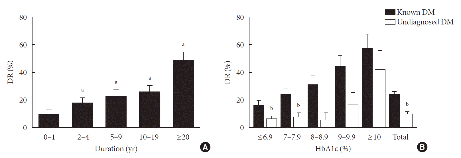
The prevalence of DR in the entire study population with diabetes was 20.2%±1.6%, which increased with increasing diabetes duration: 10.3%±3.5%, 19.0%±3.5%, 23.9%±4.2%, 27.1%±3.5%, and 49.5%±6.2% in 0–1, 2–4, 5–9, 10–19, and ≥20 years, respectively (P for trends <0.001) (Fig. 1A).
The prevalence of DR in patients with known and undiagnosed diabetes was 24.5%±2.0% and 10.7%±2.2%, respectively (P<0.001). The patients with DR had higher mean HbA1c in both patients with known diabetes and undiagnosed diabetes (known diabetes, 7.8%±0.2% vs. 7.1%±0.1%, P<0.001; undiagnosed diabetes, 8.4%±0.5% vs. 7.0%±0.1%, P=0.008) (Table 1). However, there was no difference in the prevalence of hypertension, dyslipidemia, hypertriglyceridemia, or obesity according to the presence or absence of DR in both patients with known diabetes and undiagnosed diabetes. No significant difference was found in socioeconomic status like income or education status between the groups (Table 1). In patients with known diabetes, duration of diabetes was longer (P<0.001) and treatment with insulin was more frequently observed among patients with DR compared to those without DR (P<0.001) (Table 1).

The prevalence of DR in patients according to HbA1c level

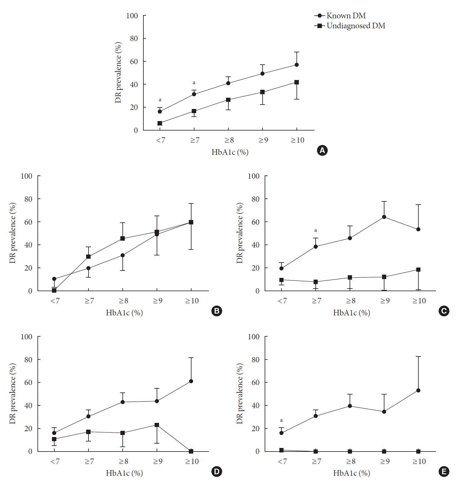
DR prevalence increased with increasing HbA1c levels both in patients with known and undiagnosed diabetes (P for trend=0.001 in both) (Fig. 1B, Supplementary Table 2). Among those with undiagnosed diabetes, the prevalence of DR was 6.9%±2.1%, 8.0%±3.4%, 5.6%±5.7%, and 16.7%±9.4% for HbA1c levels of <7.0% (53 mmol/mol), 7.0%–7.9% (53–63 mmol/mol), 8.0%–8.9% (64–74 mmol/mol), and 9.0%–9.9% (75–85 mmol/mol), respectively. And the prevalence of DR in undiagnosed diabetic patients with HbA1c levels ≥10.0% (86 mmol/mol) reached 42.6%±14.8%, which was comparable to that of known diabetic patients with ≥10 years of duration, that is, 32.7%±3.1% (Fig. 1). The prevalence of DR in undiagnosed diabetes tended to be lower than in known diabetes with same HbA1c level (Figs. 1B and 2A). This difference was statistically significant among those with HbA1c <7.0% (53 mmol/mol) and 7%–7.9% (53–63 mmol/mol) (Fig. 1B).
Given that those with undiagnosed diabetes aged 40 to 49 years had relatively high proportion of HbA1c ≥10.0% (86 mmol/mol) compared to known diabetic patients, we investigated DR prevalence in undiagnosed diabetic patients aged 40 to 49 years for each HbA1c level. In undiagnosed diabetic patients aged 40 to 49 years, the overall DR prevalence was 13.3%±5.1%. Among them, the undiagnosed diabetic patients with HbA1c ≥9% (75 mmol/mol) and HbA1c ≥10% (86 mmol/mol) had DR prevalence of 51.8%±14.3% and 60.3%±17.0%, respectively, which were comparable to those of known diabetic patients with same age and HbA1c level (49.1%±17.1% and 59.8%±22.8% in HbA1c ≥9% [75 mmol/mol] and HbA1c ≥10% [86 mmol/mol], respectively) (Fig. 2B, Supplementary Table 3). By contrast, the undiagnosed diabetic patients in the other age groups had a relatively low prevalence of DR even among those with HbA1c ≥10% (86 mmol/mol) (19.0%±17.0% in 50 to 59 years; no DR detected in ≥60 years) (Fig. 2C-E).

DISCUSSION

Using a nationwide survey database, we found that one-third of patients with diabetes was unaware of their status of diabetes in the Korean population. In addition, about one-tenth of patients with undiagnosed diabetes had already developed DR. Although the prevalence of DR in undiagnosed diabetes tended to be lower than in known diabetes with same HbA1c level, increasing the prevalence of DR according to HbA1c level was also found in patients with undiagnosed diabetes as well as those with known diabetes. Among those with undiagnosed diabetes, the prevalence of DR was 6.9% for HbA1c levels of <7.0%, which increased to 42.6% for HbA1c levels of ≥10.0%. Especially, as those with poor glycemic control with HbA1c ≥10% was frequently found in younger patients (aged 40 to 49 years) than in other age groups among the undiagnosed diabetic population, the prevalence of DR in young undiagnosed diabetic patients was seem to be comparable to that of known diabetic patients with the same age and HbA1c level in those age group with known diabetes.
To prevent the complications of diabetes, early diabetes detection and proper blood glucose management are important [21]. However, half of the patients with diabetes worldwide live without being diagnosed with diabetes [22]. In Korea, 35% of patients with diabetes aged ≥30 years are unaware of their diabetes [23], which was similar to the 30% detected in the current study. Interestingly, the current study also found that there was low awareness of the presence of diabetes among those aged 40 to 49 years, compared to the other age group (56.8% vs. 27.7%); in addition, more severe hyperglycemia, HbA1c level of ≥10% (86 mmol/mol), was prevalent in this age group. High diabetes unawareness in the young population has also been previously reported [24], which might be due to the low accessibility of health care services among younger members of the population [25]. Late detection of diabetes in the young population may also be linked to severe hyperglycemia at the period of diagnosis of diabetes. In addition, the difference in pathogenesis in developing diabetes between young and old population has been suggested. Middle-age-onset patients have lower β-cell function compared with elderly-onset diabetic patients [26], and the time from T2DM diagnosis to β-cell deterioration and/or progression to exogenous insulin use is faster when the onset age is younger [27]. Moreover, younger patients with diabetes had poorer blood glucose control compared to older patients [28], which might be from their low compliance to drugs [29], a tendency to be more obese and more insulin resistant, and consequently requiring more aggressive treatment for diabetes management than older patients [30]. Similar to patients with known diabetes, poor health management and obesity may also affect young patients with undiagnosed diabetes. It can result in individuals suffering from diabetes complications, as well as increasing the healthcare burden [31].
In the current study, patients with undiagnosed diabetes showed a tendency to have higher income and education status compared to those with known diabetes although it was not statistically significant, which was contrary to the previous studies [32,33]. It might be due to the difference in sex and age between known and undiagnosed diabetes group: the patients with undiagnosed diabetes were male-dominant (65.7% vs. 56.8%) and younger compared to the patients with known diabetes (55.8 years vs. 63.4 years).
DR progresses from mild non-proliferative abnormalities to moderate and severe non-proliferative diabetic retinopathy and proliferative diabetic retinopathy [34], leading to visual impairment and blindness [1]. Although DR treatments, such as laser photocoagulation, intravitreous injections of anti-vascular endothelial growth factor (VEGF) and steroid agents, and vitreoretinal surgery prevent vision loss and improve vision, 40% to 50% of patients still do not respond sufficiently to these treatments [35]. In large prospective randomized studies, intensive management of glucose level and associated risk factors has been shown to prevent or slow the progression of DR as well as improve patient-reported visual function [36,37]. Therefore, early detection of diabetes and DR, and management of the associated risk factors is essential in preventing vision loss due to DR [21].
In the current study, 10% of patients with undiagnosed diabetes had already developed DR. We confirmed that poor glycemic control is a significant risk factor for DR also in patients with undiagnosed diabetes as well as those with known diabetes, and the prevalence of DR reached 42.6% in undiagnosed diabetic patients with HbA1c levels ≥10.0%. In particular, as the patients with undiagnosed diabetes had higher blood glucose level in younger age groups, the prevalence of DR in undiagnosed diabetes was relatively higher in those with age 40 to 49 years compared to other age groups. Previous studies have also reported a higher risk of DR at younger ages [38,39]. Considering that the duration of diabetes would be relatively short at a young age, factors other than HbA1c levels and duration of diabetes might contribute to the high prevalence of DR in patients with undiagnosed diabetes at a young age. This may be due to poor management of metabolic factors affecting DR other than glucose levels at younger ages [40,41]. Moreover, the stronger response to VEGF in younger ages, and decrease in growth hormone and insulin-like growth factor 1 in older ages could have contributed to the higher DR prevalence in younger ages [39,42,43]. Considering that glucose level seems to be high and DR appears to be more prevalent among younger patients with undiagnosed diabetes, proper diabetes and DR screening is thought to be important, especially among younger individuals.
Nevertheless, this study has some limitations. First, we simply considered the existence of DR, and not the severity of DR. In addition, detection of DR was based on fundus photography. Although fundus photography for DR screening has become widely adopted worldwide [44] and its quality has been much improved [45], binocular slit-lamp ophthalmoscopy has remained a standard exam for DR. Second, since this study was based on cross-sectional data, it is impossible to know how the DR of patients with undiagnosed diabetes will progress in the future, compared to patients with known diabetes. Lastly, this study is difficult to generalize because our analysis was based on small number of Korean patients, within a limited period. Moreover, the unweighted number of patients with diabetes among participants aged <50 years was too small to gain a statistical significance. However, other previous studies have also reported that the risk of DR is high at a young age [38,39]. Furthermore, it is thought that this study has the strength of analyzing the prevalence of undiagnosed diabetes and DR across the population based on the KNHANES data, and in particular, identifying which patients are at high risk for undiagnosed DR.
In conclusion, we found that about one third of patients with diabetes were unaware of their diabetes, and that 10% of them have already developed DR. Considering increasing the prevalence of DR according to HbA1c level was found in patients with undiagnosed diabetes as well as those with known diabetes, screening and early detection of diabetes and DR are important.

SUPPLEMENTARY MATERIALS

Supplementary materials related to this article can be found online at https://doi.org/10.4093/dmj.2021.0099.
Supplementary Table 1.
Comparison of clinical characteristics of patients with known diabetes and undiagnosed diabetes
dmj-2021-0099-suppl1.pdf
Supplementary Table 2.
Distribution of HbA1c level in each group of known and undiagnosed diabetes
dmj-2021-0099-suppl2.pdf
Supplementary Table 3.
The prevalence of diabetic retinopathy according to HbA1c value
dmj-2021-0099-suppl3.pdf
Supplementary Fig. 1.
Flow diagram of participants for the study. Among a total of 16,119 participants in the Korean National Health and Nutrition Examination Surveys (KNHANES) 2017 to 2018, 951 individuals diagnosed with diabetes mellitus (DM) and performed a fundus exam were included in our analysis.
dmj-2021-0099-suppl4.pdf
Supplementary Fig. 2.
Distribution of patients according to glycosylated hemoglobin (HbA1c) in patients with known diabetes mellitus (DM) and undiagnosed DM. Distribution of patients according to HbA1c in patients with known DM and undiagnosed DM in age (A) ≥40 years, (B) 40 to 49 years, (C) 50 to 59 years, (D) 60 to 69 years, and (E) ≥70 years, respectively. Values are presented as prevalence±standard error and logistic linear regression was performed without correction. aP values <0.05 in comparison to the prevalence of known DM in the same HbA1c.
dmj-2021-0099-suppl5.pdf

Notes

CONFLICTS OF INTEREST

Bo Kyung Koo was editorial board member of the Diabetes & Metabolism Journal from 2020 to 2021. She was not involved in the review process of this article. Otherwise, there was no conflict of interest.

AUTHOR CONTRIBUTION

Concept or design: B.K.K.

Acquisition, analysis, or interpretation of data: H.N.J., B.K.K.

Drafting the work or revising: H.N.J., M.K.M., B.K.K.

Final approval of the manuscript: H.N.J., M.K.M., B.K.K.

FUNDING

None

ACKNOWLEDGMENTS

None

REFERENCES

1. Congdon NG, Friedman DS, Lietman T. Important causes of visual impairment in the world today. JAMA. 2003; 290:2057–60.
crossref
2. Fong DS, Aiello LP, Ferris FL 3rd, Klein R. Diabetic retinopathy. Diabetes Care. 2004; 27:2540–53.
crossref
3. Thomas RL, Halim S, Gurudas S, Sivaprasad S, Owens DR. IDF Diabetes Atlas: a review of studies utilising retinal photography on the global prevalence of diabetes related retinopathy between 2015 and 2018. Diabetes Res Clin Pract. 2019; 157:107840.
crossref
4. Zhang X, Saaddine JB, Chou CF, Cotch MF, Cheng YJ, Geiss LS, et al. Prevalence of diabetic retinopathy in the United States, 2005-2008. JAMA. 2010; 304:649–56.
crossref
5. Chung YR, Ha KH, Lee K, Kim DJ. Diabetic retinopathy and related clinical practice for people with diabetes in Korea: a 10-year trend analysis. Diabetes Metab J. 2020; 44:928–32.
crossref
6. Lima VC, Cavalieri GC, Lima MC, Nazario NO, Lima GC. Risk factors for diabetic retinopathy: a case-control study. Int J Retina Vitreous. 2016; 2:21.
crossref
7. Rema M, Deepa R, Mohan V. Prevalence of retinopathy at diagnosis among type 2 diabetic patients attending a diabetic centre in South India. Br J Ophthalmol. 2000; 84:1058–60.
crossref
8. Lee KM, Sum WM. Prevalence of diabetic retinopathy in patients with recently diagnosed diabetes mellitus. Clin Exp Optom. 2011; 94:371–5.
crossref
9. Harris MI, Klein R, Welborn TA, Knuiman MW. Onset of NIDDM occurs at least 4-7 yr before clinical diagnosis. Diabetes Care. 1992; 15:815–9.
crossref
10. American Diabetes Association. 11. Microvascular complications and foot care: standards of medical care in diabetes-2021. Diabetes Care. 2021; 44(Suppl 1):S151–67.
11. Shi Q, Zhao Y, Fonseca V, Krousel-Wood M, Shi L. Racial disparity of eye examinations among the U.S. working-age population with diabetes: 2002-2009. Diabetes Care. 2014; 37:1321–8.
crossref
12. Jones S, Edwards RT. Diabetic retinopathy screening: a systematic review of the economic evidence. Diabet Med. 2010; 27:249–56.
crossref
13. Lee YH, Armstrong EJ, Kim G, Oh J, Kang SM, Lee BW, et al. Undiagnosed diabetes is prevalent in younger adults and associated with a higher risk cardiometabolic profile compared to diagnosed diabetes. Am Heart J. 2015; 170:760–9.
crossref
14. Klein R, Sharrett AR, Klein BE, Moss SE, Folsom AR, Wong TY, et al. The association of atherosclerosis, vascular risk factors, and retinopathy in adults with diabetes: the atherosclerosis risk in communities study. Ophthalmology. 2002; 109:1225–34.
crossref
15. Niazi MK, Akram A, Naz MA, Awan S. Duration of diabetes as a significant factor for retinopathy. Pak J Ophthalmol. 2010; 26:182–6.
16. Ponto KA, Koenig J, Peto T, Lamparter J, Raum P, Wild PS, et al. Prevalence of diabetic retinopathy in screening-detected diabetes mellitus: results from the Gutenberg Health Study (GHS). Diabetologia. 2016; 59:1913–9.
crossref
17. Kim Y. The Korea National Health and Nutrition Examination Survey (KNHANES): current status and challenges. Epidemiol Health. 2014; 36:e2014002.
crossref
18. Korean Ophthalmological Society. Standardization for eye survey in Korea National Health and Nutrition Examination Survey. Seoul: Korean Ophthalmological Society;2018.
19. Lee WJ, Sobrin L, Lee MJ, Kang MH, Seong M, Cho H. The relationship between diabetic retinopathy and diabetic nephropathy in a population-based study in Korea (KNHANES V-2, 3). Invest Ophthalmol Vis Sci. 2014; 55:6547–53.
crossref
20. Seo MH, Lee WY, Kim SS, Kang JH, Kang JH, Kim KK, et al. 2018 Korean Society for the Study of Obesity guideline for the management of obesity in Korea. J Obes Metab Syndr. 2019; 28:40–5.
crossref
21. Hill L, Makaroff LE. Early detection and timely treatment can prevent or delay diabetic retinopathy. Diabetes Res Clin Pract. 2016; 120:241–3.
crossref
22. International Diabetes Federation. IDF diabetes atlas. Brussels: International diabetes Federation;2019.
23. Jung CH, Son JW, Kang S, Kim WJ, Kim HS, Kim HS, et al. Diabetes fact sheets in Korea, 2020: an appraisal of current status. Diabetes Metab J. 2021; 45:1–10.
crossref
24. Claypool KT, Chung MK, Deonarine A, Gregg EW, Patel CJ. Characteristics of undiagnosed diabetes in men and women under the age of 50 years in the Indian subcontinent: the National Family Health Survey (NFHS-4)/Demographic Health Survey 2015-2016. BMJ Open Diabetes Res Care. 2020; 8:e000965.
crossref
25. Wong A, Wouterse B, Slobbe LC, Boshuizen HC, Polder JJ. Medical innovation and age-specific trends in health care utilization: findings and implications. Soc Sci Med. 2012; 74:263–72.
crossref
26. Koo BK, Roh E, Yang YS, Moon MK. Difference between old and young adults in contribution of β-cell function and sarcopenia in developing diabetes mellitus. J Diabetes Investig. 2016; 7:233–40.
crossref
27. Barrett T, Jalaludin MY, Turan S, Hafez M, Shehadeh N; Novo Nordisk Pediatric Type 2 Diabetes Global Expert Panel. Rapid progression of type 2 diabetes and related complications in children and young people: a literature review. Pediatr Diabetes. 2020; 21:158–72.
crossref
28. Carter JS, Gilliland SS, Perez GE, Skipper B, Gilliland FD. Public health and clinical implications of high hemoglobin A1c levels and weight in younger adult Native American people with diabetes. Arch Intern Med. 2000; 160:3471–6.
crossref
29. El-Kebbi IM, Cook CB, Ziemer DC, Miller CD, Gallina DL, Phillips LS. Association of younger age with poor glycemic control and obesity in urban African Americans with type 2 diabetes. Arch Intern Med. 2003; 163:69–75.
crossref
30. Toh MP, Wu CX, Leong HS. Association of younger age with poor glycemic and cholesterol control in Asians with type 2 diabetes mellitus in Singapore. J Endocrinol Metab. 2011; 1:27–37.
crossref
31. Dall TM, Yang W, Halder P, Pang B, Massoudi M, Wintfeld N, et al. The economic burden of elevated blood glucose levels in 2012: diagnosed and undiagnosed diabetes, gestational diabetes mellitus, and prediabetes. Diabetes Care. 2014; 37:3172–9.
crossref
32. Karachaliou F, Simatos G, Simatou A. The challenges in the development of diabetes prevention and care models in low-income settings. Front Endocrinol (Lausanne). 2020; 11:518.
crossref
33. Le C, Jun D, Zhankun S, Yichun L, Jie T. Socioeconomic differences in diabetes prevalence, awareness, and treatment in rural southwest China. Trop Med Int Health. 2011; 16:1070–6.
crossref
34. Fong DS, Aiello L, Gardner TW, King GL, Blankenship G, Cavallerano JD, et al. Retinopathy in diabetes. Diabetes Care. 2004; 27 Suppl 1:S84–7.
crossref
35. Duh EJ, Sun JK, Stitt AW. Diabetic retinopathy: current understanding, mechanisms, and treatment strategies. JCI Insight. 2017; 2:e93751.
crossref
36. ACCORD Study Group; ACCORD Eye Study Group, Chew EY, Ambrosius WT, Davis MD, Danis RP, et al. Effects of medical therapies on retinopathy progression in type 2 diabetes. N Engl J Med. 2010; 363:233–44.
crossref
37. Writing Team for the DCCT/EDIC Research Group, GubitosiKlug RA, Sun W, Cleary PA, Braffett BH, Aiello LP, et al. Effects of prior intensive insulin therapy and risk factors on patient-reported visual function outcomes in the Diabetes Control and Complications Trial/Epidemiology of Diabetes Interventions and Complications (DCCT/EDIC) cohort. JAMA Ophthalmol. 2016; 134:137–45.
crossref
38. Zou W, Ni L, Lu Q, Zou C, Zhao M, Xu X, et al. Diabetes onset at 31-45 years of age is associated with an increased risk of diabetic retinopathy in type 2 diabetes. Sci Rep. 2016; 6:38113.
crossref
39. Middleton TL, Constantino MI, Molyneaux L, D’Souza M, Twigg SM, Wu T, et al. Young-onset type 2 diabetes and younger current age: increased susceptibility to retinopathy in contrast to other complications. Diabet Med. 2020; 37:991–9.
crossref
40. Zhang Y, Moran AE. Trends in the prevalence, awareness, treatment, and control of hypertension among young adults in the United States, 1999 to 2014. Hypertension. 2017; 70:736–42.
crossref
41. Kalra R, Parcha V, Patel N, Bhargava A, Booker KS, Arora G, et al. Increased awareness, inadequate treatment, and poor control of cardiovascular risk factors in American young adults: 2005-2016. Eur J Prev Cardiol. 2021; 28:304–12.
crossref
42. Heng LZ, Comyn O, Peto T, Tadros C, Ng E, Sivaprasad S, et al. Diabetic retinopathy: pathogenesis, clinical grading, management and future developments. Diabet Med. 2013; 30:640–50.
crossref
43. Bermea KC, Rodriguez-Garcia A, Tsin A, Barrera-Saldana HA. Somatolactogens and diabetic retinopathy. Growth Horm IGF Res. 2018; 41:42–7.
crossref
44. Prescott G, Sharp P, Goatman K, Scotland G, Fleming A, Philip S, et al. Improving the cost-effectiveness of photographic screening for diabetic macular oedema: a prospective, multicentre, UK study. Br J Ophthalmol. 2014; 98:1042–9.
crossref
45. Shi L, Wu H, Dong J, Jiang K, Lu X, Shi J. Telemedicine for detecting diabetic retinopathy: a systematic review and meta-analysis. Br J Ophthalmol. 2015; 99:823–31.
crossref

Fig. 1.
Prevalence of diabetic retinopathy according to duration of diabetes and glycosylated hemoglobin (HbA1c) value. (A) The prevalence of diabetic retinopathy (DR) according to duration of diabetes in the entire study population. (B) The prevalence of DR according to HbA1c levels in patients with known (black bar) and undiagnosed diabetes mellitus (DM; white bar). Values are presented as prevalence±standard error. aP values <0.05 compared with the prevalence of DR in the subjects with diabetes duration 0 to 1 year by logistic regression analysis without adjustment, bP values <0.05 compared with the prevalence of DR in known diabetic patients with the same HbA1c level by logistic regression analysis without adjustment.
dmj-2021-0099f1.tif
Fig. 2.
The cumulative prevalence of diabetic retinopathy according to glycosylated hemoglobin (HbA1c) value, in (A) total ages, (B) 40 to 49 years, (C) 50 to 59 years, (D) 60 to 69 years, and (E) ≥70 years. Values are presented as mean or prevalence±standard error. DM, diabetes mellitus; DR, diabetic retinopathy. aP values <0.05 compared with the prevalence of DR in known diabetic patients with the same HbA1c level.
dmj-2021-0099f2.tif
Table 1.
Clinical characteristics of diabetic patients according to the presence or absence of diabetic retinopathy
Characteristic Total Known DM
Undiagnosed DM
DR (–) DR (+) P valuea DR (–) DR (+) P valuea
Unweighted number 951 516 174 233 28
Male sex, % 59.6±2.1 56.8±2.8 56.8±4.2 0.989 65.0±3.5 71.6±8.3 0.478
Age, yr 61.0±0.5 63.5±0.7 63.3±0.9 0.876 56.0±0.6 53.8±1.9 0.286
BMI, kg/m² 25.2±0.1 24.9±0.2 24.9±0.3 0.846 25.6±0.2 26.5±0.6 0.192
WC, cm 88.1±0.3 87.6±0.4 87.6±0.7 0.997 89.0±0.7 90.4±1.5 0.437
SBP, mm Hg 125.8±0.7 123.7±0.8 128.1±1.7 0.022 127.3±1.3 134.9±5.1 0.153
DBP, mm Hg 76.2±0.5 73.8±0.6 74.1±0.9 0.751 80.9±0.8 86.1±2.8 0.072
HbA1c, % 7.2±0.1 7.1±0.1 7.8±0.2 <0.001 7.0±0.1 8.4±0.5 0.008
FPG, mg/dL 144.5±1.5 136.3±1.6 155.2±4.3 <0.001 148.6±2.9 184.9±13.6 0.014
TC, mg/dL 179.6±1.8 167.5±2.0 167.2±4.0 0.948 204.6±3.3 223.2±9.7 0.073
HDL-C, mg/dL 45.6±0.4 45.6±0.6 44.6±0.9 0.294 45.7±0.9 47.9±1.3 0.179
TG, mg/dL 182.9±9.8 156.2±7.2 149.4±7.7 0.514 233.1±18.1 339.6±164.3 0.517
LDL-C, mg/dL 108.8±2.9 99.3±3.2 104.3±7.3 0.557 116.2±4.3 138.3±14.2 0.141
Hypertension, % 62.3±1.9 64.8±2.6 66.0±4.4 0.808 56.4±3.9 54.3±10.5 0.848
Dyslipidemia, % 45.4±1.9 50.7±2.5 47.1±4.7 0.495 35.4±3.3 38.4±10.4 0.777
Hypertriglyceridemia, % 46.9±2.3 40.1±3.1 44.2±4.8 0.446 60.4±4.1 47.6±10.7 0.262
Obesity, % 48.0±1.9 46.2±2.7 42.8±4.3 0.496 53.3±3.6 58.3±11.2 0.673
FHx of diabetes, % 47.2±2.1 51.8±2.9 41.7±4.5 0.070 42.6±3.9 43.9±11.3 0.913
Age at the diagnosis of diabetes, yr 54.6±0.6 55.6±0.7 51.4±1.0 <0.001
Duration of diabetes, yr 6.1±0.3 7.9±0.4 11.9±0.8 <0.001
Type of treatment
 OAD 66.1±2.0 96.6±1.0 94.7±2.1 0.399
 Insulin 3.7±0.6 2.8±0.8 13.1±2.7 <0.001
Income, 10,000 KRW 346.0±10.5 335.6±16.4 329.0± 23.9 0.817 422.6±22.0 387.0±73.1 0.647
High school graduation 53.4±2.3 48.2±3.1 44.1±4.3 0.426 67.2±3.6 67.0±8.6 0.979

Values are presented as mean or prevalence±standard error. Linear regression and logistic linear regression were performed without correction.

DM, diabetes mellitus; DR, diabetic retinopathy; BMI, body mass index; WC, waist circumference; SBP, systolic blood pressure; DBP, diastolic blood pressure; HbA1c, glycosylated hemoglobin; FPG, fasting plasma glucose; TC, total cholesterol; HDL-C, high-density lipoprotein cholesterol; TG, triglyceride; LDL-C, low-density lipoprotein cholesterol; FHx, family history; OAD, oral anti-diabetic drug; KRW, Korean won.

a P values for difference between patients with diabetic retinopathy and without diabetic retinopathy.

TOOLS
Similar articles