Abstract
Vascular compression of neural tissue causing neurological symptoms is a wellknown phenomenon. This is commonly seen in trigeminal neuralgia and, less commonly, in hemifacial spasm by small arteries, which can be treated by microvascular decompression. Rarely, larger arteries, such as the vertebral arteries, may compress the brainstem. This can lead to symptoms of pontine or medullary distress like hemiparesis, dysphagia, or respiratory distress. This is treated by macrovascular decompression. Due to the rare and heterogenous nature of this disease, there is no standardized approach. We describe a novel technique whereby the vertebrobasilar system is mobilized anterolaterally towards the occipital condyle with a sling to decompress the brainstem.
We report two cases of vertebrobasilar dolichoectasia causing brainstem compression. A carotid patch graft sling with anterolateral mobilization to the occipital condyle is described as a surgical nuance to macrovascular decompressive surgery. Briefly, the vertebral artery was identified and dissected away from the brainstem and the bulbar cranial nerves. Bovine pericardium graft was used to create a sling around the artery by suturing the two ends together. The sling was then fixed either to the occipital condyle using cranial plating screws or suturing to the dura of the occipital condyle.
A novel surgical technique for management of vertebrobasilar dolichoectasia causing brainstem compression with progressive neurological deterioration is reported. Anatomical location and the offending vessel should guide neurosurgeons to select the best surgical option to achieve complete decompression of the involved neural structures.
Vascular compression of neural tissue causing symptoms is a well-known phenomenon [8]. Microvascular decompression is used to treat diseases such as trigeminal neuralgia, hemifacial spasm, and glossopharyngeal neuralgia when a vascular cause is identified [1]. Commonly, the procedure involves placement of Teflon pieces between the offending vessel and affected nerve [3]. This creates a physical separation and buffer from arterial pulsations. This technique works well for small arteries and branches of the posterior circulation.
On the other hand, large vessel compression requires a different set of tools and to achieve macrovascular decompression. A common large vessel compression comes from vertebrobasilar dolichoectasia. This often can cause symptomatic brainstem and/or cranial nerve compression. Such as hemiparesis, dysphagia, or respiratory failure. Mobilization of the vertebrobasilar system anteriorly or anteromedially toward the clivus has been described previously [2,4,6,9]. Often, however, the curve of the dolichoectactic vessel is more readily mobilized laterally. Here, we describe a novel technique whereby the vertebrobasilar system is mobilized anterolaterally via utilization of a sling to decompress the brainstem.
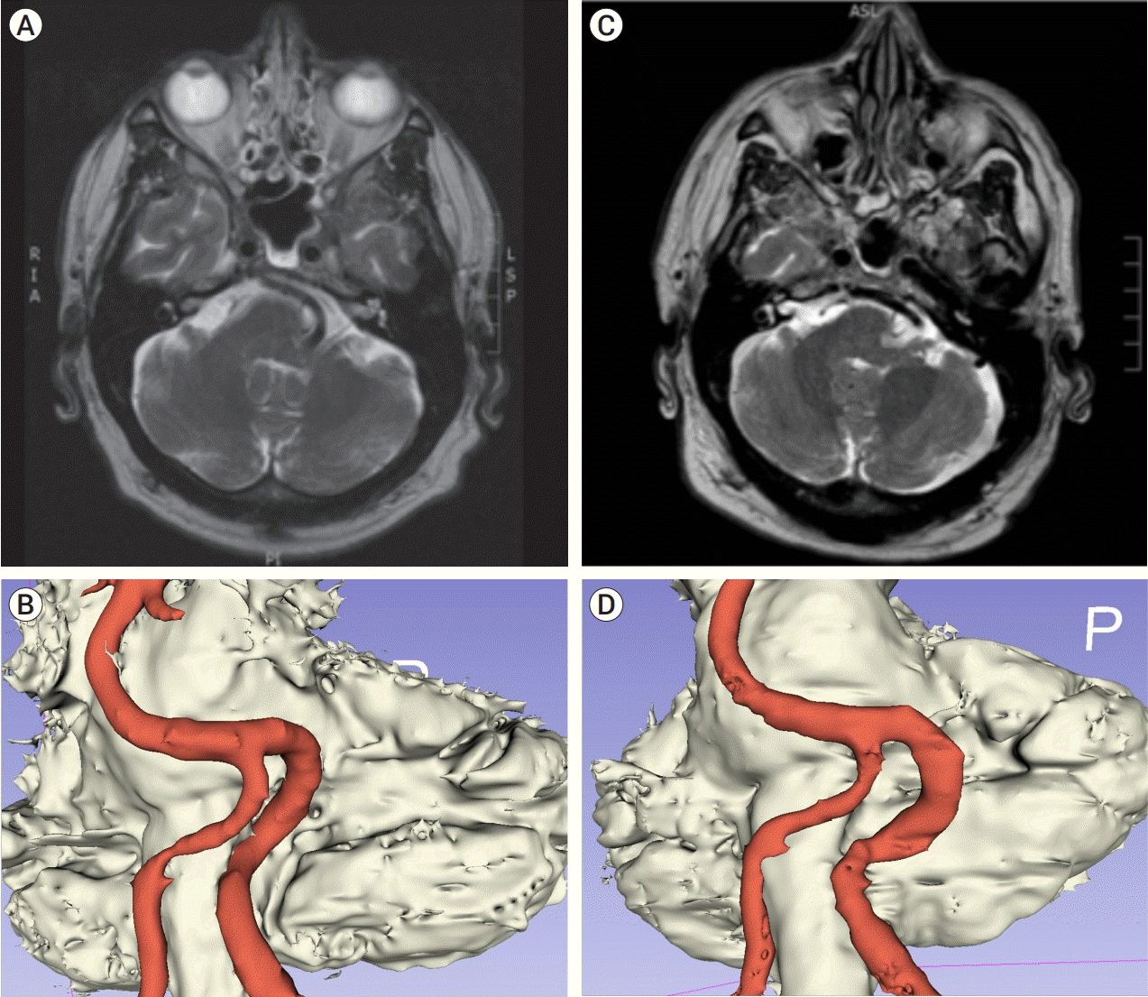
A 67-year-old male with hypertension presented with a two-and-a-half-year history of gait imbalance (described as veering to the left) and dysmetria. On examination he had endgaze nystagmus and bilateral finger-to-nose dysmetria. Brain magnetic resonance imaging (MRI) was obtained that revealed vertebrobasilar dolichoectasia with brainstem deformation of both the pons and medulla by the left vertebral artery (Fig. 1A, B). He underwent a leftsided far lateral craniectomy for mobilization of the left vertebral artery via sling that was sutured to the dura of the occipital condyle. The patient tolerated the surgery with no complications. All of his preoperative symptoms resolved. Immediate post-operative MRI confirmed decompression of the brainstem (Fig. 1C, D).
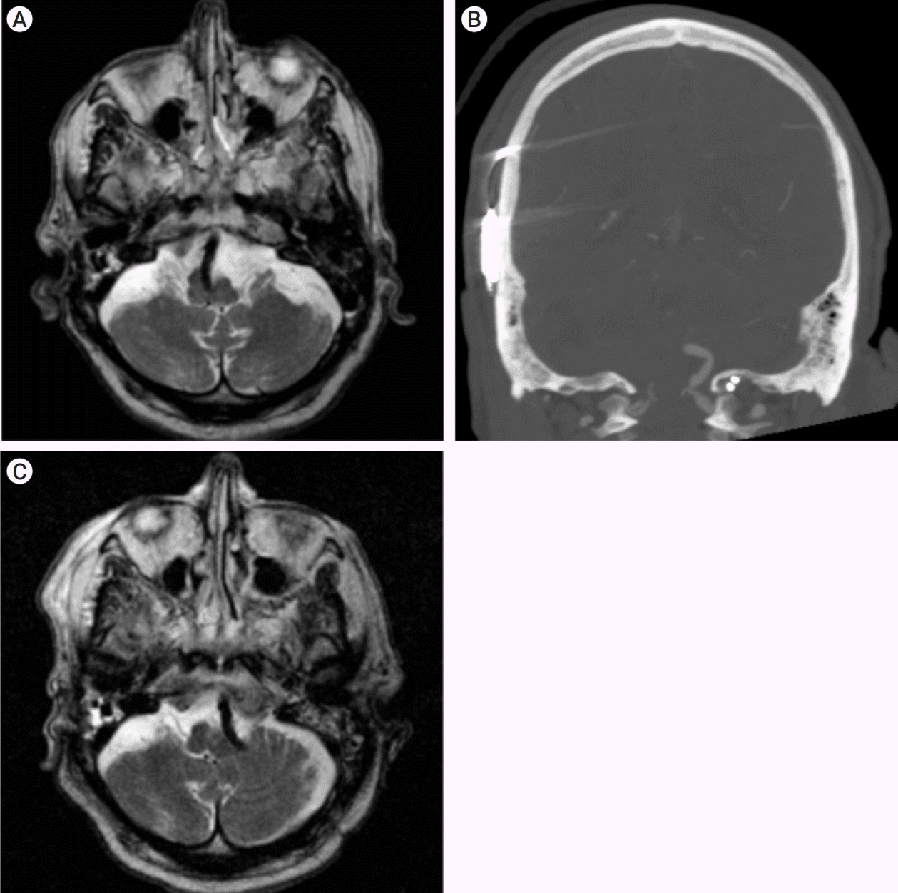
A 79-year-old male with hypertension, stage III chronic kidney disease, and sensorineural hearing loss presented with 6 months of subacute progressive gait instability, right hemiparesis, and dysphagia. He rapidly developed left facial weakness and respiratory failure. He was intubated with inability to wean of a ventilator. Imaging studies demonstrated left vertebral artery dolichoectasia causing brainstem compression at the level of the medulla (Fig. 2A). He underwent a left-sided far lateral craniectomy. The left vertebral artery was mobilized with a sling and secured to the ipsilateral occipital condyle with cranial fixation screws (Fig. 2B). The patient tolerated surgery and post-operative imaging confirmed decompression of the brainstem (Fig. 2C). Post-operatively, his symptoms improved significantly.
The patient is positioned three-quarter prone. Electrophysiology is used to monitor the lower cranial nerves, brainstem auditory evoked potentials, somatosensory evoked potentials, and motor evoked potentials throughout the operation. A lazy “S” shaped incision is utilized. The advantage of this incision, compared to a hockey stick incision, is improved visualization given that the surgical field is clear of bulky cervical musculature. A large, far-lateral craniectomy is performed. This enables access from the pons to the cervicomedullary junction with access to cranial nerves V to XII. Wide arachnoid dissection around the affected cranial nerves is crucial to decrease the possibility of a retraction injury.
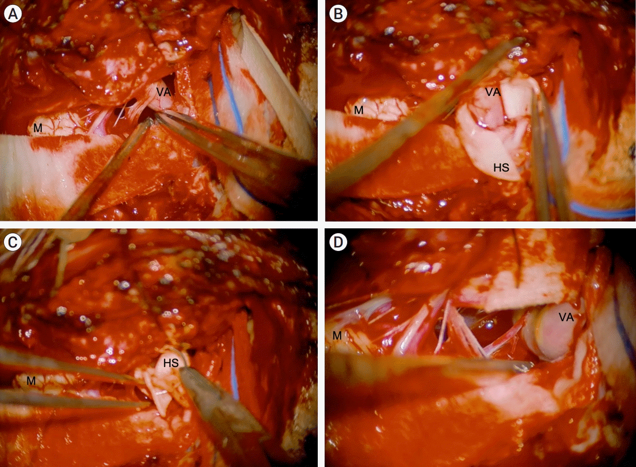
The dolichoectatic vertebral artery is identified and dissected away from the brainstem and the lower cranial nerves using sharp and blunt microsurgical techniques (Fig. 3A). Patties are placed in the epipial space to develop and preserve this plane. Hemashield bovine pericardium graft (Atrium Medical Corporation, Hudson, NH, USA) is wrapped around the offending vessel to create a sling (Fig. 3B). The two ends of the sling are sutured together using 4-0 Surgilon suture (Medtronic, Minneapolis, MN, USA) (Fig. 3C). The sling is then fixed either to the occipital condyle using cranial plating 4 mm screws (DePuy Synthes, West Chester, PA, USA) or to the dura of the occipital condyle using a 2-0 silk suture. The vertebrobasilar system, cranial nerves, and brainstem are observed for no iatrogenic compression or kinking (Fig. 3D). Doppler ultrasonography is used to confirm vessel flow after placement of the sling. Neuromonitoring is checked to confirm no changes in signals.
Closure is achieved by approximation of the dural leaflets and on lay duraplasty. Cranioplasty is performed. Cervical muscles and skin are closed in layers.
Even though rare, vertebrobasilar dolichoectasia can cause symptoms secondary to compression of the brainstem and cranial nerves. In a 2008 study, of 156 patients with vertebrobasilar dolichoectasia, 56 presented with compressive symptoms with various involvement of the third, fifth, sixth, seventh, eighth, and bulbar nerves [7]. Different surgical techniques have been described previously for management of vascular compression including an anteromedial directed muslin sling with clip-assisted tethering to the clival dura [2], a transcondylar fossa approach for laterally slinging the vertebral artery using a sling stitched to the dura of the jugular tubercle [6], and a ‘strip-clip’ technique developed by Raabe, et al., using an equine collagen strip and mini-aneurysm clips to decompress the vertebral artery off the brainstem [9]. Additionally, hemifacial spasm has been relieved with a Gore-Tex sling to the petrous ridge [5]. Also, vessel sectioning and bypass has been described to decompress cranial nerves [10]. Here, we describe a novel surgical technique of anterolateral mobilization of the vertebral artery. This technique allows for use of a suture to the dura of the occipital condyle or a cranial fixation screw to the condyle itself for anchor. This anchor is sturdy and safe operative technique when compared to other published examples. For medullary compression seen in basilar dolichoectasia, the anterolateral direction allows for decompression by placing the offending vasculature into the lateral cerebromedullary cistern as described above.
The Hemashield graft is composed of woven polyester and bovine collagen. It is a thin, knitted graft, which is easy to handle and provides sufficient tensile strength for mobilization without harming or inciting inflammation of the target vessel. This method allows the surgeon an additional tool for safe, quick and easy decompression of the cervicomedullary junction in an anterolateral direction towards the occipital condyle.
Multiple techniques are available for microvascular decompression, while less exist for macrovascular decompression. Depending on the anatomical location of the offending vessel and affected neurological structures, the neurosurgeon should select the best option to achieve complete decompression. Here, we present a novel surgical technique for management of vertebrobasilar dolichoectasia with brainstem compression.
REFERENCES
1. Baldauf J, Rosenstengel C, Schroeder HWS. Nerve compression syndromes in the posterior cranial fossa. Dtsch Arztebl Int. 2019; Jan. 116(4):54–60.

2. Choudhri O, Connolly ID, Lawton MT. Macrovascular decompression of the brainstem and cranial nerves: evolution of an anteromedial vertebrobasilar artery transposition technique. Neurosurgery. 2017; Aug. 81(2):367–76.

4. Lin CF, Chen HH, Hernesniemi J, Lee CC, Liao CH, Chen SC, et al. An easy adjustable method of ectatic vertebrobasilar artery transposition for microvascular decompression. Clin Neurol Neurosurg. 2012; Sep. 114(7):951–6.

5. Munich SA, Morcos JJ. “Macrovascular” decompression of dolichoectatic vertebral artery causing hemifacial spasm using goretex sling: 2-dimensional operative video. Oper Neurosurg (Hagerstown). 2019; Feb. 16(2):267–8.

6. Nakahara Y, Kawashima M, Matsushima T, Kouguchi M, Takase Y, Nanri Y, et al. Microvascular decompression surgery for vertebral artery compression of the medulla oblongata: 3 cases with respiratory failure and/or dysphagia. World Neurosurg. 2014; Sep-Oct. 82(3-4):535.e11–6.

7. Passero SG, Rossi S. Natural history of vertebrobasilar dolichoectasia. Neurology. 2008; Jan. 70(1):66–72.

8. Pico F, Labreuche J, Amarenco P. Pathophysiology, presentation, prognosis, and management of intracranial arterial dolichoectasia. Lancet Neurol. 2015; Aug. 14(8):833–45.

Fig. 1.
(A) Pre-operatively obtained axial, T2-weighted MRI image showing significant left brainstem compression by the left vertebral artery. (B) 3D reconstruction of the pre-operative MRI demonstrating severe pontomedullary compression with indentation. (C) Post-operative axial, T2-weighted MRI with decompression of the brainstem with the left vertebral artery iatrogenically displaced anterolaterally. (D) 3D reconstruction of the post-operative MRI demonstrating decompression with residual pontomedullary indentation. MRI, magnetic resonance imaging

Fig. 2.
(A) Pre-operative obtained axial, T2-weighted MRI showing significant medullary compression from the left vertebral artery. (B) Post-operative coronal computed tomography angiography (CTA) showing placement of cranial fixation screws into the occipital condyle with adjacent displaced vertebrobasilar system. (C) Post-operative axial, T2-weighted MRI with substantial decompression of the brainstem with the left vertebral artery iatrogenically displaced anterolaterally. MRI, magnetic resonance imaging

Fig. 3.
(A) Intra-operative image demonstrating the medulla (M) with the vertebral artery (VA) being mobilized by closed forceps prior to (B) Intra-operative image showing the VA being wrapped in the Hemashield sling (HS). (C) Intra-operative image of the loose ends of the sling being stitched together by a single suture. (D) Intra-operative picture of the sling mobilizing the VA away from the medulla after anchoring to the dura of the occipital condyle.


 PDF
PDF Citation
 Print
Print


 XML Download