Journal List > Korean J Anesthesiol > v.75(5) > 1516079499

Nam, Chin, Kang, Kim, Joung, and Choi: Prognostic value of left ventricular apical four-chamber longitudinal strain after heart valve surgery in real-world practice

Abstract

Background

Left ventricular longitudinal strain is an emerging marker of ventricular systolic function. However, the prognostic value of apical four-chamber longitudinal strain after heart valve surgery in real-world clinical practice is uncertain. The authors investigated whether left ventricular apical four-chamber longitudinal strain measured in real-world practice is helpful for predicting postoperative outcomes in patients undergoing heart valve surgery.

Methods

This observational cohort study was conducted in patients who underwent heart valve surgery between January 2014 and December 2018 at a tertiary hospital in South Korea. The exposure of interest was preoperative left ventricular apical four-chamber longitudinal strain. The primary outcome was postoperative all-cause mortality.

Results

Among 1,773 study patients (median age, 63 years; female, 45.9%), 132 (7.4%) died during a median follow-up of 27.2 months. Preoperative left ventricular apical four-chamber longitudinal strain was significantly associated with all-cause mortality (adjusted hazard ratio, 0.94 per 1% increment in absolute value; 95% CI [0.90, 0.99], P = 0.022), whereas left ventricular ejection fraction (LVEF) was not significantly associated with all-cause mortality (adjusted hazard ratio: 1.01, 95% CI [0.99, 1.03], P = 0.222). Moreover, combining left ventricular apical four-chamber longitudinal strain to the LVEF and conventional prognostic factors enhance the prognostic model for all-cause mortality (P = 0.022).

Conclusions

In patients undergoing heart valve surgery without coronary artery disease, left ventricular apical four-chamber longitudinal strain measured in real-world clinical practice was independently associated with postoperative survival. Left ventricular longitudinal strain measurement may be helpful for outcome prediction after valve surgery.

Introduction

In patients with advanced valvular heart disease, surgical treatment is one of the key management options. However, considering that cardiac surgery entails substantial operative risks, accurate prediction of both the risks and benefits of surgery in each patient is crucial. Evaluating the left ventricular systolic function carries a vital role in making treatment decisions; specifically, left ventricular ejection fraction (LVEF) has been a cornerstone in determining surgical intervention and risk prediction [14].
Left ventricular longitudinal strain has recently gained interest as a marker of left ventricular systolic function. Strain is a mechanical term representing the degree of deformation relative to the material’s reference length; accordingly, left ventricular longitudinal strain directly reflects longitudinal myocardial shortening during a cardiac cycle. Recently, left ventricular longitudinal strain has shown significant prognostic value in a variety of cardiac diseases, such as heart failure [5,6], acute myocardial infarction [7], and cardiomyopathy [8].
In terms of valvular surgery, several studies have shown that left ventricular longitudinal strain was independently associated with long-term postoperative survival [9,10]. However, as previous studies were exclusively conducted in patients with mitral regurgitation (MR), the prognostic value of left ventricular longitudinal strain in patients with other types of heart conditions is less clear. Furthermore, strain analyses in previous studies [9,10] were performed post-hoc using stored echocardiography data for research purposes. Hence, it is unclear whether left ventricular longitudinal strain can confer significant incremental prognostic values over conventional risk factors in real-world clinical settings. In addition, while the above-mentioned studies used global left ventricular longitudinal strain, several reports have demonstrated the feasibility and reliability of left ventricular apical four-chamber longitudinal strain [11,12].
Thus, we investigated whether left ventricular apical four-chamber longitudinal strain measured in clinical practice can be helpful for predicting postoperative survival in patients with various types of valvular heart diseases including MR. We also examined the predictive value of left ventricular longitudinal strain for postoperative complications.

Materials and Methods

Design and participants

This observational cohort study was conducted at a tertiary hospital (Asan Medical Center) in South Korea. All patients who underwent heart valve surgery at our institution between January 2014 and December 2018 were screened for eligibility. We excluded patients under 20 years of age, those who underwent urgent or emergent surgery, and those who underwent combined coronary artery bypass surgery. Patients who did not undergo left ventricular longitudinal strain analysis preoperatively were also excluded.
The Institutional Review Board (AMC IRB no. 2020-1630) approved the study protocol and waived the need for informed consent considering the retrospective nature of the study. Clinical data of the study population was collected from the electronic medical record and institutional echocardiography database. This study was conducted in accordance with the Strengthening the Reporting of Observational Studies in Epidemiology (STROBE) Statement [13].

Echocardiography and strain analysis

All candidates for heart valve surgery were preoperatively evaluated with transthoracic echocardiography using standard machines and techniques in accordance with the American Society of Echocardiography guidelines [14]. At our institution, the incorporation of left ventricular longitudinal strain measurement as a part of transthoracic echocardiography began in late 2013. This new policy encouraged the provision of formal left ventricular longitudinal strain reporting. In the initial phase of the left ventricular longitudinal strain reporting, speckle tracking analysis was performed with EchoPAC (GE Healthcare, USA) or QLAB (Philips Healthcare, The Netherlands) according to the availability of ultrasound machines. Afterward, they have been replaced by a vendor-independent software, Image-ArenaTM (TomTec, Germany) since 2015. In this transitional phase, only the apical four-chamber longitudinal strain was measured and reported, thus highlighting the expansion of strain reporting against resource limitation. Strain measurements were performed by experienced sonographers. After acquiring an adequate apical four-chamber view, the region of interest is automatically traced by strain software. Endocardial border tracing was manually adjusted if appropriate. A four-chamber strain curve throughout the cardiac cycle was derived and peak longitudinal strain value was calculated from the average of the six segments. This strain reporting policy was phased in over six years, and the final strain analysis implementation with global longitudinal strain was adopted in 2020; however, the data acquired in the final phase was not included in this study.

Study exposure and outcomes

The primary exposure of this study was left ventricular four-chamber longitudinal strain measured from the last echocardiography prior to heart valve surgery. In its original definition, left ventricular longitudinal strain has a negative value; however, in this study, we converted the left ventricular longitudinal strain to an absolute value for a more straightforward interpretation.
In the primary analysis, the outcome was all-cause mortality after surgery. The mortality data were obtained from our medical record and the National Health Insurance status. The data on the survival status were collected until July 31, 2020. Patients who survived over five years were censored at five years, and those who underwent redo-cardiac surgery were censored at the time of redo-surgery. The secondary outcome was operative morbidity defined by the Society of Thoracic Surgeons risk calculator (i.e., composite of operative mortality, stroke, renal failure, prolonged ventilation, mediastinitis/deep sternal wound infection, and reoperation). The detailed definitions of the secondary outcomes are provided in Supplementary Table 1.

Statistical analysis

Sample size was driven by all eligible patients from 2014 to 2018. Missing values were replaced with mode or median. Categorical variables are presented as frequency (proportion), and continuous variables are presented as mean ± SD or median (Q1, Q3). Comparison of descriptive statistics between groups was performed with chi-square test for categorical variables and Student’s t-test or Mann–Whitney U test for continuous variables according to the normality of the data. Correlation between continuous variables was assessed with Pearson’s or Spearman’s correlation test depending on the normality of the variables.
In order to determine the association between predictors and outcomes, univariate Cox proportional hazard regression analysis and univariate logistic regression were performed for the primary outcome and the secondary outcome, respectively. For continuous variables, the univariate association with outcomes was explored using the restricted cubic spline. If there were significant non-linear relationships between continuous variables and the study outcomes, the variables were transformed or categorized as appropriate.
To examine the independent associations between left ventricular longitudinal strain and outcomes, multivariable regression analyses were performed. Multivariable Cox proportional models for the primary outcome were adjusted for LVEF, age, sex, Charlson Comorbidity Index (CCI), pulmonary hypertension, mitral stenosis (MS), MR, aortic stenosis (AS), aortic regurgitation (AR), tricuspid regurgitation (TR), New York Heart Association (NYHA) classification, and atrial fibrillation. Multivariable logistic regression for the secondary outcome was adjusted for LVEF, age, sex, CCI, redo-surgery, pulmonary hypertension, body mass index, hematocrit, hypertension, MS, MR, AS, AR, TR, smoking, combined surgery, NYHA classification, and atrial fibrillation. Possible confounders from background knowledge were selected as adjusted variables.
To assess the incremental value of left ventricular longitudinal strain as a prognostic factor, the likelihood test was used to compare the prediction performance between models with and without left ventricular longitudinal strain. Additional interaction analyses were performed to evaluate the effect-modification of left ventricular longitudinal strain according to prespecified subgroups (LVEF ≥ 50% or < 50%; patients with or without MR). Two sensitivity analyses—multivariable Cox regression including other echocardiographic parameters as potential confounders and multivariable logistic regression with different outcome definitions—were performed, and their details are provided in Supplementary Table 2.
We also performed post-hoc analyses to obtain more straightforward interpretations of our results. These post-hoc analyses categorized preoperative left ventricular function according to left ventricular longitudinal strain and LVEF (LVEF ≥ 50% and left ventricular longitudinal strain ≥ 16.3% vs. LVEF ≥ 50% and left ventricular longitudinal strain < 16.3% vs. LVEF < 50%). For the comparison of patients with preserved and reduced LVEF, a cut-off value of 50% was used [15]. A cut-off value for the left ventricular longitudinal strain was based on the median value of the population (16.3%). Multivariable Cox regression, logistic regression, and Kaplan–Meier survival curve analyses were used as appropriate.
All statistical analyses were two-tailed with a significance level of 0.05. Statistical analysis was performed with R version 4.0.3 (R Foundation for Statistical Computing, Austria).

Results

Patient population and characteristics

A total of 3,666 patients underwent heart valve surgery at our institution during the study period. Of them, 1,773 were included in the final analysis (Supplementary Fig. 1A). The leading cause of exclusion was the absence of strain analysis, which was primarily due to the low availability of strain during the early study period. The proportion of strain reporting has gradually increased, with 92% of patients in 2018 having strain results (Supplementary Fig. 1B).
The baseline characteristics of the study patients are shown in Table 1. The median age was 63 years (interquartile range [IQR], 54–70), and 45.9% were female. The median LVEF was 61% (IQR, 56%–65%), and the median left ventricular longitudinal strain was 16.3% (IQR, 13.2%–19.0%). Left ventricular longitudinal strain and LVEF had a moderate degree of positive correlation (Spearman’s ρ = 0.56, P < 0.001). At each level of the LVEF, the left ventricular longitudinal strain had a broad distribution, especially in higher LVEF levels (Fig. 1). The majority (92.4%) of left ventricular longitudinal strain data were analyzed with Image-ArenaTM (TomTec). The median (Q1, Q3) time interval between preoperative transthoracic echocardiography and surgery was 23 (7, 56) days.

Primary analysis: postoperative all-cause mortality

During a median follow-up of 27.2 months (19.1, 38.9), 132 (7.4%) patients died. Patients who survived had higher preoperative left ventricular longitudinal strain values than did non-survivors (16.4 [13.4, 19.2] vs. 14.9 [11.5, 17.2], P < 0.001; Table 1); in contrast, the preoperative LVEF was not significantly different between the survivors and non-survivors (61.0 [56.0, 66.0] vs. 61.0 [52.0, 65.0], P = 0.071). Univariate associations between left ventricular longitudinal strain, LVEF, and all-cause mortality are shown in Supplementary Fig. 2. While left ventricular longitudinal strain had a statistically significant linear negative relationship with all-cause mortality (P value for univariate Cox regression with restricted cubic spline = 0.005, P value for non-linearity = 0.062), LVEF did not show a significant relationship with all-cause mortality (P value for univariate Cox regression with restricted cubic spline = 0.17).
The negative relationship between left ventricular longitudinal strain and all-cause mortality remained statistically significant in a multivariable-adjusted Cox proportional hazard model (Fig. 2A, Table 2). On the contrary, LVEF did not have a statistically significant relationship with all-cause mortality (Fig. 2B, Supplementary Table 3). Combining left ventricular longitudinal strain to the conventional prognostic factors (i.e., age, sex, CCI, PHTN, MS, MR, AS, AR, TR, NYHA class, atrial fibrillation) and LVEF significantly enhanced the prognostic model for all-cause mortality (P = 0.022; Fig. 2C).

Secondary analysis: operative morbidity

During index hospitalization or within 30 days postoperatively, 251 (14.2%) had operative morbidity; of them, 40 (2.3%) patients died, 175 (9.9%) had prolonged mechanical ventilation or reintubation, 81 (4.6%) underwent reoperation, 60 (3.5%) had renal failure, 40 (2.3%) had a stroke, and 11 (0.6%) had mediastinitis or deep sternal wound infection.
Descriptive statistics according to the occurrence of morbidity are presented in Supplementary Table 4. Patients with operative morbidity had a lower value of left ventricular longitudinal strain than those without morbidity (15.1 [12.8, 18.0] vs. 16.5 [13.4, 19.1], P < 0.001). LVEF was also lower in patients with morbidity than those without (60.0 [52.5, 64.0] vs. 62.0 [56.0, 66.0], P < 0.001).
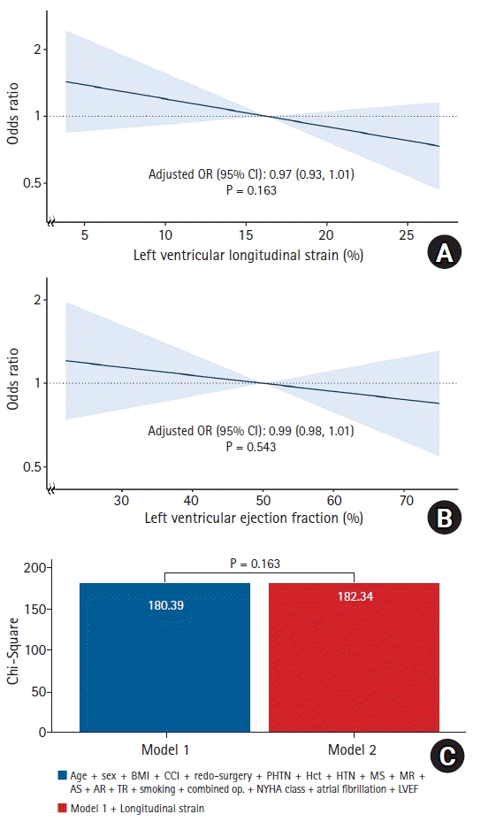
Univariate analysis showed that both left ventricular longitudinal strain and LVEF had negative linear relationships with operative morbidity (Supplementary Fig. 3, P values for univariate logistic regression with restricted cubic spline < 0.05). After adjusting for potential confounders, the negative relationship between LVEF and operative morbidity remained statistically significant (odds ratio [OR]: 0.99, 95% CI: 0.97, 1.00, P = 0.049; Supplementary Table 5); however, after further adjustment with left ventricular longitudinal strain, the association between LVEF and operative morbidity was no longer statistically significant (OR: 0.99, 95% CI: 0.98, 1.01, P = 0.543; Fig. 3B). Likewise, left ventricular longitudinal strain did not have a statistically significant relationship with operative morbidity in this final model (OR: 0.97, 95% CI: 0.93, 1.01, P = 0.163; Fig. 3A, Table 2). Furthermore, combining left ventricular longitudinal strain to the LVEF and conventional risk factors did not show a significant incremental prognostic value for predicting operative morbidity (Fig. 3C).

Subgroup and sensitivity analysis

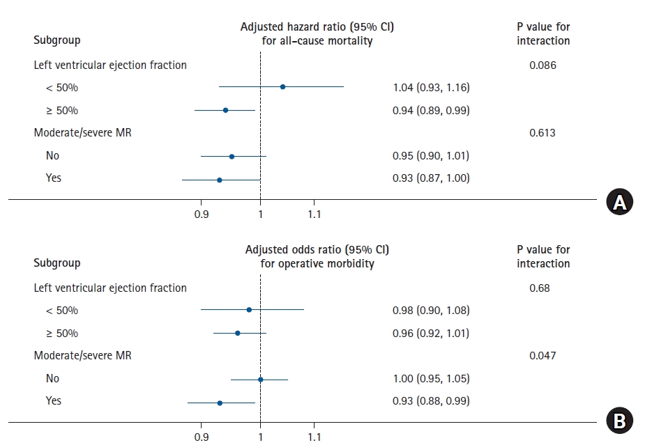
In the subgroup analysis, the association between left ventricular longitudinal strain and all-cause mortality was different across different levels of LVEF, albeit without statistical significance (P value for interaction = 0.086; Fig. 4A, Table 2). The presence of moderate/severe MR did not significantly alter the relationship between left ventricular longitudinal strain and all-cause mortality as well (P value for interaction = 0.613). On the contrary, there was a significant interaction between left ventricular longitudinal strain and the presence of moderate/severe MR in terms of operative morbidity (P value for interaction = 0.047), as a conditional negative relationship between left ventricular longitudinal strain and operative morbidity was shown in the moderate/severe MR group (Fig. 4B, Table 2).
Sensitivity analyses showed similar results to the main analyses in terms of the relationships of left ventricular longitudinal strain and LVEF with all-cause mortality and operative morbidity. The results are shown in Supplementary Table 69.

Post-hoc survival analysis according to the left ventricular longitudinal strain and LVEF strata

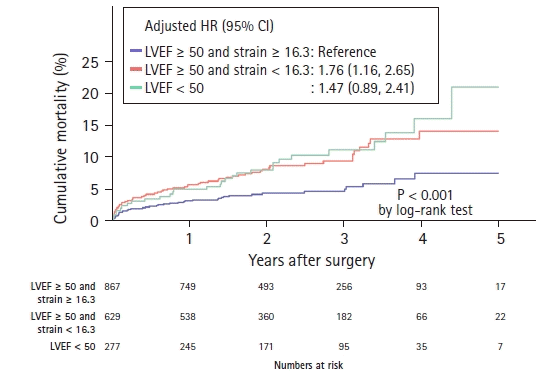
All-cause mortality according to the left ventricular longitudinal strain and LVEF strata (LVEF ≥ 50% and left ventricular longitudinal strain ≥ 16.3% vs. LVEF ≥ 50% and left ventricular longitudinal strain < 16.3% vs. LVEF < 50%) is shown in Fig. 5. Patients with preserved LVEF (≥ 50%) and normal left ventricular longitudinal strain (≥ 16.3%) had the lowest risk of death. Moreover, patients with preserved LVEF and low left ventricular longitudinal strain (< 16.3%) had a significantly high mortality rate was comparable to that of patients with low LVEF. Patients with preserved LVEF and normal left ventricular longitudinal strain also had the lowest risk of operative morbidity. Patients with preserved LVEF and low left ventricular longitudinal strain (adjusted OR: 1.56, 95% CI: 1.12, 2.16, P = 0.009) and patients with low LVEF (adjusted OR: 1.74, 95% CI: 1.15, 2.65, P= 0.009) had significantly higher risks of operative morbidity.

Discussion

In this observational study of 1,773 patients who underwent heart valve surgery, we showed that left ventricular longitudinal strain was significantly associated with all-cause mortality after surgery. Furthermore, left ventricular longitudinal strain had incremental value for predicting all-cause mortality beyond previously known risk factors including LVEF. However, left ventricular longitudinal strain did not provide a significant benefit over LVEF in predicting operative morbidity.
Left ventricular longitudinal strain is an emerging parameter of systolic function. Previous studies constantly reported that left ventricular longitudinal strain was a valuable predictor of long-term mortality in a variety of cardiac diseases. In terms of valvular surgery, left ventricular longitudinal strain was also independently associated with long-term survival and had incremental prognostic value beyond LVEF [9,10]. However, most of the existing studies only included patients who underwent surgery to correct MR. In patients with significant MR, the LVEF is a limited parameter of the systolic function because LVEF is overestimated due to the regurgitant fraction. Therefore, the impaired systolic function may be masked in the preoperative LVEF, and become overt after mitral valve surgery. In contrast, left ventricular longitudinal strain directly reflects the myocardial shortening and is less dependent on loading conditions than LVEF [16]. In this respect, left ventricular longitudinal strain may be a superior parameter of systolic function to LVEF in patients with MR. Indeed, the correlation between preoperative left ventricular longitudinal strain and immediate postoperative LVEF was stronger than that between preoperative LVEF and immediate postoperative LVEF [17,18]. The pronounced association between left ventricular longitudinal strain and operative morbidity in patients with MR in our study further supports the prognostic value of left ventricular longitudinal strain in patients with MR.
Notably, the significant relationship between left ventricular longitudinal strain and long-term mortality shown in this study was not limited to patients with MR. Left ventricular longitudinal strain is regarded to detect subtle left ventricular dysfunction, which LVEF cannot detect. Longitudinal myocardial fibers, which are predominantly presented in the subendocardial layer, are more vulnerable to injury than oblique and circumferential fibers [1921]. Thus, longitudinal shortening can deteriorate in the early stage of valve disease. In contrast, compensatory ventricular remodeling can lead to preserved LVEF until the manifestation of overt myocardial damage [2224]. Accordingly, our results also showed that a substantial proportion of patients had impaired left ventricular longitudinal strain while having an LVEF of above 50%. Furthermore, we showed that left ventricular longitudinal strain can differentiate long-term survival among patients with preserved LVEF. Thus, our results also support the current concept that left ventricular longitudinal strain can detect subtle myocardial dysfunction, which can impact the clinical outcomes.
In patients undergoing valve surgery, it should also be considered that left ventricular longitudinal strain may reflect not only the negative myocardial impact of valve diseases but also the reversibility of myocardial damage. For example, Kim et al. [10] reported that patients with preserved left ventricular longitudinal strain had a more significant reduction of left ventricular end-diastolic diameter after mitral valve surgery. Another study also showed that preoperative left ventricular longitudinal strain was associated with remodeling status three months after mitral valve replacement [25]. Thus, impairments in left ventricular longitudinal strain may imply a low likelihood of reverse remodeling after surgery. This is especially important considering that early intervention before the occurrence of irreversible myocardial damage may lead to better survival outcomes. As the aforementioned studies have been conducted in patients with MR, it is unknown whether reverse remodeling can differ according to preoperative left ventricular longitudinal strain in other valve diseases. Our results also do not provide direct evidence on this topic, and further studies are needed to test this hypothesis.
In contrast to long-term survival, left ventricular longitudinal strain did not have a significant incremental predictive value above LVEF regarding operative morbidity. Instead, the statistical significance of LVEF disappeared after left ventricular longitudinal strain was incorporated into the multivariable model. Thus, our results did not support incorporating left ventricular longitudinal strain as a predictor of operative morbidity. Left ventricular longitudinal strain may have limited role in specific situations such as the presence of significant MR.
Our study has several limitations. First, this study is from a single tertiary referral center and the study population exclusively consisted of Asian patients. Thus, our findings should be validated in different clinical settings. Second, more than one-third of the eligible patients did not have strain measurements. This is presumed to be largely due to the limited availability of strain analysis in the initial phase of the introduction of the strain measurement, but we cannot preclude the possibility of other patient-specific reasons, such as suboptimal endocardial tracing, atrial fibrillation, and tachycardia. Finally, the strain analysis in our study had a few practical limitations. All values were from the strain adaptation period in clinical practice; accordingly, a tradeoff between clinical feasibility and measurement precision was inevitable. There were heterogeneities in the vendors and versions of the strain softwares used. Also, the experience levels of the sonographers might have been different and interobserver variability also existed. However, in the mid-2010s, inter-vendor variability decreased to a level similar to conventional echocardiogram parameters [26,27]. Also, the reproducibility of left ventricular longitudinal strain was better than that of LVEF, and competency could be achieved with a short learning curve [26,28,29].
Nevertheless, the most critical limitation of our strain measurement was that left ventricular longitudinal strain values were not a global one and obtained from apical four-chamber view. Left ventricular global longitudinal strain is the standard method that averages four, two, and three-chamber longitudinal strain. Thus, some valuable prognostic information from other views might have been ignored in the apical four-chamber longitudinal strain. Nevertheless, a few studies advocated the apical four-chamber longitudinal strain. For example, Alenzi et al. [12] reported that there was little difference between apical four-chamber longitudinal strain with global longitudinal strain in patients with heart failure without regional wall motion abnormality (median difference, -0.03%; interquartile range, -0.3% to 0.27%; 95% pairwise difference < 2% in absolute magnitude). Similarly, a study of patients with moderate to severe AS showed that apical four-chamber longitudinal strain was in good agreement with global longitudinal strain (mean bias: –0.09%, 95% limits: –3.6 to 3.4%) [11]. Moreover, apical four-chamber longitudinal strain was independently associated with mortality, suggesting it may serve as a new prognostic factor for patients with AS [11]. Considering the above-mentioned studies and our findings, the apical four-chamber longitudinal strain measurement could be a useful alternative. As an example, apical four-chamber longitudinal strain could be implemented more easily in routine clinical practice, as we have experienced. It may be also useful as a substitute for global longitudinal strain when the imaging quality from apical two or three-chamber view is poor. Nevertheless, it should be noted that studies investigating apical four-chamber longitudinal strain excluded patients with regional wall motion abnormality or significant coronary artery disease. Also, the apical four-chamber longitudinal strain needs to be validated in other cardiac diseases, such as amyloidosis, congenital heart disease, etc.
Although the value of left ventricular longitudinal strain for risk prediction in a broad spectrum of cardiac diseases has been repeatedly studied, the widespread clinical implementation of left ventricular longitudinal strain has been slow [20]. This may be due to the lack of availability and concern about standardization [21,22]. However, our study showed that the gradual implementation of left ventricular longitudinal strain may be feasible. Moreover, even in its limited form, left ventricular longitudinal strain provided additional prognostic information in the actual clinical setting. Although further validation is needed, our results suggest that it may be worthwhile to implement strain analysis according to the availability of each institution while considering the current limitation of strain measurement described above. Newer systems that allow fully automated analysis may facilitate the clinical implementation of left ventricular longitudinal strain in the future. Along with the widespread clinical implementation of left ventricular longitudinal strain, a future pragmatic trial can answer whether strain-based surgical decisions can improve the clinical outcomes in patients with valve disease.
In patients undergoing heart valve surgery without coronary artery disease, apical four-chamber left ventricular longitudinal strain measured in a real-world clinical practice was independently associated with postoperative survival. Left ventricular longitudinal strain may be successfully implemented in clinical practice and aid the outcome prediction after valve surgery.

ACKNOWLEDGMENTS

We would like to thank Sehee Kim, Ph.D., from the Department of Clinical Epidemiology and Biostatistics of Asan Medical Center, for the professional help with the statistical analysis. We also thank Joon Seo Lim, Ph.D., ELS, from the Scientific Publications Team at Asan Medical Center for his editorial assistance in the preparation of the manuscript.

Notes

Funding

None.

Conflicts of Interest

No potential conflict of interest relevant to this article was reported.

Author Contributions

Jae-Sik Nam (Conceptualization; Data curation; Formal analysis; Investigation; Methodology; Writing – original draft)

Ji-Hyun Chin (Conceptualization; Investigation; Methodology; Supervision; Writing – review & editing)

Hyun-Uk Kang (Investigation)

Juyoun Kim (Data curation)

Kyoung-Woon Joung (Data curation)

In-Cheol Choi (Conceptualization; Supervision)

Supplementary Materials

Supplementary Fig. 1.
(A) Flow diagram of the study and (B) surgery case volume with or without preoperative strain analysis.
kja-22201-suppl1.pdf
Supplementary Fig. 2.
Unadjusted relationship between longitudinal strain, ejection fraction, and all-cause mortality using restricted cubic spline.
kja-22201-suppl2.pdf
Supplementary Fig. 3.
Unadjusted relationship between longitudinal strain, ejection fraction, and operative morbidity using restricted cubic spline.
kja-22201-suppl3.pdf
Supplementary Table 1.
Definitions of outcomes
kja-22201-suppl4.pdf
Supplementary Table 2.
Sensitivity analyses
kja-22201-suppl5.pdf
Supplementary Table 3.
Multivariate Cox regression analyses for all-cause mortality
kja-22201-suppl6.pdf
Supplementary Table 4.
Descriptive statistics according to the occurrence of the operative morbidity
kja-22201-suppl7.pdf
Supplementary Table 5.
Multivariate logistic regression analyses for all-cause mortality
kja-22201-suppl8.pdf
Supplementary Table 6.
Sensitivity analyses for all-cause mortality
kja-22201-suppl9.pdf
Supplementary Table 7.
Sensitivity analyses for MACCE
kja-22201-suppl10.pdf
Supplementary Table 8.
Sensitivity analyses for all-cause mortality
kja-22201-suppl11.pdf
Supplementary Table 9.
Sensitivity analyses for operative morbidity
kja-22201-suppl12.pdf

References

1. Nishimura RA, Otto CM, Bonow RO, Carabello BA, Erwin JP 3rd, Guyton RA, et al. 2014 AHA/ACC guideline for the management of patients with valvular heart disease: a report of the American College of Cardiology/American Heart Association Task Force on Practice Guidelines. J Am Coll Cardiol. 2014; 63:e57–185.
2. Baumgartner H, Falk V, Bax JJ, De Bonis M, Hamm C, Holm PJ, et al. 2017 ESC/EACTS Guidelines for the management of valvular heart disease. Eur Heart J. 2017; 38:2739–91.
3. Shahian DM, Jacobs JP, Badhwar V, Kurlansky PA, Furnary AP, Cleveland JC Jr, et al. The Society of Thoracic Surgeons 2018 Adult Cardiac Surgery Risk Models: part 1-background, design considerations, and model development. Ann Thorac Surg. 2018; 105:1411–8.
4. Nashef SA, Roques F, Sharples LD, Nilsson J, Smith C, Goldstone AR, et al. EuroSCORE II. Eur J Cardiothorac Surg. 2012; 41:734–44.
5. Park JJ, Park JB, Park JH, Cho GY. Global longitudinal strain to predict mortality in patients with acute heart failure. J Am Coll Cardiol. 2018; 71:1947–57.
6. Tröbs SO, Prochaska JH, Schwuchow-Thonke S, Schulz A, Müller F, Heidorn MW, et al. Association of global longitudinal strain with clinical status and mortality in patients with chronic heart failure. JAMA Cardiol. 2021; 6:448–56.
7. Ersbøll M, Valeur N, Mogensen UM, Andersen MJ, Møller JE, Velazquez EJ, et al. Prediction of all-cause mortality and heart failure admissions from global left ventricular longitudinal strain in patients with acute myocardial infarction and preserved left ventricular ejection fraction. J Am Coll Cardiol. 2013; 61:2365–73.
8. Liu H, Pozios I, Haileselassie B, Nowbar A, Sorensen LL, Phillip S, et al. Role of global longitudinal strain in predicting outcomes in hypertrophic cardiomyopathy. Am J Cardiol. 2017; 120:670–5.
9. Hiemstra YL, Tomsic A, van Wijngaarden SE, Palmen M, Klautz RJ, Bax JJ, et al. Prognostic value of global longitudinal strain and etiology after surgery for primary mitral regurgitation. JACC Cardiovasc Imaging. 2020; 13:577–85.
10. Kim HM, Cho GY, Hwang IC, Choi HM, Park JB, Yoon YE, et al. Myocardial strain in prediction of outcomes after surgery for severe mitral regurgitation. JACC Cardiovasc Imaging. 2018; 11:1235–44.
11. Salaun E, Casalta AC, Donal E, Bohbot Y, Galli E, Tribouilloy C, et al. Apical four-chamber longitudinal left ventricular strain in patients with aortic stenosis and preserved left ventricular ejection fraction: analysis related with flow/gradient pattern and association with outcome. Eur Heart J Cardiovasc Imaging. 2018; 19:868–78.
12. Alenezi F, Ambrosy AP, Phelan M, Chiswell K, Abudaqa L, Alajmi H, et al. Left ventricular global longitudinal strain can reliably be measured from a single apical four-chamber view in patients with heart failure. J Am Soc Echocardiogr. 2019; 32:317–8.
13. von Elm E, Altman DG, Egger M, Pocock SJ, Gøtzsche PC, Vandenbroucke JP; STROBE Initiative. The Strengthening the Reporting of Observational Studies in Epidemiology (STROBE) statement: guidelines for reporting observational studies. Int J Surg. 2014; 12:1495–9.
14. Mitchell C, Rahko PS, Blauwet LA, Canaday B, Finstuen JA, Foster MC, et al. Guidelines for performing a comprehensive transthoracic echocardiographic examination in adults: recommendations from the American Society of Echocardiography. J Am Soc Echocardiogr. 2019; 32:1–64.
15. Heidenreich PA, Bozkurt B, Aguilar D, Allen LA, Byun JJ, Colvin MM, et al. 2022 AHA/ACC/HFSA guideline for the management of heart failure: a report of the American College of Cardiology/American Heart Association Joint Committee on clinical practice guidelines. J Am Coll Cardiol. 2022; 79:e263–421.
16. Namazi F, van der Bijl P, Hirasawa K, Kamperidis V, van Wijngaarden SE, Mertens B, et al. Prognostic value of left ventricular global longitudinal strain in patients with secondary mitral regurgitation. J Am Coll Cardiol. 2020; 75:750–8.
17. de Isla LP, de Agustin A, Rodrigo JL, Almeria C, del Carmen Manzano M, Rodríguez E, et al. Chronic mitral regurgitation: a pilot study to assess preoperative left ventricular contractile function using speckle-tracking echocardiography. J Am Soc Echocardiogr. 2009; 22:831–8.
18. Pandis D, Sengupta PP, Castillo JG, Caracciolo G, Fischer GW, Narula J, et al. Assessment of longitudinal myocardial mechanics in patients with degenerative mitral valve regurgitation predicts postoperative worsening of left ventricular systolic function. J Am Soc Echocardiogr. 2014; 27:627–38.
19. Smiseth OA, Torp H, Opdahl A, Haugaa KH, Urheim S. Myocardial strain imaging: how useful is it in clinical decision making? Eur Heart J. 2016; 37:1196–207.
20. Liu JE, Barac A, Thavendiranathan P, Scherrer-Crosbie M. Strain imaging in cardio-oncology. JACC CardioOncol. 2020; 2:677–89.
21. Marwick TH, Shah SJ, Thomas JD. Myocardial strain in the assessment of patients with heart failure: a review. JAMA Cardiol. 2019; 4:287–94.
22. Fortuni F, Bax JJ, Delgado V. Changing the paradigm in the management of valvular heart disease: in addition to left ventricular ejection fraction, focus on the myocardium. Circulation. 2021; 143:209–11.
23. Kearney LG, Lu K, Ord M, Patel SK, Profitis K, Matalanis G, et al. Global longitudinal strain is a strong independent predictor of all-cause mortality in patients with aortic stenosis. Eur Heart J Cardiovasc Imaging. 2012; 13:827–33.
24. Ng AC, Delgado V, Bertini M, Antoni ML, van Bommel RJ, van Rijnsoever EP, et al. Alterations in multidirectional myocardial functions in patients with aortic stenosis and preserved ejection fraction: a two-dimensional speckle tracking analysis. Eur Heart J. 2011; 32:1542–50.
25. Cho EJ, Park SJ, Yun HR, Jeong DS, Lee SC, Park SW, et al. Predicting left ventricular dysfunction after surgery in patients with chronic mitral regurgitation: assessment of myocardial deformation by 2-dimensional multilayer speckle tracking echocardiography. Korean Circ J. 2016; 46:213–21.
26. Farsalinos KE, Daraban AM, Ünlü S, Thomas JD, Badano LP, Voigt JU. Head-to-head comparison of global longitudinal strain measurements among nine different vendors: the EACVI/ASE inter-vendor comparison study. J Am Soc Echocardiogr. 2015; 28:1171–81, e2.
27. Yang H, Marwick TH, Fukuda N, Oe H, Saito M, Thomas JD, et al. Improvement in strain concordance between two major vendors after the strain standardization initiative. J Am Soc Echocardiogr. 2015; 28:642–8.e7.
28. Chan J, Shiino K, Obonyo NG, Hanna J, Chamberlain R, Small A, et al. Left ventricular global strain analysis by two-dimensional speckle-tracking echocardiography: the learning curve. J Am Soc Echocardiogr. 2017; 30:1081–90.
29. Negishi T, Negishi K, Thavendiranathan P, Cho GY, Popescu BA, Vinereanu D, et al. Effect of experience and training on the concordance and precision of strain measurements. JACC Cardiovasc Imaging. 2017; 10:518–22.
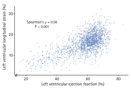
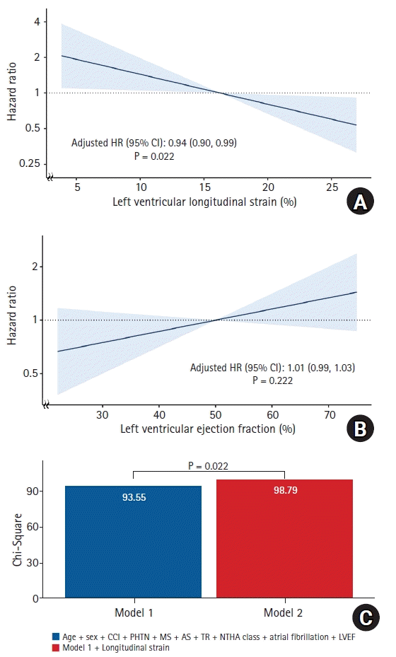
Fig. 1.
Relationship between left ventricular longitudinal strain and ejection fraction. A scatter plot showing the relationship between left ventricular longitudinal strain and ejection fraction. Spearman’s coefficient indicated a moderate correlation between left ventricular longitudinal strain and ejection fraction.
kja-22201f1.tif
Fig. 2.
Adjusted relationship of (A) left ventricular longitudinal strain, (B) LVEF with all-cause mortality, and (C) incremental value of left ventricular longitudinal strain for predicting all-cause mortality. (A, B) Solid lines represent adjusted hazard ratios and the shaded areas indicate 95% CIs. Left ventricular longitudinal strain of 16.3% and LVEF of 50% were used as references. Hazard ratios were estimated per 1% increase in left ventricular longitudinal strain or LVEF. (C) Bar plots represent the Chi-Square statistics of each model. P values are from the likelihood ratio test to compare the nested models (including conventional risk factors with or without left ventricular longitudinal strain). HR: hazard ratio, CCI: Charlson Comorbidity Index, PHTN: pulmonary hypertension, MS: mitral stenosis, AS: aortic stenosis, TR: tricuspid regurgitation, NYHA class: New York Heart Association Functional Classification, LVEF: left ventricular ejection fraction.
kja-22201f2.tif
Fig. 3.
Adjusted relationship between (A) left ventricular longitudinal strain, (B) LVEF with operative morbidity, and (C) incremental value of left ventricular longitudinal strain for predicting operative morbidity. (A, B) Solid lines represent the adjusted odds ratios, and the shaded areas indicate the 95% CIs. Left ventricular longitudinal strain of 16.3% and LVEF of 50% were used as references. The odds ratios were estimated per 1% increase in left ventricular longitudinal strain or LVEF. (C) Bar plots represent the Chi-Square statistics of each model. P values are from the likelihood ratio test to compare the nested models (including conventional risk factors with or without left ventricular longitudinal strain). BMI: body mass index, CCI: Charlson Comorbidity Index, PHTN: pulmonary hypertension, Hct: hematocrit, HTN: hypertension, MS: mitral stenosis, MR: mitral regurgitation, AS: aortic stenosis, AR: aortic regurgitation, TR: tricuspid regurgitation, NYHA class: New York Heart Association Functional Classification, LVEF: left ventricular ejection fraction.
kja-22201f3.tif
Fig. 4.
Subgroup analyses for (A) all-cause mortality and (B) operative morbidity. Dots indicate the (A) adjusted hazard ratio and (B) odds ratio. Horizontal lines represent 95% CI. MR: mitral regurgitation.
kja-22201f4.tif
Fig. 5.
Kaplan–Meier survival curve according to left ventricular longitudinal strain and LVEF strata. Kaplan–Meier curve for all-cause mortality. Preoperative left ventricular systolic function is categorized into three strata (EF ≥ 50% and left ventricular longitudinal strain ≥ 16.3% vs. EF ≥ 50% and left ventricular longitudinal strain < 16.3% vs. EF < 50%). The median value of left ventricular longitudinal strain (16.3%) was used as the cut-off value. HR: hazard ratio, LVEF: left ventricular ejection fraction.
kja-22201f5.tif
Table 1.
Baseline Characteristics of the Study Patients
Characteristics Total population (n = 1,773) Survivors (n = 1,641) Non-survivors (n = 132) P value
Age (yr) 63.0 (54.0, 70.0) 62.0 (53.0, 70.0) 69.0 (60.5, 75.0) < 0.001
Sex (F) 814 (45.9) 756 (46.1) 58 (43.9) 0.703
Body mass index (kg/m2) 24.0 (21.9, 26.2) 24.1 (22.0, 26.3) 22.5 (20.2, 25.2) < 0.001
Current smoker* 181 (10.2) 166 (10.1) 15 (11.4) 0.759
CCI 3.0 (1.0, 4.0) 3.0 (1.0, 4.0) 4.0 (3.0, 6.0) < 0.001
Hypertension 750 (42.3) 684 (41.7) 66 (50.0) 0.077
Pulmonary hypertension 722 (41.0) 649 (39.8) 73 (55.3) 0.001
Atrial fibrillation 640 (36.1) 578 (35.2) 62 (47.0) 0.009
NYHA class ≥ 2 1263 (73.7) 1157 (72.9) 106 (83.5) 0.013
Hematocrit (%) 39.0 (35.5, 42.2) 39.1 (35.8, 42.5) 35.0 (30.4, 39.8) < 0.001
Redo-surgery 216 (12.2) 189 (11.5) 27 (20.5) 0.004
Combined surgery 262 (14.8) 233 (14.2) 29 (22.0) 0.022
LVEF (%) 61.0 (56.0, 65.0) 61.0 (56.0, 66.0) 61.0 (52.0, 65.0) 0.071
Longitudinal strain (%) 16.3 (13.2, 19.0) 16.4 (13.4, 19.2) 14.9 (11.5, 17.2) < 0.001
MS§ 276 (15.6) 256 (15.6) 20 (15.2) 0.990
MR§ 608 (34.3) 564 (34.4) 44 (33.3) 0.884
AS§ 693 (39.1) 641 (39.1) 52 (39.4) 1.000
AR§ 497 (28.0) 458 (27.9) 39 (29.5) 0.763
TR§ 369 (20.8) 323 (19.7) 46 (34.8) < 0.001
LVEDD (mm) 54.0 (48.0, 61.0) 54.0 (48.0, 61.0) 53.0 (47.5, 59.0) 0.284
LVESD (mm) 35.0 (29.0, 42.0) 35.0 (29.0, 42.0) 35.0 (30.0, 42.0) 0.869
LAD (mm) 47.0 (40.0, 54.0) 47.0 (40.0, 54.0) 48.0 (42.5, 58.0) 0.017
Strain software vendor 0.857
 TomTec (ARENA) 1638 (92.4) 39 (2.4) 3 (2.3)
 Philips (Qlab) 93 (5.2) 85 (5.2) 8 (6.1)
 General Electric (EchoPAC) 42 (2.4) 1517 (92.4) 121 (91.7)

Values are presented as the number of patients (%) or median (Q1, Q3). CCI: Charlson Comorbidity Index, NYHA class: New York Heart Association Functional Classification, LVEF: left ventricular ejection fraction, MS: mitral stenosis, MR: mitral regurgitation, AS: aortic stenosis, AR: aortic regurgitation, TR: tricuspid regurgitation, LVEDD: left ventricular end-diastolic dimension, LVESD: left ventricular end-systolic dimension, LAD: left atrial dimension.

* Smoking history within eight weeks before surgery.

Variables with missing values: pulmonary hypertension (10/1773; 0.6%), atrial fibrillation (1/1773; 0.1%), NYHA class (59/1773; 3.3%), LAD (3/1773; 0.2%).

Mean pulmonary artery pressure ≥ 25 mmHg assessed by right heart catheterization, peak TR velocity ≥ 2.9 m/s, or early diastolic pulmonary regurgitation velocity > 2.2 m/s on preoperative echocardiography.

§ More than or equal to the moderate grade.

Table 2.
Relationship between Left Ventricular Longitudinal Strain and Clinical Outcomes
All-cause mortality Operative morbidity
Hazard ratio (95% CI) Odds ratio (95% CI)
Adjusted 0.94 (0.90, 0.99) 0.97 (0.93, 1.01)
Subgroup
 LVEF < 50% 1.04 (0.93, 1.16) 0.98 (0.90, 1.08)
 LVEF ≥ 50% 0.94 (0.89, 0.99) 0.96 (0.92, 1.01)
 Moderate/severe MR 0.93 (0.87, 1.00) 0.93 (0.88, 0.99)
 No moderate/severe MR 0.95 (0.90, 1.01) 1.00 (0.95, 1.05)

LVEF: left ventricular ejection fraction, MR: mitral regurgitation.

TOOLS
Similar articles