Journal List > Korean J Anesthesiol > v.75(4) > 1516079495

Cho, Lee, Hong, Kang, Kim, Kim, Kim, Jeon, and Hwang: Microvascular reactivity as a predictor of major adverse events in patients with on-pump cardiac surgery

Abstract

Background

Microcirculatory disturbances are typically most severe during cardiopulmonary bypass (CPB), which occurs during cardiac surgeries. If microvascular reactivity compensates for microcirculatory disturbances during CPB, tissue hypoxemia can be minimized. The primary aim of this study was to assess whether microvascular reactivity during CPB could predict major adverse events (MAE) after cardiac surgery.

Methods

This prospective observational study included 115 patients who underwent elective on-pump cardiac surgeries. A vascular occlusion test (VOT) with near-infrared spectroscopy was performed five times for each patient: before the induction of general anesthesia, 30 min after the induction of general anesthesia, 30 min after applying CPB, 10 min after protamine injection, and post-sternal closure. The postoperative MAE was recorded. The area under the receiver operating characteristic (AUROC) curve analysis was performed for the prediction of MAE using the recovery slope.

Results

Of the 109 patients, MAE occurred in 32 (29.4%). The AUROC curve for the recovery slope during CPB was 0.701 (P < 0.001; 95% CI [0.606, 0.785]). If the recovery slope during CPB was < 1.08%/s, MAE were predicted with a sensitivity of 62.5% and specificity of 72.7%.

Conclusions

Our study demonstrated that the recovery slope of the VOT during CPB could predict MAE after cardiac surgery. These results support the idea that disturbances in microcirculation induced by CPB can predict the development of poor clinical outcomes, thereby demonstrating the potential role of microvascular reactivity as an early predictor of MAE after cardiac surgery.

Introduction

Within the body’s circulatory system, the desynchronization between macrocirculation and microcirculation, such that the restoration of systemic circulation does not ensure the restoration of microcirculatory perfusion and oxygenation of the tissues, is called ‘microcirculatory shock’ [1,2]. Numerous studies have shown that microcirculatory shock is significantly associated with poor clinical outcomes in patients with sepsis [35]. However, most studies related to microcirculation in cardiac surgeries are insufficient to conclusively establish the associations between microcirculatory dysfunction and clinical outcomes. Notably, some studies have shown a correlation between microcirculatory dysfunction and major adverse events (MAE) such as stroke [6], myocardial infarction (MI) [7], heart failure [8], acute kidney injury (AKI) [9], gastrointestinal (GI) bleeding [10], and acute respiratory distress syndrome (ARDS) [11].
Ample studies have been conducted on microcirculation in patients undergoing cardiac surgeries and sepsis patients, because they are at an increased risk of microcirculatory shock [12,13]. The patients undergoing cardiac surgeries are typically older adults with comorbidities. Cardiac surgery causes systemic inflammation and endothelial dysfunction induced by the production of free radicals and inflammatory mediators, complement activation, and ischemia/reperfusion injuries [4,14]. Excessive transfusions, fluids, and vasopressors can negatively affect microcirculation [15,16]. Cardiopulmonary bypass (CPB)-induced microcirculatory perfusion disturbances have been demonstrated in several studies [13,17]. The observed microcirculatory perfusion disturbance was largely absent in the patients undergoing off-pump surgeries [17]. Another study showed that microcirculatory alterations decreased with time but persisted for > 24 h post on-pump cardiac surgeries [13]. Interestingly, the extent of the microvascular changes seems related to tissue ischemia and correlated with the peak lactate levels post-surgery. This data suggests that impaired microvascular perfusion contributes to organ dysfunction following CPB. The preservation of microcirculatory perfusion may thus improve clinical outcomes.
Therefore, several studies have investigated whether microcirculatory shock in cardiac surgeries correlates with the postoperative outcomes using a vascular occlusion test (VOT) with near-infrared spectroscopy (NIRS) [1821]. Some studies have found that the recovery slope of VOT (representing the microvascular reactivity) in the intensive care unit (ICU) post-surgery was associated with postoperative outcomes [19,20]. However, predicting the postoperative outcomes as early as possible is beneficial for adopting intraoperative preventive strategies.
We hypothesized that if the microvascular reactivity is preserved during CPB, tissue hypoxemia can be minimized. In contrast, in the patients with disturbed microvascular reactivity during CPB, tissue hypoxemia develops, which may lead to poor clinical outcomes. The primary aim of this study was to assess whether the microvascular reactivity during CPB can predict MAE post-cardiac surgery. Additionally, the association of these microcirculatory parameters with other postoperative clinical outcomes, including the Sequential Organ Failure Assessment (SOFA) score, Acute Physiologic and Chronic Health Evaluation (APACHE) II score, duration of ventilator care, length of ICU stay, and length of hospital stay, was investigated. Finally, the additional risk factors for MAE after cardiac surgery were investigated.  

Materials and Methods

Patients

This study was approved by the Institutional Review Board (Pusan National University Hospital, Busan, Republic of Korea, No. 1702-003-051, date of approval 10/02/2017), and written informed consent was obtained from all participants on the day before surgery. This study was conducted in accordance with the ethical principles of the Helsinki Declaration-2013 and followed good clinical practice guidelines. This prospective, observational, single-center study was conducted between February 2017 and May 2019 at a tertiary university hospital. The study adhered to the applicable STROBE statement. Adult patients aged > 18 years who were scheduled for elective on-pump cardiac surgeries were included. The exclusion criteria were pregnancy, the inability to tolerate VOT (e.g., patients with arm deformities, arteriovenous shunts, and burns), or refusal to participate in the study.

Anesthesia

The standard monitoring for cardiac surgery was performed in the operating room, which consisted of the following: electrocardiography, arterial blood pressure, pulse oximetry, rectal temperature, bispectral index (BIS), cerebral oximetry, pulmonary artery catheterization (PAC), and transesophageal echocardiography. General anesthesia was induced using 1.0–1.5 mg/kg propofol and a continuous infusion of 0.1 μg/kg/min remifentanil. After the injection of 0.8 mg/kg rocuronium, tracheal intubation was performed, and mechanical ventilation was commenced with an inspired oxygen fraction (FiO2) of 0.5, tidal volume of 6–8 ml/kg, and respiratory rate adjusted to maintain an end-tidal CO2 of 30–35 mmHg. The general anesthesia was maintained using sevoflurane and remifentanil to sustain a BIS ranging between 40 and 60 during the surgical procedure, including CPB. The hemodynamic parameters were maintained within 20% of the baseline values. A packed red blood cell (pRBC) transfusion was administered to maintain a hemoglobin level of 8–10 g/dl. Treatments using fresh frozen plasma, cryoprecipitate, and platelets were considered in case of insufficient surgical coagulation following the normalization of activated clotting time (ACT). The decision to administer vasopressors and inotropes was made by the attending physicians based on the hemodynamic status evaluation.
A Sorin Stockert C5 or S5 heart‐lung machine with a centrifugal blood pump and heater‐cooler device (Sorin Stockert Instrumente GMBH, Germany) was used for bypass with a phosphorylcholine-coated extracorporeal circuit (Phisio, LivaNova, Italy). The non-pulsatile CPB technique was used with a membrane oxygenator primed with 1,000 ml plasma solution, 250 ml 15% mannitol, 40 ml 8.4% sodium bicarbonate, 100 ml 20% albumin, 20 ml 10% magnesium sulfate, 2 g tranexamic acid, 120,000 units Ulinastatin, 1 g methylprednisolone, 5,000 IU bovine heparin, and 1 g cefazolin. CPB was initiated after heparin administration (300 IU/kg), when the target ACT exceeded 480 s. Myocardial protection was achieved using a 4°C Del Nido solution. The pump flow rate was calculated based on the patient’s body surface area and subsequently maintained to ensure a mean arterial pressure (MAP) of 60–80 mmHg with mild-to-moderate hypothermia (28–35°C). After weaning from CPB, protamine was administered in a 1 : 1 ratio, in addition to 2 g of tranexamic acid to achieve normal ACT.
In the ICU, patients were sedated with dexmedetomidine and remifentanil until the following extubation criteria were met: the patient could obey commands, was stable, and had adequate hemodynamics, insignificant arrhythmia, chest tube drainage < 100 ml/h for two consecutive hours, an arterial partial pressure of carbon dioxide (PaCO2) < 50 mmHg, arterial partial pressure of oxygen (PaO2) ≥ 70 mmHg with FiO2 ≤ 50%, and urine output ≥ 1 ml/kg/h.

VOT

The VOT was performed five times in each patient: before the induction of general anesthesia (baseline, T0), 30 min after the induction of general anesthesia (T1), 30 min after application of CPB (T2), 10 min after protamine injection (T3), and after sternal closure (T4). Before the induction of anesthesia, an NIRS sensor (INVOSTM 5100C Cerebral/Somatic Oximeter; Medtronic, USA) was placed on the thenar eminence and an automated tourniquet (A.T.S® 3000 Automatic Tourniquet System; Zimmer Inc., USA) was placed around the upper arm. The arterial catheter was placed in the contralateral radial artery, and the baseline blood pressure was measured. When the baseline tissue oxygen saturation (StO2) stabilized, the automatic tourniquet was inflated to 50 mmHg over the patient’s baseline systolic blood pressure and maintained for 5 min. After a 5 min ischemic period, the tourniquet deflated rapidly. The StO2 data was recorded continuously during the VOT procedure. The baseline StO2, minimum StO2 during the 5 min tourniquet inflation, and maximum StO2 during tourniquet deflation were obtained. The occlusion and recovery slopes were calculated based on the measured StO2 data. The occlusion slope, related to the oxygen extraction, was defined as the slope of the StO2 descent to the lowest value. The recovery slope, related to the microvascular reactivity, was calculated from the deflation of the tourniquet until the StO2 recovery reached the highest value. As the range of StO2 measurable by the INVOSTM is 15–95%, the occlusion and recovery slope calculations were made using 15% as the lowest value and 95% as the highest value, even when the actual values exceeded this range.

Intraoperative data collection

All the data was recorded at five distinct time points, as described previously. The hemodynamic data was obtained using standard monitoring. The systemic vascular resistance (SVR) was calculated using the measured MAP, central venous pressure (CVP), and cardiac output (CO) using the following equation: SVR = (MAP − CVP) / CO × 80. Arterial blood gas analysis was performed to obtain the PaO2, PaCO2, hemoglobin, and lactate levels. After weaning from CPB at T3 and T4, the vasoactive-inotropic score (VIS) was calculated as follows: dopamine dose (mg/kg/min) + dobutamine dose (mg/kg/min) + 100 × epinephrine dose (mg/kg/min) + 10 × milrinone dose (mg/kg/min) + 10,000 × vasopressin dose (unit/kg/min) + 100 × norepinephrine dose (mg/kg/min) [22]. Post-CPB vasoplegia was defined as MAP < 65 mmHg with a cardiac index > 2.2 L/kg/m2 and SVR < 800 dyne/s/cm5 [23].

Postoperative outcomes

The SOFA and APACHE II scores were assessed in the ICU. Both were calculated within 24 h after ICU admission, and the SOFA score was calculated daily until ICU discharge. Additionally, the duration of ventilator care, ICU stay, and hospital stay were recorded. All postoperative MAE within 30 days post-surgery were recorded. The patients were routinely scheduled to visit the clinic two weeks post-discharge and instructed to visit the emergency center at the occurrence of any MAE. The MAE included death, aggravated heart failure, MI, AKI, stroke or seizure, ARDS, and GI bleeding. Aggravated heart failure was defined as an increase in the functional class or left ventricular ejection fraction of < 35%. MI was defined by elevated cardiac troponin (cTn) values > 10 × 99th percentile upper reference limit (URL) in patients with normal baseline cTn values (< 99th percentile URL), in addition to either: (a) new pathological Q waves or new left bundle branch block, (b) angiography-documented new graft or new native coronary artery occlusion, or (c) imaging evidence of recent loss of viable myocardium or new regional wall motion abnormalities [24]. AKI was defined in accordance with the Kidney Disease Improving Global Outcomes definition, i.e., 0.3 mg/dl increase in serum creatinine over 48 h or 1.5 times higher than the baseline value within 7 days post-surgery [25]. ARDS was defined as the PaO2/FiO2 ≤ 300 mmHg with a positive end-expiratory pressure or continuous positive airway pressure ≥ 5 cmH2O. These pressures were associated with bilateral opacities on chest imaging, not fully explained by effusions and lobar/lung collapse or nodules and not fully explained by cardiac failure or fluid overload [26].

Statistical analysis

The primary outcome of this study was the predictive value of the recovery slope of VOT during CPB. Based on our pilot study consisting of 20 patients before enrollment, three patients had MAE (15%), and the area under the receiver operating characteristic (AUROC) curve was 0.723. A power calculation determined a sample size of 104 patients with two-sided α risk of 5%, β risk of 20%, and positive/negative ratio of 5.5. Considering the dropout rate of 10%, 115 patients were required for the study.
The data is expressed as frequencies (%), median (Q1, Q3), or mean ± SD. All the continuous variables were tested for normality using a Q–Q plot and Kolmogorov–Smirnov test. Continuous variables were compared using independent t-tests or Mann–Whitney U tests. Categorical variables were compared using the chi-squared or Fisher’s exact tests. The intraoperative VOT, hemodynamic, and laboratory data were compared using two-way repeated-measures analysis of variance (ANOVA). For the intra-subject factor analysis, either ANOVA or Friedman analysis was performed. For post hoc tests of inter-subject factors, the Bonferroni correction was used. Appropriate adjustments for Bonferroni correction are stated in the tables with multiple P value comparisons.
The patients were divided according to post-surgery MAE. The VOT variables that were significantly different between the two groups were tested for accuracy of predicting MAE using the ROC curve with a 95% CI. The AUROC was compared using a method previously used by DeLong et al. [27]. The optimal cutoff value was selected to maximize the Youden index. Patients were divided according to the optimal cutoff. The incidence of post-CPB vasoplegia and postoperative outcomes were compared. The association between the occurrences of MAE was analyzed using univariate and multivariate logistic regression analyses. The factors significantly associated with MAE were selected for the univariate analysis (P < 0.05). The Spearman’s correlation coefficients were computed to evaluate collinearity (r > 0.7). If correlation was found between the variables, the ones most relevant from a clinical perspective were chosen. The candidate variables were entered into a forward stepwise multivariate logistic regression model. The model was evaluated using a −2 log-likelihood ratio, Nagelkerke R2, Hosmer–Lemeshow test, and predicted probabilities. Statistical significance was set at P < 0.05. The data analyses were performed using MedCalc® Statistical Software version 19.5.3 (MedCalc Software Ltd., Ostend, Belgium) and SPSS statistics software (IBM SPSS Statistics for Windows, Version 25.0, Armonk, NY, USA).

Results

Of the 219 eligible patients, 115 were included in the study. Six patients were excluded for the following reasons: could not tolerate the VOT (n = 1), technical problems with NIRS (n = 3), failed weaning from CPB (n = 1), and changed to off-pump surgery (n = 1) (Fig. 1). The patient demographics and intraoperative characteristics are summarized in Table 1. Of the 109 patients, MAE occurred in 32 (29.4%). The patients who experienced MAE underwent valvular surgery more frequently. Hypertension and chronic kidney disease were more common, and the EuroSCORE II was significantly higher in the patients with MAE. The surgery durations, anesthesia, CPB, and aortic cross-clamping were significantly longer in the patients with MAE. Additional pRBC transfusions were required, and the urine output was lower in patients with MAE. After weaning from CPB, more vasopressors and inotropes were administered. Additionally, post-CPB vasoplegia developed more frequently in the patients with MAE.
The hemodynamic and laboratory data are presented in Supplementary Table 1. Baseline hemoglobin and lactate levels were significantly lower in the patients with MAE. The MAP decreased after CPB application, and the HR, CI, and serum lactate levels increased after weaning from CPB. The SVR decreased significantly after weaning from CPB. The rectal temperature was markedly lower and PaO2 was markedly higher during CPB. Table 2 summarizes the VOT variables at the five specified time points. The MAE were as follows: death (3.7%), aggravated heart failure (4.6%), MI (1.8%), AKI (22.9%), stroke or seizure (4.6%), ARDS (1.8%), and GI bleeding (1.0%). Most MAE developed within 7 days post-cardiac surgery. Nearly 30 days after surgery, one case of aggravated heart failure and two cases of death occurred.
For the primary outcome, the AUROC of the VOT variables for predicting the post-surgery MAE has been presented in Table 3. The AUROC for the recovery slope during CPB was 0.701 (P < 0.001; 95% CI [0.606, 0.785]). If the recovery slope during CPB was ≤ 1.08%/s, MAE were predicted with a sensitivity of 62.5% and specificity of 72.7%. The AUROC of the recovery slope during CPB was not significantly different from other AUROC associated with the other VOT variables.
The patients were divided according to the cutoff value of the recovery slope during CPB, and the incidences of post-CPB vasoplegia and postoperative outcomes were compared (Table 4). The patients with a recovery slope < 1.08%/s during CPB had a higher maximum SOFA score and longer duration of ventilator care. MAE, particularly postoperative AKI, developed more frequently in the patients with lower recovery slopes during CPB.
As shown in Table 5, we identified the risk factors for post-cardiac surgery MAE using logistic regression analysis. The P value of the Hosmer–Lemeshow test of the model was 0.676. The model had −2 log-likelihood of 77.1, Nagelkerke R2 of 0.564, and predicted probabilities of 85.3%. According to the multivariate analysis, the independent risk factors for MAE were hemoglobin at T1 (odds ratio [OR]: 0.604, 95% CI [0.433, 0.842], P = 0.003), CPB duration (OR: 1.022, 95% CI [1.007, 1.038], P = 0.005), intraoperative urine output × 0.1 (OR: 0.988, 95% CI [0.978, 0.998], P = 0.015), VIS at T3 (OR: 1.097, 95% CI [0.978, 0.998], P = 0.012), and recovery slope at T2 (OR: 0.220, 95% CI [0.076, 0.635], P = 0.005).

Discussion

This study found that the microvascular reactivity during CPB could be a predictive factor for the occurrence of postoperative MAE. Patients with recovery slopes ≤ 1.08%/s during CPB had higher maximum SOFA scores in the ICU and longer durations of ventilator care. Additionally, MAE developed more frequently in these patients.
Only one previous study investigated the predictive value of VOT variables during CPB for postoperative outcomes, such as the length of ICU stay and SOFA scores, and reported negative results [18]. However, since they only analyzed 40 patients, the study may have lacked sufficient statistical power. Moreover, VOT was performed using the StO2-targeted method, wherein the cuff was deflated upon reaching 40% of the baseline. We used the 5 min time-targeted VOT method, wherein most patients reached StO2 values of 15%, the lowest possible value measurably by INVOSTM. The difference in the recovery slopes between patients with and without MAE was significant in our study because smaller minimum StO2 values after ischemia result in more defined recovery slopes [28,29].
Other studies have performed VOT at the end of surgery or after ICU admissions [1921]. The reason for the inconsistent results might be the timing at which the VOT was performed. Microcirculatory alterations occur most severely during CPB via complex mechanisms, including hemodilution, production of free radicals, hypothermia, low flow, low pressure, non-pulsatile perfusion, and ischemia/reperfusion injuries [30,31]. However, the microcirculatory system has microvascular reactivity to recruit capillary reserves, wherein 70% of the capillaries can be opened under stress conditions [32,33]. If the patient has sufficient microvascular reactivity to compensate for the microcirculatory alterations induced by CPB, tissue hypoxemia would be minimized. If not, the tissue hypoxemia progresses, leading to poor clinical outcomes. Another reason the recovery slope during the CPB period was chosen as the primary outcome was the difference in perfusion pressure or oxygen delivery, which might influence the VOT parameters, making it easier to observe true microvascular reactivity.
In our study, the microcirculatory alterations progressively deteriorated from the induction of anesthesia until the CPB period, following a slow recovery after weaning from CPB. The differences in the VOT recovery slopes between patients with and without MAE might be progressively smaller, as the microcirculatory alterations are restored after weaning from CPB. Similarly, the AUROC for MAE predictions of the recovery slope at T4 was 0.637, which was less accurate than that of T2 or T3. No significant differences were seen in the AUROCs between them, and the specific point wherein the microvascular reactivity was the best MAE predictor could not be pinpointed.
Another notable point in this study was that the maximum StO2 values differed significantly between the patients with and without MAE throughout the study period. The maximum StO2 reveals hyperemic areas, which are also representative of microvascular reactivity. However, the maximum StO2 value was 95% in most cases, which is the upper limit of the INVOSTM measurement range [34]. Because of this ‘ceiling effect,’ the level of hyperoxygenation remained uncertain, potentially limiting the reliability of microvascular reactivity assessment. On the other hand, the recovery slope more accurately reflects the microvascular reactivity as compared to the maximum StO2 value, because the maximum StO2 is divided by the time taken regardless of a ceiling effect.
The knowledge and prediction of the postoperative outcomes is best obtained as early as possible to ensure the application of appropriate pre- or intraoperative preventive strategies. Moreover, the preventive strategies should be applied to select patients. Unfortunately, till date, the strategies to protect microcirculation are still under debate, with insufficient clinical evidence. Therefore, even if the prediction of postoperative outcomes is possible at early stages, there is currently a limit to improving the postoperative outcomes. Rather, accurate hemodynamic therapy for microcirculation protection, including precise fluid resuscitation and adjustment of organ perfusion pressure should be applied [35]. Importantly, they must be guided by microcirculatory monitoring, such as NIRS, side-stream or incident dark-field imaging, or peripheral perfusion index [35,36]. Additionally, the implementation of preventive strategies is desirable only in the high-risk groups. Implementing the preventive strategies in all patients is not feasible because the necessary tests or treatments can be delayed and are cost intensive.
Post-CPB vasoplegia is a common complication of on-pump cardiac surgery, with an estimated incidence of 5–25% [37]. Inflammatory mediators released during cardiac surgery result in the derangement of vascular reactivity [38]. A recent study showed that endothelial glycocalyx alterations after CPB were associated with postoperative vasoplegia in the CBP-treated patients [39]. In our study, the patients with MAE experienced post-CPB vasoplegia more frequently. The incidence of post-CPB vasoplegia did not differ between the patients with recovery slopes slope ≤1.08%/s at T2, which may be partially explained by the limited statistical power of the study.
In addition to the recovery slope at T2, additional factors like the hemoglobin at T1, duration of CPB, VIS at T3, and intraoperative urine output were also selected as risk factors for MAE in the multivariate regression model. Preoperative anemia is a well-known risk factor for postoperative complications in cardiac surgery [40]. Anemia may compromise the oxygen delivery, leading to tissue hypoxemia and increased pRBC transfusion, also an independent risk factor [41]. A longer CPB duration leads to longer exposure to inflammatory mediators, longer tissue hypoperfusion, higher risk of coagulopathy, and requirement for transfusion support [42]. A previous study showed that intraoperative inotropic therapy is associated with increased mortality and major postoperative morbidity after cardiac surgery [43]. The higher VIS after weaning from CPB may be related to the higher incidence of post-CPB vasoplegia in our study. A few studies have demonstrated that the urine output during CPB is associated with CSA-AKI [44]. In our study, only three patients had oliguria. Therefore, the clinical influence of intraoperative urine output on postoperative MAE remains unclear.
There are some limitations of the interpretation of our study results. First, INVOSTM was used to perform the VOT in our study. Our results are not comparable to those of previous studies that used InSpectraTM because of inter-device differences [34]. The baseline StO2 value was lower, and the recovery and occlusion slopes during VOT were faster on INVOSTM than on InSpectraTM. Second, the VOT was performed only once at the beginning of the CPB period. According to previous studies, the recovery slope progressively decreases during CPB and starts to increase after weaning from CPB [18,45]. In our study, the recovery slope at 10 min post-protamine injection was also a reliable predictor for MAE development. However, we assume that the VOT variables at this time point should be interpreted cautiously because the protamine may affect microcirculatory reactivity. Considering that microcirculatory disturbances may occur most severely during the CPB period, we cannot exclude the possibility that the recovery slope measured at the end of the CPB period may have been a better predictor.
In conclusion, our study demonstrated that the recovery slope of VOT during CPB could predict the occurrence of MAE post-cardiac surgery. Additionally, the patients with a lower recovery slope during CPB had higher SOFA scores and longer durations of ventilator care. These results support those of previous studies that showed that the disturbances in microcirculation induced by CPB can predict poor clinical outcomes. Furthermore, the results highlight the potential role of microvascular reactivity as an early predictor of MAE after cardiac surgery.

Notes

Funding

This work was supported by the National Research Foundation of Korea (NRF) grant funded by the Korean government (MEST) (No. NRF-2018R1C1B5040928).

Conflicts of Interest

No potential conflict of interest relevant this article was reported.

Author Contributions

Ah-Reum Cho (Conceptualization; Data curation; Formal analysis; Funding acquisition; Writing – original draft)

Hyeon-Jeong Lee (Conceptualization; Methodology; Software; Supervision; Writing – review & editing)

Jeong-Min Hong (Investigation; Methodology; Writing – review & editing)

Christine Kang (Data curation; Validation; Writing – review & editing)

Hyae-Jin Kim (Conceptualization; Supervision; Validation; Writing – review & editing)

Eun-Jung Kim (Conceptualization; Validation; Writing – review & editing)

Min Su Kim (Methodology; Supervision; Writing – review & editing)

Soeun Jeon (Data curation; Formal analysis; Validation; Writing – review & editing)

Hyewon Hwang (Data curation; Investigation; Writing – review & editing)

Supplementary Material

Supplementary Table 1.
Hemodynamic and laboratory variables
kja-22097-suppl.pdf

References

1. Kanoore Edul VS, Ince C, Dubin A. What is microcirculatory shock? Curr Opin Crit Care. 2015; 2:245–52.
2. Kara A, Akin S, Ince C. Monitoring microcirculation in critical illness. Curr Opin Crit Care. 2016; 22:444–52.
3. van Genderen ME, Lima A, Akkerhuis M, Bakker J, van Bommel J. Persistent peripheral and microcirculatory perfusion alterations after out-of-hospital cardiac arrest are associated with poor survival. Crit Care Med. 2012; 40:2287–94.
4. De Backer D, Donadello K, Sakr Y, Ospina-Tascon G, Salgado D, Scolletta S, et al. Microcirculatory alterations in patients with severe sepsis: impact of time of assessment and relationship with outcome. Crit Care Med. 2013; 41:791–9.
5. Vellinga NA, Boerma EC, Koopmans M, Donati A, Dubin A, Shapiro NI, et al. International study on microcirculatory shock occurrence in acutely ill patients. Crit Care Med. 2015; 43:48–56.
6. Pérez-Bárcena J, Goedhart P, Ibáñez J, Brell M, García R, Llinás P, et al. Direct observation of human microcirculation during decompressive craniectomy after stroke. Crit Care Med. 2011; 39:1126–9.
7. Wijntjens GW, Fengler K, Fuernau G, Jung C, den Uil C, Akin S, et al. Prognostic implications of microcirculatory perfusion versus macrocirculatory perfusion in cardiogenic shock: a CULPRIT-SHOCK substudy. Eur Heart J Acute Cardiovasc Care. 2020; 9:108–19.
8. De Backer D, Creteur J, Dubois MJ, Sakr Y, Vincent JL. Microvascular alterations in patients with acute severe heart failure and cardiogenic shock. Am Heart J. 2004; 147:91–9.
9. Le Dorze M, Legrand M, Payen D, Ince C. The role of the microcirculation in acute kidney injury. Curr Opin Crit Care. 2009; 15:503–8.
10. Tarnawski AS, Ahluwalia A, Jones MK. The mechanisms of gastric mucosal injury: focus on microvascular endothelium as a key target. Curr Med Chem. 2012; 19:4–15.
11. Orbegozo Cortés D, Rahmania L, Irazabal M, Santacruz C, Fontana V, De Backer D, et al. Microvascular reactivity is altered early in patients with acute respiratory distress syndrome. Respir Res. 2016; 17:59.
12. Koning NJ, Atasever B, Vonk AB, Boer C. Changes in microcirculatory perfusion and oxygenation during cardiac surgery with or without cardiopulmonary bypass. J Cardiothorac Vasc Anesth. 2014; 28:1331–40.
13. De Backer D, Dubois MJ, Schmartz D, Koch M, Ducart A, Barvais L, et al. Microcirculatory alterations in cardiac surgery: effects of cardiopulmonary bypass and anesthesia. Ann Thorac Surg. 2009; 88:1396–403.
14. Dekker NA, Veerhoek D, Koning NJ, van Leeuwen AL, Elbers PW, van den Brom CE, et al. Postoperative microcirculatory perfusion and endothelial glycocalyx shedding following cardiac surgery with cardiopulmonary bypass. Anaesthesia. 2019; 74:609–18.
15. Dubin A, Pozo MO, Casabella CA, Pálizas F Jr, Murias G, Moseinco MC, et al. Increasing arterial blood pressure with norepinephrine does not improve microcirculatory blood flow: a prospective study. Crit Care. 2009; 13:R92.
16. Tanaka S, Escudier E, Hamada S, Harrois A, Leblanc PE, Vicaut E, et al. Effect of RBC transfusion on sublingual microcirculation in hemorrhagic shock patients: a pilot study. Crit Care Med. 2017; 45:e154–60.
17. Koning NJ, Vonk AB, Meesters MI, Oomens T, Verkaik M, Jansen EK, et al. Microcirculatory perfusion is preserved during off-pump but not on-pump cardiac surgery. J Cardiothorac Vasc Anesth. 2014; 28:336–41.
18. Morel J, Bouchet JB, Vola M, Béraud AM, Clerc M, Awad S, et al. Tissue near infra red spectroscopy change is not correlated with patients' outcome in elective cardiac surgery. Acta Anaesthesiol Scand. 2014; 58:835–42.
19. Butler E, Mahendran S, Nguyen J, Aneman A. Microvascular reactivity, assessed by near-infrared spectroscopy and a vascular occlusion test, is associated with patient outcomes following cardiac surgery: a prospective observational study. Eur J Anaesthesiol. 2018; 35:356–64.
20. Kim TK, Cho YJ, Min JJ, Murkin JM, Bahk JH, Hong DM, et al. Microvascular reactivity and clinical outcomes in cardiac surgery. Crit Care. 2015; 19:316.
21. Scolletta S, Franchi F, Damiani E, Cennamo A, Domizi R, Meola A, et al. Tissue oxygen saturation changes and postoperative complications in cardiac surgery: a prospective observational study. BMC Anesthesiol. 2019; 19:229.
22. Gaies MG, Gurney JG, Yen AH, Napoli ML, Gajarski RJ, Ohye RG, et al. Vasoactive-inotropic score as a predictor of morbidity and mortality in infants after cardiopulmonary bypass. Pediatr Crit Care Med. 2010; 11:234–8.
23. Dayan V, Cal R, Giangrossi F. Risk factors for vasoplegia after cardiac surgery: a meta-analysis. Interact Cardiovasc Thorac Surg. 2019; 28:838–44.
24. Thygesen K, Alpert JS, Jaffe AS, Simoons ML, Chaitman BR, White HD, et al. Third universal definition of myocardial infarction. Eur Heart J. 2012; 33:2551–67.
25. Khwaja A. KDIGO clinical practice guidelines for acute kidney injury. Nephron Clin Pract. 2012; 120:c179–84.
26. ARDS Definition Task Force, Ranieri VM, Rubenfeld GD, Thompson BT, Ferguson ND, Caldwell E, et al. Acute respiratory distress syndrome: the Berlin Definition. JAMA. 2012; 307:2526–33.
27. DeLong ER, DeLong DM, Clarke-Pearson DL. Comparing the areas under two or more correlated receiver operating characteristic curves: a nonparametric approach. Biometrics. 1988; 44:837–45.
28. Lipcsey M, Eastwood GM, Woinarski NC, Bellomo R. Near-infrared spectroscopy of the thenar eminence: comparison of dynamic testing protocols. Crit Care Resusc. 2012; 14:142–7.
29. Iannetta D, Inglis EC, Soares RN, McLay KM, Pogliaghi S, Murias JM; CAPES scholarship holder. Reliability of microvascular responsiveness measures derived from near-infrared spectroscopy across a variety of ischemic periods in young and older individuals. Microvasc Res. 2019; 122:117–24.
30. Wan S, LeClerc JL, Vincent JL. Inflammatory response to cardiopulmonary bypass: mechanisms involved and possible therapeutic strategies. Chest. 1997; 112:676–92.
31. Laffey JG, Boylan JF, Cheng DC. The systemic inflammatory response to cardiac surgery: implications for the anesthesiologist. Anesthesiology. 2002; 97:215–52.
32. Chade AR. Renal vascular structure and rarefaction. Compr Physiol. 2013; 3:817–31.
33. Levy BI, Schiffrin EL, Mourad JJ, Agostini D, Vicaut E, Safar ME, et al. Impaired tissue perfusion: a pathology common to hypertension, obesity, and diabetes mellitus. Circulation. 2008; 118:968–76.
34. Lee JH, Park YH, Kim HS, Kim JT. Comparison of two devices using near-infrared spectroscopy for the measurement of tissue oxygenation during a vascular occlusion test in healthy volunteers (INVOS® vs. InSpectra™). J Clin Monit Comput. 2015; 29:271–8.
35. Pan P, Su L, Liu D, Wang X. Microcirculation-guided protection strategy in hemodynamic therapy. Clin Hemorheol Microcirc. 2020; 75:243–53.
36. Ince C. Hemodynamic coherence and the rationale for monitoring the microcirculation. Crit Care. 2015; 19 Suppl 3(Suppl 3):S8.
37. Fischer GW, Levin MA. Vasoplegia during cardiac surgery: current concepts and management. Semin Thorac Cardiovasc Surg. 2010; 22:140–4.
38. Shaefi S, Mittel A, Klick J, Evans A, Ivascu NS, Gutsche J, et al. Vasoplegia after cardiovascular procedures-pathophysiology and targeted therapy. J Cardiothorac Vasc Anesth. 2018; 32:1013–22.
39. Abou-Arab O, Kamel S, Beyls C, Huette P, Bar S, Lorne E, et al. Vasoplegia after cardiac surgery is associated with endothelial glycocalyx alterations. J Cardiothorac Vasc Anesth. 2020; 34:900–5.
40. Karkouti K, Wijeysundera DN, Beattie WS; Reducing Bleeding in Cardiac Surgery (RBC) Investigators. Risk associated with preoperative anemia in cardiac surgery: a multicenter cohort study. Circulation. 2008; 117:478–84.
41. Loor G, Koch CG, Sabik JF 3rd, Li L, Blackstone EH. Implications and management of anemia in cardiac surgery: current state of knowledge. J Thorac Cardiovasc Surg. 2012; 144:538–46.
42. Salis S, Mazzanti VV, Merli G, Salvi L, Tedesco CC, Veglia F, et al. Cardiopulmonary bypass duration is an independent predictor of morbidity and mortality after cardiac surgery. J Cardiothorac Vasc Anesth. 2008; 22:814–22.
43. Nielsen DV, Hansen MK, Johnsen SP, Hansen M, Hindsholm K, Jakobsen CJ. Health outcomes with and without use of inotropic therapy in cardiac surgery: results of a propensity score-matched analysis. Anesthesiology. 2014; 120:1098–108.
44. Song Y, Kim DW, Kwak YL, Kim BS, Joo HM, Ju JW, et al. Urine output during cardiopulmonary bypass predicts acute kidney injury after cardiac surgery: a single-center retrospective analysis. Medicine (Baltimore). 2016; 95:e3757.
45. Smith RS, Murkin JM. A novel assessment of peripheral tissue microcirculatory vasoreactivity using vascular occlusion testing during cardiopulmonary bypass. J Cardiothorac Vasc Anesth. 2014; 28:1217–20.
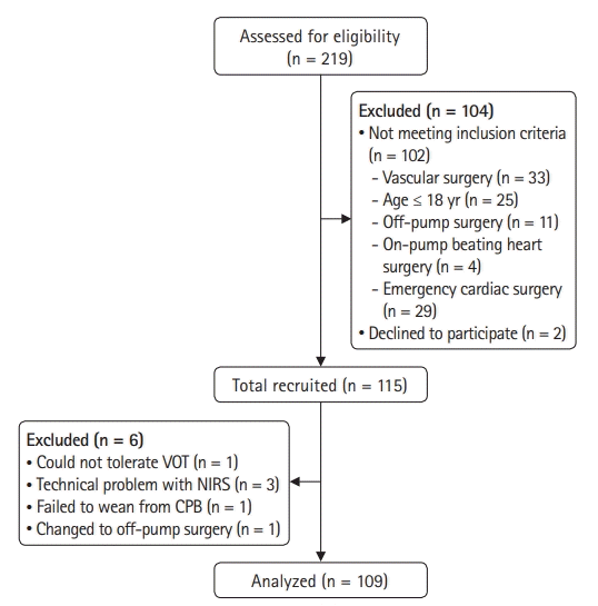
Fig. 1.
A CONSORT flow diagram. VOT: vascular occlusion test, NIRS: near-infrared spectroscopy, CPB: cardiopulmonary bypass.
kja-22097f1.tif
Table 1.
A Summary of the Patient Demographics and Intraoperative Characteristics
Variable Total (n = 109) Patients with MAE (n = 32) Patients without MAE (n = 77) P value
Age (yr) 63.7 ± 11.2 66.6 ± 8.9 62.6 ± 11.9 0.086
Sex (Male) 74 (67.9) 19 (59.4) 55 (71.4) 0.220
Height (cm) 162.2 ± 8.6 160.5 ± 9.2 163.0 ± 8.2 0.167
Weight (kg) 63.1 ± 12.0 61.4 ± 12.4 63.8 ± 11.9 0.333
BMI (kg/m2) 24.1 ± 3.6 23.9 ± 3.6 24.2 ± 3.6 0.679
BSA (m2) 1.7 ± 0.2 1.64 ± 0.19 1.68 ± 0.18 0.238
ASA classification 0.141
 II 8 (7.3) 0 (0) 8 (10.4)
 III 99 (90.8) 31 (96.9) 68 (88.3)
 IV 2 (1.8) 1 (3.1) 1 (1.3)
Smoker 29 (26.6) 7 (21.9) 22 (28.6) 0.471
Type of surgery 0.023
 CABG 37 (33.9) 6 (18.8) 31 (40.3)
  Valvular surgery 61 (56.0) 24 (75.0) 37 (48.1)
  CABG + Valvular surgery 1 (0.9) 1 (3.1) 0 (0)
  ASD closure 7 (6.4) 0 (0) 7 (9.1)
  Myxoma removal 3 (2.8) 1 (3.1) 2 (2.6)
 Comorbidities
  Hypertension 72 (66.1) 26 (81.3) 46 (59.7) 0.031
  Diabetes mellitus 37 (33.9) 14 (43.8) 23 (29.9) 0.163
  Stroke 7 (6.4) 4 (12.5) 3 (3.9) 0.191
  Congestive heart failure 4 (3.7) 2 (6.3) 2 (2.6) 0.579
  Chronic kidney disease 8 (7.3) 5 (15.6) 3 (3.9) 0.046
  Chronic obstructive pulmonary disease 7 (6.4) 2 (6.3) 5 (6.5) 1.000
EuroSCORE II 3.6 (2.4, 4.7) 3.4 (2.3, 7.5) 1.8 (0.9, 2.4) < 0.001
Surgery time (min) 346.2 (331.1, 361.4) 380 (322.5, 437.5) 330 (280.0, 385.0) 0.005
Anesthesia time (min) 397.7 (382.2, 413.2) 435 (36.0, 480.0) 390 (345.0, 420.0) 0.029
Cardiopulmonary bypass time (min) 135 (105.0, 160.0) 170 (127.0, 229.0) 125 (105.0, 145.0) < 0.001
Aortic cross clamping time (min) 100 (74.5, 120.0) 120 (79.0, 165.0) 90 (73.0, 113.0) 0.023
Transfusion
 Red blood cell (unit) 4 (2, 6) 5.0 (4.0, 6.0) 3.0 (2.0, 5.0) < 0.001
 Fresh frozen plasma (unit) 3 (2, 3) 3.0 (2.0, 3.0) 3.0 (2.0, 3.0) 0.575
 Platelet (unit) 8 (8, 8) 8.0 (8.0, 8.0) 8.0 (8.0, 8.0) 0.079
 Cryoprecipitate (unit) 6 (6, 6) 6.0 (6.0, 6.0) 6.0 (6.0, 6.0) 0.059
Infused crystalloid (ml) 2700 (2100.0, 3500.0) 3000 (2200.0, 3630.0) 2700 (2000.0, 3300.0) 0.307
Urine output (ml) 1300 (947.0, 1720.0) 1000 (820.0, 1345.0) 1370 (1130.0, 1820.0) 0.001
VIS at T3 12.0 (8.0, 20.0) 20.0 (10.0, 24.0) 12.0 (6.0, 16.0) < 0.001
VIS at T4 12.0 (6.0, 20.5) 20.0 (11.0, 27.0) 10.0 (4.0, 18.0) 0.002
Post-CPB vasoplegia 36 (33.0) 17 (53.1) 19 (24.7) 0.004

Values are presented as mean ± SD, mean (95% CI), and frequency (%). BMI: body mass index, BSA: body surface area, ASA: American Society of Anesthesiologists, CABG: coronary artery bypass graft, ASD: atrial septal defect, MAE: major adverse events, VIS: vasoactive-inotropic score, T3: 10 min after protamine injection, T4: after sternal closure.

Table 2.
The Vascular Occlusion Test Variables
Parameter Total (n = 109) Patients with MAE (n = 32) Patients without MAE (n = 77) P value
Baseline StO2 (%)
 T0 56.0 (48.0, 65.0) 52.0 (44.0, 63.0) 59.0 (52.0, 66.0) 0.014
 T1 54.0 (48.5, 62.0) 51.0 (46.5, 57.5) 55.0 (50.0, 64.0) 0.056
 T2 64.0 (52.5, 72.0)*† 62.0 (49.0, 68.5) 66.0 (54.0, 74.0)*† 0.086
 T3 71.0 (64.0, 82.0)*†‡ 70.0 (62.0, 78.5)*†‡ 74.0 (65.0, 85.0)*†‡ 0.120
 T4 72.0 (64.0, 81.0)*†‡ 74.0 (65.5, 82.0)*†‡ 71.0 (62.0, 81.0)*†‡§ 0.670
Occlusion slope (%/s)
 T0 0.20 (0.15, 0.25) 0.20 (0.15, 0.24) 0.20 (0.15, 0.25) 0.926
 T1 0.18 (0.13, 0.23)* 0.16 (0.09, 0.21) 0.18 (0.13, 0.23)* 0.188
 T2 0.17 (0.12, 0.22)* 0.15 (0.09, 0.21) 0.19 (0.12, 0.23) 0.034
 T3 0.24 (0.16, 0.30)†‡ 0.20 (0.13, 0.28)†‡ 0.25 (0.16, 0.30)*†‡ 0.421
 T4 0.23 (0.16, 0.29) 0.19 (0.15, 0.29)†‡ 0.24 (0.16, 0.30) 0.309
Minimum StO2 (%)
 T0 15.0 (15.0, 15.0) 15.0 (15.0, 15.0) 15.0 (15.0, 16.0) 0.471
 T1 15.0 (15.0, 21.5) 15.0 (15.0, 20.3) 15.0 (15.0, 22.0) 0.919
 T2 15.0 (15.0, 25.0)* 17.0 (15.0, 30.0)* 15.0 (15.0, 24.0) 0.067
 T3 15.0 (15.0, 23.0)* 15.0 (15.0, 26.8) 15.0 (15.0, 22.5) 0.651
 T4 15.0 (15.0, 21.5) 15.0 (15.0, 28.5)* 15.0 (15.0, 19.5) 0.246
Recovery slope (%/s)
 T0 2.96 (2.17, 3.60) 3.02 (2.14, 3.45) 2.96 (2.19, 3.81) 0.272
 T1 2.32 (1.82, 2.86)* 2.03 (1.61, 2.51)* 2.50 (1.90, 2.94)* 0.006
 T2 1.23 (0.97, 1.91)*† 1.01 (0.69, 1.44)*† 1.48 (1.03, 2.00)*† 0.001ΙΙ
 T3 1.42 (1.02, 1.96)*† 1.11 (0.72, 1.46)*† 1.54 (1.08, 2.13)*† 0.001ΙΙ
 T4 1.57 (1.06, 2.16)*†‡§ 1.44 (0.91, 1.75)*†‡§ 1.63 (1.15, 2.46)*†‡§ 0.024
Maximum StO2 (%)
 T0 85.0 (78.0, 90.0) 80.5 (70.8, 86.3) 87.0 (80.0, 93.3) 0.001ΙΙ
 T1 88.0 (82.0, 95.0)* 84.0 (76.0, 88.0)* 91.0 (84.5, 95.0)* < 0.001ΙΙ
 T2 87.0 (79.5, 94.0) 80.0 (73.8, 87.3) 90.0 (83.0, 94.0) 0.001ΙΙ
 T3 89.0 (82.0, 95.0)* 82.0 (79.8, 89.0) 92.0 (87.0, 95.0)* 0.009ΙΙ
 T4 90.0 (83.0, 95.0)*‡ 87.5 (79.8, 92.3)* 92.5 (86.0, 95.0) < 0.001ΙΙ

Values are presented as median (Q1, Q3). *P < 0.05 compared to T0; P < 0.05, compared to T1; P < 0.05, compared to T2; §P < 0.05, compared to T3. ΙΙP < 0.010 indicates a significant difference between the patients with and without MAE following the Bonferroni correction. T0: prior to anesthesia induction, T1: 30 min post-anesthesia induction, T2: 30 min after CPB initiation, T3: 10 min post-protamine injection, T4: after sternal closure, MAE: major adverse events, StO2: tissue oxygen saturation.

Table 3.
The ROC Curve Analysis for the Prediction of MAE after Surgery
Variable AUC 95% CI P value Threshold Sensitivity (%) Specificity (%) PPV (%) NPV (%)
Recovery slope at T1 0.668 0.572, 0.756 0.002 ≤ 2.26 68.8 61.0 42.4 82.4
Recovery slope at T2 0.701 0.606, 0.785 < 0.001 ≤ 1.08 62.5 72.7 48.8 82.4
Recovery slope at T3 0.705 0.610, 0.788 < 0.001 ≤ 1.54 84.4 50.7 41.5 88.6
Recovery slope at T4 0.637 0.540, 0.727 0.013 ≤ 1.89 90.6 36.4 37.2 90.3
Maximum StO2 at T0 0.707 0.612, 0.790 < 0.001 ≤ 88 90.6 45.5 40.9 92.1
Maximum StO2 at T1 0.713 0.619, 0.796 < 0.001 ≤ 88 78.1 62.3 46.3 87.3
Maximum StO2 at T2 0.697 0.601, 0.782 < 0.001 ≤ 86 71.9 67.1 47.6 81.1
Maximum StO2 at T3 0.721 0.627, 0.803 < 0.001 ≤ 87 68.8 71.1 49.7 84.5
Maximum StO2 at T4 0.658 0.561, 0.746 0.003 ≤ 93 87.5 42.9 38.9 89.2

ROC: receiver operating characteristic, AUC: area under the curve, PPV: positive predictive value, NPV: negative predictive value, T0: prior to anesthesia induction, T1: 30 min post-anesthesia induction, T2: 30 min after cardiopulmonary bypass initiation, T3: 10 min post-protamine injection, T4: after sternal closure, StO2: tissue oxygen saturation.

Table 4.
Post-CPB Vasoplegia and Postoperative Outcomes
Variable Total (n = 109) Recovery slope at T2
≤ 1.08 (n = 40) > 1.08 (n = 69) P value
Post-CPB vasoplegia 36 (33.0) 14 (35.0) 22 (31.9) 0.739
Initial SOFA 5.0 (4.0, 7.0) 6.0 (4.3, 7.0) 5.0 (4.0, 6.0) 0.105
Maximum SOFA 6.0 (4.0, 7.0) 6.0 (4.3, 8.0) 5.0 (4.0, 6.5) 0.047
APACHE II 14.0 (11.0, 17.0) 14.5 (11.0, 17.8) 12.0 (11.0, 16.5) 0.654
Duration of ventilator care (days) 2.0 (2.0, 2.0) 2.0 (2.0, 2.0) 2.0 (2.0, 2.0) 0.008
Length of ICU stay (days) 3.0 (3.0, 4.0) 3.0 (3.0, 5.8) 3.0 (3.0, 4.0) 0.111
Length of hospital stay (days) 20 (15, 28) 20.5 (15.0, 27.0) 19.0 (15.0, 28.5) 0.669
Major adverse events 19 (47.5) 13 (18.8) 0.002
 Death 4 (3.7) 3 (7.5) 0 (0) 0.139
 Aggravated heart failure 5 (4.6) 3 (7.5) 2 (2.9) 0.354
 Myocardial infarction 2 (1.8) 1 (2.5) 1 (1.4) 1.000
 Acute kidney injury 25 (22.9) 15 (37.5) 10 (14.5) 0.006
 Stroke or seizure 5 (4.6) 2 (5.0) 3 (4.3) 1.000
 ARDS 2 (1.8) 1 (2.5) 1 (1.4) 1.000
 Gastrointestinal bleeding 1 (1.0) 1 (1.6) 0(0) 1.000

Values are presented as mean ± SD, mean (95% CI), and frequency (%). T2: 30 min after CPB initiation, CPB: cardiopulmonary bypass, SOFA: Sequential Organ Failure Assessment, APACHE: Acute Physiologic and Chronic Health Evaluation, ICU: intensive care unit, ARDS: acute respiratory distress syndrome.

Table 5.
The Univariate and Multivariate Logistic Regression Analysis of Variables Associated with MAE after Cardiac Surgery
Variable Unadjusted OR (95% CI) P value Adjusted OR (95% CI) P value
Hemoglobin at T1 (g/dl) 0.659 (0.506, 0.859) 0.002 0.604 (0.433, 0.842) 0.003
Surgery (CABG)
 Valvular surgery 3.243 (1.297, 8.111) 0.012
  CABG + valvular surgery
  ASD closure
  Myxoma removal
Comorbidities
 Hypertension 2.920 (1.077, 7.920) 0.035
 CKD 4.568 (1.022, 20.425) 0.047
Surgery time 1.009 (1.003, 1.015) 0.003
CPB time (min) 1.018 (1.001, 1.028) < 0.001 1.022 (1.007, 1.038) 0.005
Intraoperative RBC transfusion 1.409 (1.139, 1.742) 0.002
Intraoperative urine output/10 (ml) 0.991 (0.984, 0.999) 0.022 0.988 (0.978, 0.998) 0.015
VIS at T3 1.083 (1.033, 1.135) < 0.001 1.097 (0.978, 0.998) 0.012
Recovery slope at T2 (%/s) 0.322 (0.152, 0.683) 0.003 0.220 (0.076, 0.635) 0.005

Values are presented as mean (95% CI). MAE: major adverse event, T1: 30 min post-anesthesia induction, CABG: coronary artery bypass graft, ASD: atrial septal defect, CKD: chronic kidney disease, CPB: cardiopulmonary bypass, RBC: red blood cell, VIS: vasoactive-inotropic score, T2: 30 min after CPB initiation, T3: 10 min post-protamine injection.

TOOLS
Similar articles