Journal List > Clin Endosc > v.55(5) > 1516079470

Netinatsunton, Cheewasereechon, Pattarapuntakul, Sottisuporn, Kanjanapradit, and Ovartlarnporn: Optical diagnosis by near-focus versus normal-focus narrow band imaging colonoscopy in colorectal polyps based on combined NICE and WASP classification: a randomized controlled trial

Abstract

Background/Aims

Narrow Band Imaging (NBI) International Colorectal Endoscopic (NICE) and Workgroup Serrated Polyps and Polyposis (WASP) classifications were developed for optical diagnosis of neoplastic and sessile serrated polyps, respectively. Near-focus NBI with NICE combined with WASP criteria for optical diagnosis of colonic polyps has not yet been evaluated. We aimed to compare the accuracy of near-focus NBI (group A) with normal-focus NBI (group B) in real-time optical diagnosis of colorectal polyps using combined NICE and WASP criteria.

Methods

Among 362 patients, 118 with 227 polyps were recruited. Groups A and B included 62 patients with 130 polyps (three lost polyps) and 56 patients with 106 polyps (six lost polyps), respectively. Optical diagnoses were compared with pathological reports.

Results

The accuracy of optical diagnosis of neoplastic polyps in groups A and B was not significantly different (76% vs. 71%, p=0.52). WASP criteria provided all false positive diagnoses of sessile polyps as serrated polyps in 31 (16.2%) patients.

Conclusions

Near-focus NBI was not superior to normal-focus NBI in optical diagnostics of neoplastic polyps using NICE criteria. In our study, WASP classification yielded all false positives in the diagnosis of sessile serrated adenomas/polyps. Routine real-life optical diagnosis of polyps is still unadvisable.

INTRODUCTION

Colorectal cancer is the third most common cancer with substantial contribution to cancer-related mortality worldwide. Screening colonoscopy can reduce the risk and mortality to approximately 53% to 80%.1,2 However, nearly half of detected small polyps are non-neoplastic with a negligible risk of transformation to cancer; therefore, polypectomies of these polyps are unnecessary.2,3 Narrow band imaging (NBI) is an image-enhancing technology that utilizes narrow-band light filters to improve visualization of mucosal surface architecture and vascular patterns.4 Many studies, including multiple meta-analyses, show the efficacy of NBI for the optical diagnosis of polyps, with reported sensitivities of 85% to 92.2%.5-14
The NBI International Colorectal Endoscopic Society has proposed the NBI International Colorectal Endoscopic (NICE) criteria to differentiate neoplastic lesions, namely, adenoma and deep submucosal invasive cancer from hyperplastic polyps.15,16 This classification system showed a good correlation between the optical and histopathological diagnoses in some pilot studies by expert endoscopists, while other studies in community settings showed inferior results.3,17,18 Sessile serrated adenomas/polyps (SSAPs) are difficult to differentiate from hyperplastic polyps by colonoscopy, even when using NBI and the NICE criteria.19-21 A meta-analysis of 10 studies (eight with magnifying NBI and two with standard NBI) showed a pooled sensitivity and specificity of 0.6 and 0.75 for optical diagnosis of SSAPs, respectively.22 Endoscopic features of SSAPs, as validated by international NBI experts, include (1) a cloud-like surface, (2) indistinct borders, (3) irregular shapes, and (4) dark spots inside the crypts, with the combination of all four characters yielding an accuracy of 93%.23 The Workgroup Serrated Polyps and Polyposis (WASP) developed a classification combining the NICE classification and the criteria for SSAPs to distinguish hyperplastic polyps from SSAPs, and the WASP criteria significantly improved the accuracy of optical diagnosis despite the use of only two features of SSAPs.24 One study evaluated the accuracy of optical diagnosis of NBI without optic magnification using combined NICE and WASP classification as assessed by non-expert endoscopists and showed moderate accuracy for the real-time optical diagnosis of colorectal lesions.25
The introduction of a novel dual-focus capability (Olympus Co., Tokyo, Japan) enables optical magnification of up to 65× in near-focus (NF) compared with 52× in standard focus (SF).26 Several case series and comparative studies have used SF-NBI to assess the diagnostic accuracy of available NF-NBI technology,26-30 which reported high accuracies in series reports and better or similar results to SF-NBI in comparative studies. To the best of our knowledge, no study on NF-NBI has incorporated the NICE and WASP criteria in the optical diagnosis of colonic polyps. Therefore, we aimed to compare the accuracy of optical diagnosis between NF-NBI and SF-NBI by NBI-non-expert endoscopists using the combined NICE and WASP criteria.

METHODS

Patient selection

This prospective randomized trial was conducted at the NKC Institute, a tertiary health center in affiliation with the Prince of Songkla University. Patients aged 18 to 75 years who were scheduled for colonoscopy at the NKC Institute from April 2019 to January 2020 were enrolled. Patients with inflammatory bowel disease, polyposis syndrome, colonic masses, active gastrointestinal bleeding, pregnancy, inadequate bowel preparation (Boston bowel preparation score <5).31 or any contraindications for polypectomy were excluded.

Study design and colonoscopy procedure

Randomization codes were generated by a computer using a block of two and concealed in envelopes. Patients who consented to the study were randomized to either the NF-NBI (group A) or the SF-NBI (group B) group, according to the randomization codes in sealed envelopes selected at the start of the procedure.

Bowel preparation

A split dose of 90 mL was used. A solution of monobasic sodium phosphate+dibasic sodium phosphate (Swiff, Berlin, Osoth Inter Laboratories Co.,Ltd., Bangkok, Thailand) or a hospital-prepared formula of polyethylene glycol solution was used for bowel preparation. Bowel preparation quality was assessed using the Boston bowel preparation score scale.31

Colonoscopy procedure

All procedures were performed with a high-definition colonoscope (Evis Exera III CF HQ 190 L/I with DF capability; Olympus Co.). The NF mode measured a 2 to 6-mm depth of field with 65× magnification at a 2-mm distance, and the SF mode provides a 5 to 100-mm depth of field with 52× magnification at 5 mm.26 No prior formal training for NBI image interpretation was completed by the endoscopists participating in the study, and none of the endoscopists were blinded to the NBI colonoscope model. All staff endoscopists were familiar with the NICE criteria but not the WASP criteria during routine colonoscopy. The colonoscopy was performed under conscious sedation using midazolam and pethidine or fentanyl by three trainees under the supervision of staff members according to our institute’s sedation protocol. All colonoscopies were performed initially with high-definition white light, and if a polyp was detected, NBI mode was employed. The optical diagnosis of polyps was aided using color image charts of the NICE and WASP classifications as reference standards.16,24 The confidence of endoscopists in making an optical diagnosis was not assessed in this study. In group A, the optical diagnosis of polyps was performed using the NF-NBI mode, and in group B, using the SF-NBI mode. Each polyp was evaluated in real time by the best image captured and stored on a computer using the Endo-Smart Program ver. 7 (Kainatic Engineering Co., Ltd., Bangkok, Thailand). The polyp size was estimated using the width of the opened biopsy forceps as the reference. The polyp morphology was described as either pedunculated (O-Ip), sessile (O-Is), or flat (O-IIb).32 Diminutive polyps were defined as polyps <5 mm.26 The location, size, number, morphology, and optical diagnosis of the polyps were recorded in a data recording form.

Polyp characterization

Optical diagnosis of each polyp was made according to the NICE and WASP classifications. Based on the NICE classification,16 polyps were classified as type I, hyperplastic; type II, adenomatous or superficial mucosal invasive cancer (depth <1,000 µm); and type III, deep mucosal invasive cancer (depth ≥1,000 µm). For sessile and flat polyps, regardless of the NICE classification, additional assessments were performed based on the WASP classification.24 SSAPs were diagnosed if two of the following features were present: (1) a clouded surface, (2) indistinctive borders, (3) irregular shapes, and (4) dark spots inside the crypts.24

Pathological polyp evaluation

All polyps were removed using biopsy forceps, cold snares, or hot snares at the discretion of the attending staff, and the resected specimens were sent for histopathological examination. All histological assessments were performed by a single pathologist (KK) according to the World Health Organization criteria.33 No validation of the pathologist’s accuracy by an independent pathologist was performed in this study.

Endpoints

The primary outcome was the performance of NF-NBI compared with SF-NBI in the following: (1) optical diagnosis of neoplastic polyps and (2) differentiation of serrated adenomas from hyperplastic polyps for sessile polyps, compared with histologic evaluation. The performance was assessed by accuracy, sensitivity, specificity, positive predictive value (PPV), and negative predictive value (NPV) of NF-NBI compared to SF-NBI.

Sample size calculation

The sample size was calculated with the assumption that NF-NBI was 10% superior to SF-NBI. The number of polyps required was 252 in each group in order to detect a difference with an accuracy with a power of 80% at the significance level α=0.05, calculated by using the comparison of two proportions based on the accuracy of SF-NBI=75%.34,35

Statistical analysis

Continuous variables are presented as means±standard deviations, and statistical analysis was performed using Student t-test. The discrete variables are presented as percentages. The accuracy, sensitivity, specificity, PPV, and NPV were presented as percentages, and the chi-square test or Fisher exact test were performed for analysis as appropriate. Statistical significance was set at p<0.05. All statistical analysis was conducted using R (ver. 3.6.1; Vienna, Austria).

Ethical statements

The study protocol was approved by the Ethics Committee of the Faculty of Medicine, Prince of Songkla University (Registration No. 61355143) and was registered with Clinical Trials.gov (NCT 04831814) on 04/01/2021. This study was conducted in accordance with the principles of the Declaration of Helsinki. Informed consent was obtained from each patient prior to study entry.

RESULTS

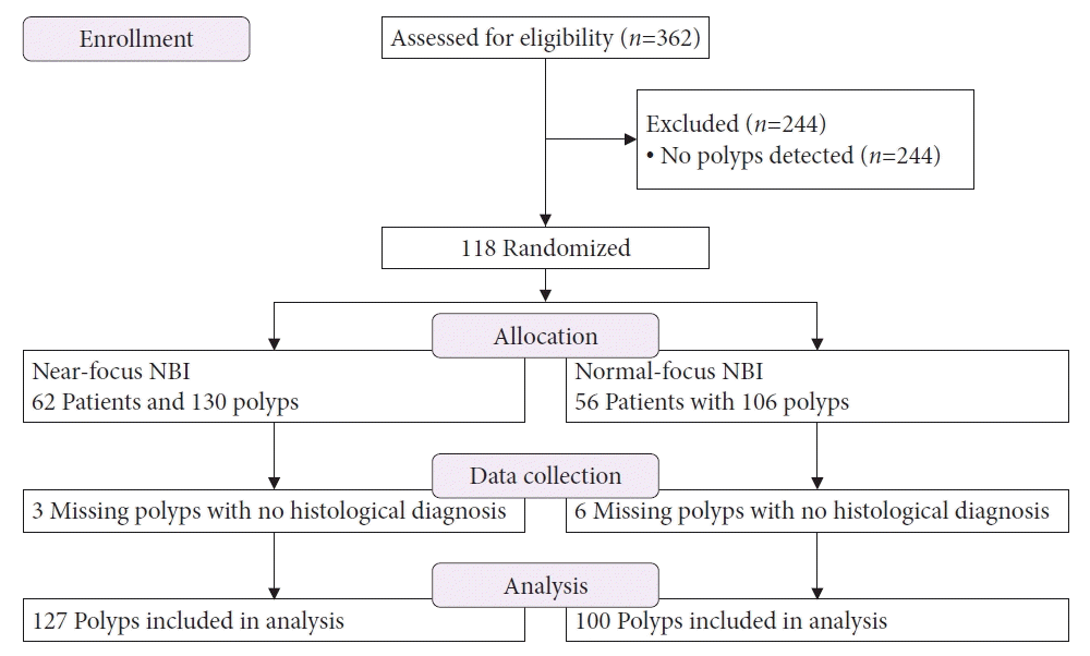
In total, 362 patients were recruited between April 2019 and January 2020, and 118 of them had polyps on colonoscopy and were eventually randomized into the study groups (Fig. 1). A total of 236 polyps were detected in the whole group, but nine polyps (3 of 130 polyps in group A, 6 of 106 polyps in group B) were lost during the retrieval process, leaving only 227 polyps eventually available for pathological assessment. The number of missing polyps was not significantly different between the two groups (p=0.35).
Of the 227 total polyps, 144 (63.4%) of the polyps were adenomas, 79 (34.8%) were hyperplastic polyps, and 4 (1.8%) were cancers according to histological results. There were 191 sessile and flat polyps, but no SSAPs were detected. The most common site of polyps was in the sigmoid colon (30%), and 188 of 227 (82.8%) polyps were diminutive in type. The polyp detection rate and adenoma detection rate for the entire group of 362 patients were 118 of 362 (32.6%) and 86 of 362 (23.8%), respectively.
Sixty-two patients with 130 polyps, including three polyps lost before histologic evaluation, were assigned to group A, and 56 patients with 106 polyps, including six polyps lost before histologic evaluation, were allocated to group B (Fig. 1). There were no significant differences between the two groups regarding age, sex, indications for colonoscopy, total procedure time, withdrawal time, cecal intubation rate, complication rate, polyp size, morphology, histology, and location (Table 1). The quality of bowel preparation in group A was significantly better than that in group B (p=0.01). There were 76 adenomatous polyps and two cancers by histology in group A, and 68 adenomatous polyps and two cancers in group B, but the differences were not statistically significant (p=0.43).

Primary end point

The accuracy, sensitivity, specificity, PPV, and NPV of NICE in optical diagnosis of neoplastic polyps were 76%, 72%, 81%, 86%, 64%, respectively, in group A, and 71%, 66%, 83%, 90%, 51%, respectively, in group B. The performance of NF-NBI was not better than that of SF-NBI for the optical diagnosis of neoplastic polyps (p=0.52, p=0.45, p=0.85, p=0.51, and p=0.09, respectively) (Table 2). The NPV in NF-NBI was greater than that in SF-NBI, although the difference was not statistically significant (p=0.09).

Outcome measures according to interobserver variations

The capability of each endoscopist in optical diagnosis of polyps using the NICE classification (excluding WASP for SSAPs) compared with histological analysis by using Cohen’s K-coefficient were 79.21% (kappa=0.579) for endoscopist I, 64.58% (kappa=0.431) for endoscopist II, 54.55% (kappa=–0.009) for endoscopist III, and 50% (kappa=0.213) for endoscopist IV.
The accuracy of the NICE classification for optical diagnosis of neoplastic polyps in both groups by the endoscopists with greater kappa values (kappa=0.579 and 0.431) was 76%. This was significantly greater than the 57% score by the endoscopists with lower kappa values (kappa=0.0213 and –0.0009) (p=0.007). The accuracy of the NICE classification in the optical diagnosis of neoplastic polyps by the endoscopists with higher kappa values was 79% in group A, and was greater than the 71% accuracy in group B, however the difference was not significant (p=0.25) (Supplementary Table 1), whereas the accuracy of optical diagnosis using the NICE classification for neoplastic polyps by the endoscopists with lower kappa values was 55% in group A, which was lower than the 60% accuracy in group B, but the difference failed to reach statistical significance (p=0.57) (Supplementary Table 2).
The sensitivity and NPV of using the NICE classification in predicting neoplastic polyps of both groups by the endoscopists with higher kappa values were 72% and 62%, which were significantly higher than the sensitivity and NPVs of 48% and 49%, respectively, by the endoscopists with lower kappa values (p=0.002 and p=0.004, respectively), but the specificity and PPV were not significantly different between the two groups of endoscopists, namely 83% and 88% for the endoscopists with higher kappa compared with 77% and 82% for the endoscopists with lower kappa; (p=0.207 and p=0.228, respectively).
The sensitivity, specificity, PPV, and NPV using the NICE classification in predicting neoplastic polyps by the endoscopists with higher kappa values were 75%, 86%, 90%, 67% in group A, respectively, and 65%, 83%, 89%, 54% in group B, respectively, with no significant difference (p=0.165, p=0.696, p=1.000, and p=0.083, respectively). The sensitivity, specificity, PPV, and NPV using the NICE classification in predicting neoplastic polyps by the endoscopists with lower kappa values were 41%, 80%, 78%, 44% in group A, respectively, and 58%, 67%, 88%, 29% in group B, respectively. The sensitivity in group A was significantly lower than that in group B (p=0.023). In contrast, the NPV of 44% in group A was significantly higher than that of 29% in group B (p=0.039); however, there were no significant differences in specificity and PPV between the two groups (p=0.054 and p=0.090, respectively).

For diminutive polyps

There were no statistical differences in accuracy, sensitivity, specificity, PPV, and NPV in the optical diagnosis of diminutive neoplastic polyps between groups A and B (Table 3). When performance was assessed by comparing both groups with both diminutive groups, the accuracy, sensitivity, specificity, PPV, and NPV of using the NICE classification in predicting neoplastic polyps were 74%, 69%, 82%, 88%, and 58% for the whole group and were 68%, 59%, 82%, 82%, and 58% for the diminutive group, respectively; however, the differences were not significant (p=0.44, p=0.19, p=1.00, p=0.32, and p=1.00, respectively).

Outcome of WASP criteria in sessile polyps

In 191 sessile and flat polyps, the WASP criteria classified polyps as serrated polyps in 31 (16.2%) cases; however, histology did not confirm the diagnosis in all 31 polyps. Pathological examination of polyps classified serrated polyps according to the WASP criteria revealed hyperplastic polyps in 11 cases and adenomatous polyps in 20 cases. In group A, the WASP criteria falsely diagnosed 22.5% (25 out of 111 sessile or flat polyps) SSAPs, whereas in group B, the WASP criteria falsely diagnosed 7.5% (6 out of 80 sessile or flat polyps) SSAPs, and the difference was statistically significant (p=0.0285). In group A, nine of 59 (15.3%) NICE I were designated as SSAPs using the WASP criteria and 16 of 50 (32%) NICE II were designated as SSAPs using the WASP criteria, but the difference between NICE I and NICE II was not significant (p=0.1575); in group B, 2 of 47 (4.3%) NICE I were designated as SSAPs using the WASP criteria and 4 of 31 (12.9%) NICE II were designated as SSAPs using the WASP criteria, and the difference between NICE I and NICE II was not statistically significant (p=0.39). There were no differences in false diagnosis rates between NICE I and NICE II in group A compared with group B (p=0.11 and p=0.20, respectively). Of the 31 polyps classified as serrated polyps according to the WASP criteria, 25 polyps had two features and six polyps had four features of SSAPs.

Optical diagnosis missed classification

NICE I classification misclassified adenomatous polyps as hyperplastic polyps in 20 (32.8%) in group A and 24 (49.0%) patients in group B, and one cancer as hyperplastic polyps (1.6%) in group A (p=0.16) (Table 4).
NICE II classification misclassified hyperplastic polyps as adenomatous polyps in nine (14.5%) polyps in group A and five (10.6%) polyps in group B (p=0.76).
NICE III classification misclassified three adenomatous polyps as malignant lesions in group A and misclassified two adenomatous polyps as malignant lesions in group B. There was no statistically significant difference between the two groups (p=0.82).

Complications

Post-polypectomy bleeding requiring the application of hemostatic clips occurred in one patient in group B, and the complication rate was similar between the two groups (p=0.97).

DISCUSSION

Preliminary analysis in our study showed that the diagnostic accuracy of NF-NBI was not significantly higher than that of SF-NBI (76% vs. 71%) in distinguishing neoplastic and non-neoplastic polyps based on the NICE criteria by the non-expert endoscopists, but findings must be interpreted with caution as we recruited patients with polyps of less than half the calculated sample size, so this may be underpowered to detect the difference. The efficacy of NF-NBI in the optical diagnosis of polyps has been reported in multiple studies, with variable results. Two series reports from academic centers showed a high accuracy in NF-NBI; one report of 63 polyps in 55 patients showed that combination NF-NBI with acetic acid had 85.5% accuracy in the prediction of polyp histology when the confidence in optical diagnosis of the endoscopists was high27; another report using NF-NBI with a transparent cap combined with digital magnification of 164 polyps in 87 patients with an overall accuracy of 97% if the optical diagnosis was made with high confidence.30 In comparative studies, one prospective multi-center randomized study required all participating endoscopists trained for NBI to attain 90% accuracy prior to the study and compared NF-NBI with of SF-NBI (180 series) and showed that NF-NBI yielded a higher confidence optical diagnosis than did SF-NBI (85.1% vs. 72.6%).28 Another series study comparing white light, SF-NBI, and NF-NBI sequentially in the same patient demonstrated that NF-NBI improved accuracy more than SF-NBI,29 whereas another study reported similar accuracy of NF-NBI (79%) compared of SF-NBI (180 series) (78%) in the optical diagnosis of colorectal polyps, despite prior training of endoscopists to attain 90% accuracy.26 Our study showed an accuracy of 76% for NF-NBI in the optical diagnosis of neoplastic polyps that was comparable to that reported by Wallace et al.26 using NF-NBI, but lower than that reported by Hewett et al.15 (89%) using SF-NBI. No formal training for endoscopists was required in our study, and the endoscopists relied on the color image chart of the NICE criteria as a reference. The NPV of 51% to 64% reported in our study was below the 90% Preservation and Incorporation of Valuable endoscopic Innovations, as recommended by American Society for Gastrointestinal Endoscopy as an acceptable capability to implement optical diagnosis.14 We did not assess confidence level in optical diagnosis; a high-confidence optical diagnosis may have improved the outcomes of this study.27-30
This study showed no difference between NF-NBI and SF-NBI in assessing diminutive polyps ≤5 mm compared with those >5 mm; however, the number of polyps >5 mm was low. In one study, NF-NBI performance was better in diminutive polyps ≤5 mm, with an accuracy of 89% and NPV of 96% when the optical diagnosis was made with high confidence.28 In contrast, another retrospective study by Hattori et al.36 using NBI with magnification showed that a NICE I optical diagnosis for small polyps ≤1 cm misclassified 20% of adenomatous polyps and 42% of diminutive adenomas as hyperplastic polyps.
The accuracy of endoscopists with high capability (higher kappa) was superior to that of endoscopists with lower capability (lower kappa), implying that the skill of the endoscopist contributes to the accuracy in our study, which is in line with the findings of one meta-analysis.14 However, the number of cases performed by endoscopists with lower kappa values was low. The Detect Inspect Characterise Resect and Discard 2 study that used NICE criteria in non-academic centers showed a sensitivity of 83% for the presence of adenomas;3 in contrast to our study, endoscopist expertise in the Detect Inspect Characterise Resect and Discard 2 study did not improve performance.
Histologic and optical diagnostic discrepancies have been reported to vary from 3.4% to 19.3%.3,28 We did not assess the accuracy of our pathologists; therefore, the effect on the outcome due to variation in pathological diagnosis accuracy could not be assessed.
The WASP criteria provide all false optical diagnoses for SSAPs. In this study, the WASP criteria were applied to sessile and flat polyps, regardless of NICE stratification. For NICE I lesions, the WASP criteria provided fewer false positives than NICE II in both groups A and B, although the differences were not statistically significant. The WASP criteria to detect SSAPs for non-expert endoscopists without prior training were unreliable in this study, and NF-NBI did not provide additional advantages. In our study, only two of the four WASP criteria were used for the diagnosis of SSAPs, while one study that validated the NBI for SSAPs using all four features showed an accuracy of 93%23; however, in our study, 25 out of 31 polyps had only two features, which may account for the low accuracy using the WASP criteria. Surprisingly, NF-NBI led to significantly more false positives (25/111) than SF-NBI (6/80) did. This may be due to the narrow focal length of NF-NBI compared to the longer focal length in SF-NBI and that no measures to stabilize the distance from the tip of the endoscope to the polyp were taken, leading to a more blurred margin and/or blurred surface in NF-NBI than in SF-NBI, which may contribute to more false positives in the NF-NBI group. Further studies with a larger number of polyps and prior training of the endoscopists with measures to stabilize the distance between the endoscope and polyp, as well as using all four features of the WASP criteria, may clarify the role of the WASP criteria in using NF-NBI in differentiating SSAPs from hyperplastic polyps.
There were some limitations in our study as follows: (1) the inadequate sample size, as this was a preliminary analysis, (2) the lack of assessment of endoscopist confidence in optical diagnosis that may alter the outcome in our study, (3) the lack of prior training and assessment of endoscopists participating in the study, (4) the small number of polyps included in the assessment of WASP criteria, (5) the lack of validation of the accuracy of pathological reports of sessile serrated polyps, and (6) the lack of measures to maintain constant focal length from the tip of the endoscope to the polyp that may affect the image sharpness of NF-NBI.
In conclusion, our preliminary data showed that NF-NBI was not superior to SF-NBI in the optical diagnosis of polyps using the NICE criteria. The WASP classification yielded all false positives for the diagnosis of SSAPs in our study. With the current capability of image-enhancing technology, the routine use of optical diagnosis of polyps in real-life practice is still unadvisable.

Supplementary Material

Supplementary Table 1. Accuracy, sensitivity, specificity, PPV and NPV by NICE criteria of the endoscopists with higher Kappa in optical diagnosis of polyps.
ce-2022-048-suppl1.pdf
Supplementary Table 2. Accuracy, sensitivity, specificity, PPV and NPV by NICE criteria of the endoscopists with lower Kappa in optical diagnosis of polyps.
ce-2022-048-suppl2.pdf
Supplementary materials related to this article can be found online at https://doi.org/10.5946/ce.2022.048.

Notes

Conflicts of Interest

The authors have no potential conflicts of interest.

Funding

None.

Acknowledgments

The authors would like to thank Miss Nannapat Pruphetkaew in the Department of Epidemiology for statistical analysis.

The manuscript titled “Near-focus versus normal-focus narrow band imaging colonoscopy in diagnosis of colorectal polyps based on combined NICE and WASP classification in routine colonoscopy: a randomized controlled trial” was preprinted in www.researchsquare.com (rs-809507).

Author Contributions

Conceptualization: NC, NN, BO; Data curation: all authors; Formal analysis: NC, NN, BO; Investigation: all authors; Methodology: NC, NN, BO; Project administration: NC, NN, Supervision: BO; Writing-originl draft: NC, NN, BO; Writing-review & editing: all authors.

REFERENCES

1. Zauber AG, Winawer SJ, O’Brien MJ, et al. Colonoscopic polypectomy and long-term prevention of colorectal-cancer deaths. N Engl J Med. 2012; 366:687–696.
2. Ignjatovic A, East JE, Suzuki N, et al. Optical diagnosis of small colorectal polyps at routine colonoscopy (Detect InSpect ChAracterise Resect and Discard; DISCARD trial): a prospective cohort study. Lancet Oncol. 2009; 10:1171–1178.
3. Rees CJ, Rajasekhar PT, Wilson A, et al. Narrow band imaging optical diagnosis of small colorectal polyps in routine clinical practice: the Detect Inspect Characterise Resect and Discard 2 (DISCARD 2) study. Gut. 2017; 66:887–895.
4. ASGE Technology Committee, Manfredi MA, Abu Dayyeh BK, et al. Electronic chromoendoscopy. Gastrointest Endosc. 2015; 81:249–261.
5. Komeda Y, Kashida H, Sakurai T, et al. Magnifying narrow band imaging (NBI) for the diagnosis of localized colorectal lesions using the Japan NBI Expert Team (JNET) Classification. Oncology. 2017; 93 Suppl 1:49–54.
6. Repici A, Hassan C, Radaelli F, et al. Accuracy of narrow-band imaging in predicting colonoscopy surveillance intervals and histology of distal diminutive polyps: results from a multicenter, prospective trial. Gastrointest Endosc. 2013; 78:106–114.
7. Machida H, Sano Y, Hamamoto Y, et al. Narrow-band imaging in the diagnosis of colorectal mucosal lesions: a pilot study. Endoscopy. 2004; 36:1094–1098.
8. Hirata M, Tanaka S, Oka S, et al. Evaluation of microvessels in colorectal tumors by narrow band imaging magnification. Gastrointest Endosc. 2007; 66:945–952.
9. Hirata M, Tanaka S, Oka S, et al. Magnifying endoscopy with narrow band imaging for diagnosis of colorectal tumors. Gastrointest Endosc. 2007; 65:988–995.
10. Wanders LK, East JE, Uitentuis SE, et al. Diagnostic performance of narrowed spectrum endoscopy, autofluorescence imaging, and confocal laser endomicroscopy for optical diagnosis of colonic polyps: a meta-analysis. Lancet Oncol. 2013; 14:1337–1347.
11. Backes Y, Moss A, Reitsma JB, et al. Narrow band imaging, magnifying chromoendoscopy, and gross morphological features for the optical diagnosis of T1 colorectal cancer and deep submucosal invasion: a systematic review and meta-analysis. Am J Gastroenterol. 2017; 112:54–64.
12. Picot J, Rose M, Cooper K, et al. Virtual chromoendoscopy for the real-time assessment of colorectal polyps in vivo: a systematic review and economic evaluation. Health Technol Assess. 2017; 21:1–308.
13. Mason SE, Poynter L, Takats Z, et al. Optical technologies for endoscopic real-time histologic assessment of colorectal polyps: a meta-analysis. Am J Gastroenterol. 2019; 114:1219–1230.
14. ASGE Technology Committee, Abu Dayyeh BK, Thosani N, et al. ASGE Technology Committee systematic review and meta-analysis assessing the ASGE PIVI thresholds for adopting real-time endoscopic assessment of the histology of diminutive colorectal polyps. Gastrointest Endosc. 2015; 81:502.
15. Hewett DG, Kaltenbach T, Sano Y, et al. Validation of a simple classification system for endoscopic diagnosis of small colorectal polyps using narrow-band imaging. Gastroenterology. 2012; 143:599–607.
16. Hayashi N, Tanaka S, Hewett DG, et al. Endoscopic prediction of deep submucosal invasive carcinoma: validation of the narrow-band imaging international colorectal endoscopic (NICE) classification. Gastrointest Endosc. 2013; 78:625–632.
17. Rastogi A, Bansal A, Wani S, et al. Narrow-band imaging colonoscopy: a pilot feasibility study for the detection of polyps and correlation of surface patterns with polyp histologic diagnosis. Gastrointest Endosc. 2008; 67:280–286.
18. Uraoka T, Saito Y, Matsuda T, et al. Detectability of colorectal neoplastic lesions using a narrow-band imaging system: a pilot study. J Gastroenterol Hepatol. 2008; 23:1810–1815.
19. Snover DC. Update on the serrated pathway to colorectal carcinoma. Hum Pathol. 2011; 42:1–10.
20. Leggett B, Whitehall V. Role of the serrated pathway in colorectal cancer pathogenesis. Gastroenterology. 2010; 138:2088–2100.
21. Kumar S, Fioritto A, Mitani A, et al. Optical biopsy of sessile serrated adenomas: do these lesions resemble hyperplastic polyps under narrow-band imaging? Gastrointest Endosc. 2013; 78:902–909.
22. Parikh ND, Chaptini L, Njei B, et al. Diagnosis of sessile serrated adenomas/polyps with image-enhanced endoscopy: a systematic review and meta-analysis. Endoscopy. 2016; 48:731–739.
23. Hazewinkel Y, López-Cerón M, East JE, et al. Endoscopic features of sessile serrated adenomas: validation by international experts using high-resolution white-light endoscopy and narrow-band imaging. Gastrointest Endosc. 2013; 77:916–924.
24. IJspeert JE, Bastiaansen BA, van Leerdam ME, et al. Development and validation of the WASP classification system for optical diagnosis of adenomas, hyperplastic polyps and sessile serrated adenomas/polyps. Gut. 2016; 65:963–970.
25. Castela J, Mão de Ferro S, Rosa I, et al. Real-time optical diagnosis of colorectal polyps in the routine clinical practice using the NICE and WASP classifications in a nonacademic setting. GE Port J Gastroenterol. 2019; 26:314–323.
26. Wallace MB, Crook JE, Coe S, et al. Accuracy of in vivo colorectal polyp discrimination by using dual-focus high-definition narrow-band imaging colonoscopy. Gastrointest Endosc. 2014; 80:1072–1087.
27. Wiessner JR, Brown H, Haller B, et al. Near focus NBI endoscopy plus acetic acid for optical polyp characterization in the colorectum: a proof of principle study. Scand J Gastroenterol. 2019; 54:377–383.
28. Kaltenbach T, Rastogi A, Rouse RV, et al. Real-time optical diagnosis for diminutive colorectal polyps using narrow-band imaging: the VALID randomised clinical trial. Gut. 2015; 64:1569–1577.
29. Szura M, Pasternak A, Bucki K, et al. Two-stage optical system for colorectal polyp assessments. Surg Endosc. 2016; 30:204–214.
30. Singh R, Jayanna M, Navadgi S, et al. Narrow-band imaging with dual focus magnification in differentiating colorectal neoplasia. Dig Endosc. 2013; 25 Suppl 2:16–20.
31. Lai EJ, Calderwood AH, Doros G, et al. The Boston bowel preparation scale: a valid and reliable instrument for colonoscopy-oriented research. Gastrointest Endosc. 2009; 69(3 Pt 2):620–625.
32. The Paris endoscopic classification of superficial neoplastic lesions: esophagus, stomach, and colon: November 30 to December 1, 2002. Gastrointest Endosc. 2003; 58(6 Suppl):S3–S43.
33. Bosman FT, Carneiro F, Hruban RH, et al. WHO classification of tumours of the digestive system. 4th ed. Geneva: World Heath Organization;2010.
34. Kuiper T, Marsman WA, Jansen JM, et al. Accuracy for optical diagnosis of small colorectal polyps in nonacademic settings. Clin Gastroenterol Hepatol. 2012; 10:1016–1020.
35. Ciocâlteu AM, CârŢână ET, Florescu DN, et al. Narrow band imaging with near-focus mode for colorectal polyps’ characterization. Rom J Morphol Embryol. 2016; 57(2 Suppl):619–626.
36. Hattori S, Iwatate M, Sano W, et al. Narrow-band imaging observation of colorectal lesions using NICE classification to avoid discarding significant lesions. World J Gastrointest Endosc. 2014; 6:600–605.

Fig. 1.
Flow diagram of the study.
ce-2022-048f1.tif
Table 1.
Baseline characteristics, colonoscopic procedure details, and polyp’s characteristics of groups A and B
Characteristic Near-focus (n=62) (group A) Normal-focus (n=56) (group B) p-value
Age (yr) 59.3±8.7 60±8 0.63
Sex (male/female) 40/22 34/22 0.82
Indication 0.56
 CRC screening 38 (61.3) 29 (51.8)
 Diagnosis 24 (38.7) 27 (48.2)
Boston bowel preparation score (mean) 0.01
 6 0 (0) 2 (3.6)
 7 1 (1.6) 0 (0)
 8 0 (0) 4 (7.1)
 9 61 (98.4) 50 (89.3)
Total procedure time (min) 52.9±22 54.5±27.3 0.73
Withdrawal time (min) 19.6±11.3 24.2±20.9 0.13
Cecal intubation 59 (95.2) 52 (92.9) 0.71
Complications 0 (0) 1 (1.8) 0.48
Polyp numbera) 127 100 0.13
Polyp size (mm) 3.8± 2.1 4.6±5.2 0.32
Polyp morphology 0.31
 Sessile 113 (89.0) 82 (82.0)
 Pedunculated 12 (9.4) 17 (17.0)
 Flat 2 (1.6) 1 (1.0)
Polyp location 0.65
 Cecum 10 (7.9) 11 (11.0)
 Ascending colon 23 (18.1) 14 (14.0)
 Transverse colon 24 (18.9) 16 (16.0)
 Descending colon 18 (14.2) 17 (17.0)
 Sigmoid colon 35 (27.6) 33 (33.0)
 Rectum 17 (13.4) 9 (9.0)
Histopathology 0.43
 Hyperplastic 49 (38.6) 30 (30.0)
 Adenomatous 76 (59.8) 68 (68.0)
 Cancer 2 (2.4) 2 (2.0)

Values are presented as mean±standard deviation or number (%).

CRC, colorectal cancer.

a) Available for histologic assessment.

Table 2.
Accuracy, sensitivity, specificity, PPV and NPV of NICE criteria in groups A and B
The whole group Near-focus (n=127) (group A) 95% CI Normal-focus (n=100) (group B) 95% CI p-value
Neoplastic (n=149) 79 70
 Accuracy (%) 76 67–83 71 61–80 0.52
 Sensitivity (%) 72 61–82 66 53–77 0.45
 Specificity (%) 81 67–91 83 65–94 0.85
 PPV (%) 86 76–94 90 79–97 0.51
 NPV (%) 64 51–76 51 36–66 0.09
Hyperplastic polyp (n=78) 48 30
 Accuracy (%) 76 67–83 71 61–80 0.52
 Sensitivity (%) 81 67–91 83 65–94 0.85
 Specificity (%) 72 61–82 66 53–77 0.45
 PPV (%) 64 51–76 51 36–66 0.09
 NPV (%) 86 76–94 90 79–97 0.51

PPV, positive predictive value; NPV, negative predictive value; NICE, Narrow Band Imaging International Colorectal Endoscopic; CI, confidence interval.

Table 3.
Accuracy, sensitivity, specificity, PPV and NPV by NICE criteria in the polyps ≤5 mm
The diminutive group Near-focus (n=109) (group A) 95% CI Normal-focus (n=78) (group B) 95% CI p-value
Neoplastic (n=110) 61 49
 Accuracy (%) 72 62–79 64 52–74 0.29
 Sensitivity (%) 64 51–76 53 38–67 0.15
 Specificity (%) 81 67–91 83 64–94 0.85
 PPV (%) 81 67–91 84 66–95 0.71
 NPV (%) 64 51–76 51 36–66 0.09
Hyperplastic polyp (n=77) 48 29
 Accuracy (%) 72 62–79 64 52–75 0.29
 Sensitivity (%) 81 67–91 83 64–94 0.85
 Specificity (%) 64 51–76 53 38–67 0.15
 PPV (%) 64 51–76 51 66–95 0.71
 NPV (%) 72 62–79 64 52–75 0.29

PPV, positive predictive value; NPV, negative predictive value; NICE, Narrow Band Imaging International Colorectal Endoscopic; CI, confidence interval.

Table 4.
Optical diagnostic accuracy of polyps by NICE classification in groups A and B
Hyperplastic polyp Adenomatous polyp Deep invasive cancer p-value
Group A NICE I (n=61) 40 (65.6) 20 (32.8) 1 (1.6) 0.16
Group B NICE I (n=49) 25 (51.0) 24 (49.0) 0 (0)
Group A NICE II (n=62) 9 (14.5) 53 (85.5) 0 (0) 0.76
Group B NICE II (n=47) 5 (10.6) 42 (89.4) 0 (0)
Group A NICE III (n=4) 0 (0) 3 (75.0) 1 (25.0) 0.82
Group B NICE III (n=4) 0 (0) 2 (50.0) 2 (50.0)

Values are presented as number (%).

NICE, Narrow Band Imaging International Colorectal Endoscopic.

TOOLS
Similar articles