Abstract
Background/Aims
Intralesional steroid injections have been administered as prophylaxis for stenosis after esophageal endoscopic submucosal dissection. However, this method carries a risk of potential complications such as perforation because a fine needle is used to directly puncture the postoperative ulcer. We devised a new method of steroid intralesional infusion using a spray tube and evaluated its efficacy and safety.
Methods
Intralesional steroid infusion using a spray tube was performed on 27 patients who underwent endoscopic submucosal dissection for superficial esophageal cancer with three-quarters or more of the lumen circumference resected. The presence or absence of stenosis, complications, and the number of endoscopic balloon dilations (EBDs) performed were evaluated after treatment.
Endoscopic submucosal dissection (ESD) for superficial esophageal cancer allows for en bloc resection of lesions of almost any size, contributing to its widespread use. However, postoperative stenosis of the esophagus can be a potential complication after wide resection using ESD. This condition is particularly likely to occur in resections involving more than three-quarters of the lumen.1 Severe stenosis requires frequent endoscopic balloon dilation (EBD), leading to complications and a significant decrease in quality of life.2
Intralesional steroid injection for post-ESD ulcers3,4 and oral steroids5 have been reported to be effective as prophylactic treatments for stenosis. However, fatal complications such as brain abscesses have been reported with prolonged use of oral steroids.6 The method of steroid injection is technically difficult and carries risks of perforation, drug leakage outside the esophageal wall, and abscess formation7,8 because a fine needle is used to directly puncture the postoperative ulcer. We devised an intralesional steroid infusion method using a spray tube and evaluated its efficacy and safety in a case series study.
This study included patients who underwent ESD for superficial esophageal cancer at Yamaguchi University Hospital between November 2018 and May 2021 and had a mucosal defect involving more than three-quarters of the lumen after resection. When a lesion with a tumor diameter >5 cm was resected in its entire circumference, a combination of steroid injection and oral steroids was deemed necessary, and the patient was excluded from the study.9 Patients who underwent additional surgical resections after ESD were also excluded because of their need for rapid treatment.
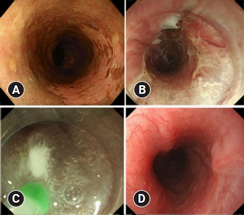
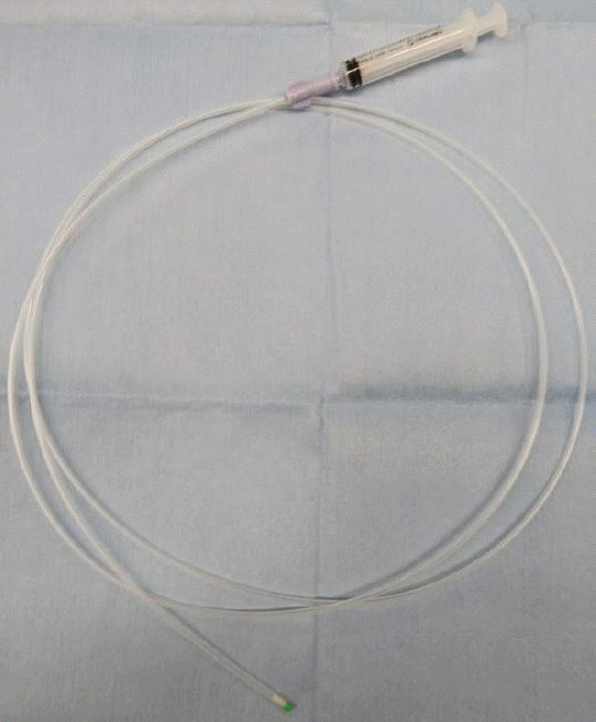
Immediately after resection of the lesion, intralesional steroid infusion using a spray tube (fine jet spraying type; Top Co., Tokyo, Japan) (Fig. 1) was performed, in which 40 mg of triamcinolone acetonide diluted in 20 mL of saline was locally infused (the product was not labeled for this use). By gently pressing the spray tube toward the base of the ulcer, 0.5 mL of the solution was infused with a 2.5-mL syringe. A white, cloudy bulge was formed when the triamcinolone acetonide solution was infused into the remaining submucosal layer. Triamcinolone acetonide was used in the range of 40–80 mg, depending on the size of the ulcer, and infusions were performed to cover the entire ulcer. The details of the procedure are shown in Figure 2 and Supplementary Video 1.
Endoscopic examination was performed before treatment, and follow-up examinations were performed 1 week, 1 month, and 2 months after treatment to test scope passage. The dysphagia score (on a scale of 0–5) was assessed at the same time as follows: 0, can eat a normal diet; 1, can swallow some solid foods; 2, can swallow only semi-solid foods; 3, can swallow liquids only; and 4, cannot swallow anything.10 Stenosis was determined if an upper gastrointestinal endoscope (GIF-H290; Olympus Medical System Co., Tokyo, Japan) with an 8.9-mm tip diameter could not be passed through the lesion after ESD or if the dysphagia score increased by 1 point or more compared to the pretreatment score. EBD was performed if stenosis was determined. The EBD procedure was performed until the endoscope could be passed and the dysphagia was improved, and the number of EBDs performed until the stenosis was relieved was recorded.
Complications of ESD such as bleeding, perforation, and mediastinal abscess formation were evaluated based on endoscopic findings and clinical symptoms at the time of treatment and 1 week, 1 month, and 2 months after treatment.
Twenty-seven patients underwent intralesional steroid infusion with a spray tube after en bloc resection by ESD for superficial esophageal cancer. Twenty-six patients underwent sub-circumferential resection, and one patient underwent circumferential resection. Table 1 shows the clinicopathological characteristics of the patients. The intralesional steroid infusion required an average time of 4.0±0.7 minutes.
Table 2 shows the posttreatment course of each patient. Among the 27 patients, 22 (81.5%) had a favorable outcome without stenosis. Stenosis was identified in five patients (18.5%), and EBD was performed. After a maximum of four EBD procedures, the scope could be passed, and the dysphagia symptoms improved in all patients. Only one patient had post-ESD bleeding, and no complications related to intralesional steroid infusion were observed.
Intralesional steroid injection is an effective method for preventing stenosis after extensive resection via esophageal ESD. Hashimoto et al.3 reported that when triamcinolone acetonide was injected locally for 3 days for resections involving more than three-quarters of the lumen, the rate of stenosis was 19%. Hanaoka et al.4 reported that when 100 mg of triamcinolone acetonide was injected only once immediately after ESD for resection involving more than three-quarters but less than the entire circumference of the lumen, the rate of stenosis was 10%. The rate of stenosis was 18.5% (5/27) with our intralesional steroid infusion method, which was not inferior to that reported in previous studies. None of the patients suffered refractory stenosis. The symptoms of stenosis in the five patients with stenosis were relieved and did not recur after a maximum of four EBD procedures.
Serious complications associated with steroid injections have been reported. In animal models, when triamcinolone acetonide was injected into the muscle layer of a post-ESD artificial ulcer, healing was prolonged, and perforations and abscess formation involving the lungs and aorta were reported.11 Delayed perforation and abscess formation after steroid injection have also been reported in patients.7 These injection methods require needle insertion into the very thin, remaining submucosal layer after ESD. It takes great care to avoid penetrating the muscular layers, and the mental stress on the endoscopist is enormous. In contrast, with the spray tube method, intralesional steroid infusion can be performed by lightly pressing the spray tube on the ulcer bed. Because a white, cloudy bulge can be recognized when triamcinolone acetonide solution is infused into the remaining submucosal layer, our method may be effective in infusing sufficient triamcinolone acetonide into this layer. The risk of injection into the muscle layer is low; thus, no complications related to this technique were recognized in this study. We believe that intralesional steroid infusion is easier and safer than direct injection.
Pathological diagnosis after ESD showed that 11 patients had an invasion depth deeper than the muscularis mucosa, which should be considered for additional treatment. Ten patients did not want to undergo additional treatment because of old age or comorbidities. One patient was treated with chemoradiation after the postoperative stenosis resolved.
The mean tumor diameter in patients with and without stenosis was 40.8±5.7 mm and 34.6±11.6 mm, respectively. Although the difference was not statistically significant, the tumor size tended to be larger in patients with stenosis. We excluded patients with a tumor diameter >5 cm in whom the entire circumference was resected; however, tumors with a diameter close to 5 cm also require attention.
In this study, we performed intralesional steroid infusion only once after ESD, and the average duration of the procedure was 4 minutes, making our technique a time-saving procedure. Another non-needle method, which involves filling the esophagus with a steroid solution, has been reported to have a prophylactic effect on stenosis.12 However, this procedure had to be performed at least twice. Moreover, stenosis developed in 45.5% of patients and required one to 13 repeat procedures.
The limitation of this study is that it was a single-center study that did not compare the present technique with conventional needle-based methods. Future randomized studies comparing these two methods are needed. Moreover, this study evaluated patients only treated with steroid infusion, and the effect on patients requiring combination therapy with oral steroids was not evaluated.
In conclusion, intralesional steroid infusion using a spray tube is a simple and safe technique and may have a comparable effect to that of the conventional injection method for preventing stenosis.
Supplementary Material
Supplementary materials related to this article can be found online at https://doi.org/10.5946/ce.2021.262.
Supplementary Video 1. Intralesional steroid infusion procedure using a spray tube (patient no. 5 in Table 2) (https://doi.org/10.5946/ce.2021.262.v001).
REFERENCES
1. Katada C, Muto M, Manabe T, et al. Esophageal stenosis after endoscopic mucosal resection of superficial esophageal lesions. Gastrointest Endosc. 2003; 57:165–169.
2. Hagel AF, Naegel A, Dauth W, et al. Perforation during esophageal dilatation: a 10-year experience. J Gastrointestin Liver Dis. 2013; 22:385–389.
3. Hashimoto S, Kobayashi M, Takeuchi M, et al. The efficacy of endoscopic triamcinolone injection for the prevention of esophageal stricture after endoscopic submucosal dissection. Gastrointest Endosc. 2011; 74:1389–1393.
4. Hanaoka N, Ishihara R, Takeuchi Y, et al. Intralesional steroid injection to prevent stricture after endoscopic submucosal dissection for esophageal cancer: a controlled prospective study. Endoscopy. 2012; 44:1007–1011.
5. Yamaguchi N, Isomoto H, Nakayama T, et al. Usefulness of oral prednisolone in the treatment of esophageal stricture after endoscopic submucosal dissection for superficial esophageal squamous cell carcinoma. Gastrointest Endosc. 2011; 73:1115–1121.
6. Ishida T, Morita Y, Hoshi N, et al. Disseminated nocardiosis during systemic steroid therapy for the prevention of esophageal stricture after endoscopic submucosal dissection. Dig Endosc. 2015; 27:388–391.
7. Yamashina T, Uedo N, Fujii M, et al. Delayed perforation after intralesional triamcinolone injection for esophageal stricture following endoscopic submucosal dissection. Endoscopy. 2013; 45 Suppl 2 UCTN:E92.
8. Rajan E, Gostout C, Feitoza A, et al. Widespread endoscopic mucosal resection of the esophagus with strategies for stricture prevention: a preclinical study. Endoscopy. 2005; 37:1111–1115.
9. Kadota T, Yoda Y, Hori K, et al. Prophylactic steroid administration against strictures is not enough for mucosal defects involving the entire circumference of the esophageal lumen after esophageal endoscopic submucosal dissection (ESD). Esophagus. 2020; 17:440–447.
10. Knyrim K, Wagner HJ, Bethge N, et al. A controlled trial of an expansile metal stent for palliation of esophageal obstruction due to inoperable cancer. N Engl J Med. 1993; 329:1302–1307.
11. Yamashita S, Kato M, Fujimoto A, et al. Inadequate steroid injection after esophageal ESD might cause mural necrosis. Endosc Int Open. 2019; 7:E115–E121.
12. Shibagaki K, Ishimura N, Oshima N, et al. Esophageal triamcinolone acetonide-filling method: a novel procedure to prevent stenosis after extensive esophageal endoscopic submucosal dissection (with videos). Gastrointest Endosc. 2018; 87:380–389.
Fig. 2.
(A) A patient with early esophageal cancer in the upper esophagus (patient no. 22 in Table 2). (B) Sub-circumferential resection was performed by endoscopic submucosal dissection. (C) Immediately after resection of the lesion, intralesional steroid infusion using a spray tube was performed. (D) The resected surface was completely re-epithelialized without stenosis 2 months after endoscopic submucosal dissection.

Table 1.
Clinicopathological characteristics of the patients
Table 2.
Posttreatment course of the patients


 PDF
PDF Citation
 Print
Print


 XML Download