INTRODUCTION
METHODS
Objectives
Methods
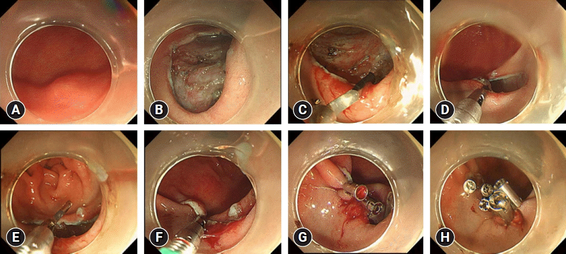
1) Group A (fishhook traction clip suture group)
 | Fig. 2.The procedure of endoscopic submucosal dissection using fishhook traction clips to suture the wound. (A) A submucosal tumor on the greater curvature of the stomach, approximately 1.0×1.0 cm. (B) After removing the lesion, the wound area was approximately 2.0×3.0 cm. (C) A fishhook traction clip was used to clamp the mucosa on the side edge of the wound. (D) The mucosa was lifted so that the hook-like device penetrated the mucosa. (E) The fishhook traction clip was opened, and the mucosa was pushed from the oral side to the anal side. (F) The anal mucosa of the wound was clamped, and the wound orifice and anal side were seamed. (G) The reduced wound was sutured with ordinary metal clips. (H) The wound was sutured well. |
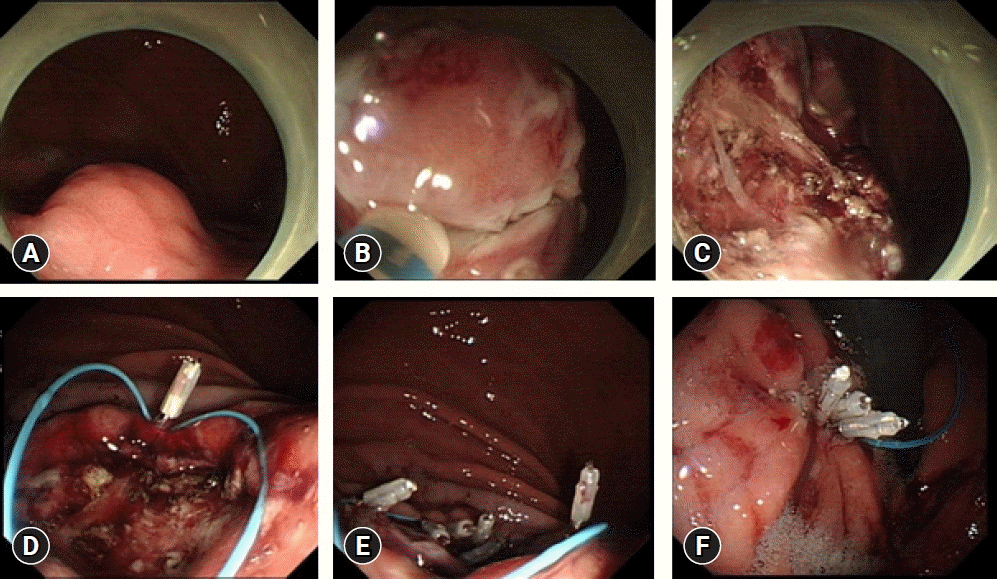
2) Group B (purse-string suture group)
 | Fig. 3.The procedure of endoscopic submucosal dissection using purse-string suture. (A) A submucosal tumor about 1.8×1.5 cm in the posterior wall of the upper gastric body. (B) Make circular incision of the mucosa, expose the lesion, and peel off the tumor. (C) The wound after resection was about 2.5×3.0 cm, with 2 mm small perforations locally. (D) The first metal clip fixes the nylon rope on the distal side of the wound. (E) Use metal clips to fix the nylon rope around the wound several times. (F) Finally tighten the nylon rope to suture the wound. |
Observation index
Statistical analyses
Ethical statements
RESULTS
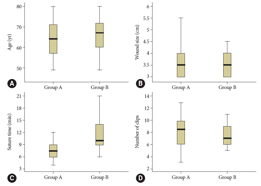
 | Fig. 4.Box plots of observation index comparison. These four groups of data can be statistically compared, and box plots are used to compare the data more intuitively. (A) Comparison of the age of the two groups. (B) Comparison of the wound size of the two groups. (C) Comparison of the suture time of the two groups. (D) Comparison of the number of clips of the two groups. |


 PDF
PDF Citation
 Print
Print


 XML Download