Journal List > J Korean Med Sci > v.37(39) > 1516079297

Kim, Kim, and Chung: Missed Ipsilateral Adrenal Adenoma With Recurrent Hypercortisolism After Prior Left Adrenalectomy
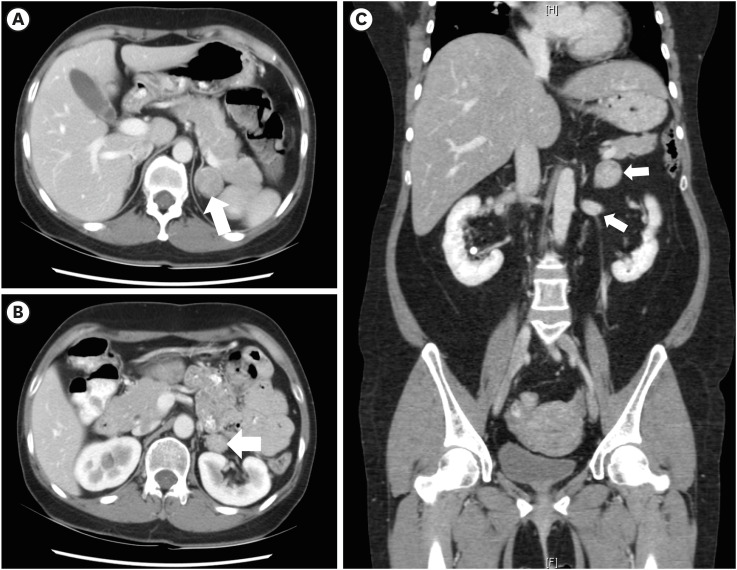
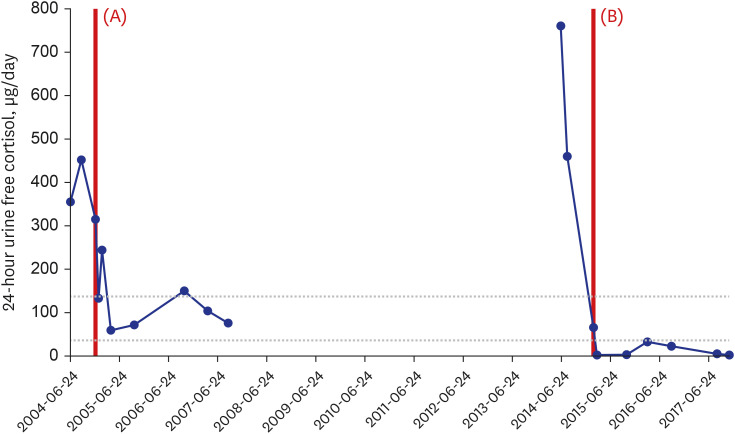
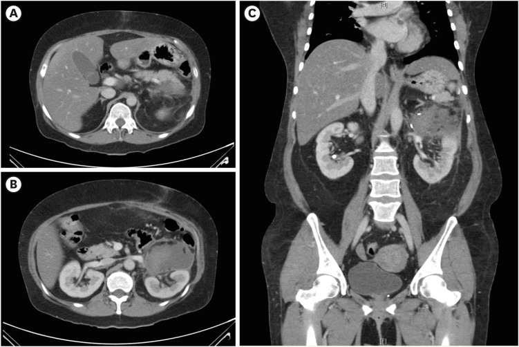
A 42-year-old woman with history of diabetes mellitus (DM), hyperlipidemia and subclinical hypercortisolism with previous left adrenalectomy was admitted for the evaluation of recurrent hypercortisolism due to gain of weight (8 kg over 3 months).1 The woman was on oral diabetic and hypertension medication (metformin, glimepiride, losartan, and amlodipine), without exposure of exogenous glucocorticoid. The woman had no familial history of hypercortisolism. Plasma ACTH levels at 8 A.M. and 4 P.M. were within normal range (16.2 pg/mL and 18.0 pg/mL, respectively), but 24-hour urine free cortisol was elevated (760.5 μg/day). A 3 × 2 cm mass on lower surgical bed level of previously resected left adrenal region was found in abdominal CT and showed increased uptake in 131I-iodocholesterol (NP-59) scan. Suggestive of adrenocortical adenoma, left adrenal mass was resected and pathology was concordant with adrenocortical adenoma. Follow-up abdominal CT 1 week after second left adrenalectomy showed left adrenal gland removed state with hematoma.
In this case, ipsilateral adrenal mass beside primary lesion was noticed in CT (Fig. 1) after the patient developed overt hypercortisolism after 10 years of previous left partial adrenalectomy.2 The 24-hour urine free cortisol was normalized after first adrenalectomy but increased after 10 years as shown in Fig. 2, suggesting that initially undetected adrenal mass changed its nature from subclinical to overt hypercortisolism over 10 years. Clinical presentation of hypercortisolism was resolved after second adrenalectomy (Fig. 3) and postoperative glucocorticoid treatment (hydrocortisone 40mg/day PO) was required.3 The level of 24-hour urine free cortisol decreased to 32.5 μg/day (normal range; 37–136 μg/day) in 1 year after second left adrenalectomy and the level of 8 A.M. plasma ACTH level increased to 52.8 pg/mL (normal range; 7.2–63.3 pg/mL) in 3 years after second left adrenalectomy.

ACKNOWLEDGMENTS

The authors thank Professor Seung-Whan Cha, Department of Radiology, Yonsei University Wonju College of Medicine, Wonju, Korea, for his technical assistance for this study.

Notes

Disclosure: No potential conflict of interest relevant to this article was reported.

Author Contributions:

  • Conceptualization: Chung CH.

  • Data curation: Chung CH.

  • Formal analysis: Kim J, Kim HK.

  • Investigation: Kim J.

  • Methodology: Kim J, Kim HK.

  • Visualization: Kim J.

  • Writing – original draft: Kim J.

  • Writing - review & editing: Kim J, Kim HK, Chung CH.

References

1. Kim KH, Kim KH, Ryu HY, Nam SM, Lee MY, Koh JH, et al. A case of adrenocortical adenoma causing subclinical cushing’s syndrome mistaken for Liddle’s syndrome. J Korean Endocr Soc. 2006; 21(1):58–62.
crossref
2. Ko OS, Kim JY, Kim HJ, Jeong YB. Laparoscopic partial adrenalectomy: surgical technique and outcome. Korean J Urol Oncol. 2019; 17(2):103–109.
crossref
3. Raffaelli M, De Crea C, D’Amato G, Gallucci P, Lombardi CP, Bellantone R. Outcome of adrenalectomy for subclinical hypercortisolism and Cushing syndrome. Surgery. 2017; 161(1):264–271. PMID: 27865591.
crossref
Fig. 1

Computed tomography images before the first adrenalectomy. (A) Axial view of first adrenal mass, which underwent surgical resection. (B) Axial view of ipsilateral additional adrenal mass. (C) Coronal view.

jkms-37-e278-g001
Fig. 2

Level of 24-hour urine free cortisol (μg/dL). (A) First left adrenalectomy (December 12, 2004). (B) Second left adrenalectomy (February 17, 2015).

jkms-37-e278-g002
Fig. 3

Following CT images 1 week after second adrenalectomy showing left adrenal gland removed state with hematoma (8.2 × 4.9 × 7.8 cm) in left anterior pararenal space. (A) Axial view of post-operation state of second adrenalectomy. (B) Axial view of post-operation state of second adrenalectomy. (C) Coronal view.

jkms-37-e278-g003
TOOLS
Similar articles