

 PDF
PDF Citation
 Print
Print


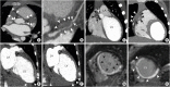
A 6-year-old male presented with acute epigastric pain. Coronary computed tomography angiography (CCTA) was performed to exclude the possibility of a coronary artery anomaly after consecutive echocardiograms showing inconspicuous signals in the middle left anterior descending (LAD) and right coronary arteries (RCA) and suspicious findings of dilated cardiomyopathy. Giant coronary artery aneurysms (CAA) with thrombotic occlusion and recanalization (Figure 1) were identified in the proximal LAD and RCA on the CCTA, leading to a diagnosis of incomplete Kawasaki disease (KD) complicated by acute coronary syndrome. One month previously, the patient presented to the emergency department due to acute pancreatitis, but without fever and classic mucocutaneous manifestations of KD, consistent with incomplete KD.
Complete occlusion of CAA leading to ACS in this patient may have been caused by both the large size of the CAA (i.e., turbulent and stagnant flow) and delayed diagnosis (i.e., no prior use of immunoglobulin therapy, aspirin, or anticoagulation).1)2) Using cutting edge techniques that minimize radiation exposure, CCTA was performed with very low radiation dose (<1 mSv) in this patient.1)2) The primary imaging tool to evaluate CAA in patients with KD is echocardiography. However, echocardiography has important drawbacks such as a limited acoustic window and operator dependency.2) Initial diagnosis of KD was made by CCTA in this patient rather than echocardiography. Thus, use of CCTA should be considered in patients with non-visualization of coronary flow on echocardiography or clinical suspicion of incomplete KD to avoid delayed diagnosis.
The patient’s informed consent was waived by Institutional Review Board approval (CHA University, IRB-2022-02-020).
 XML Download