Transarterial radioembolization (TARE) has recently emerged as a new therapeutic option for patients with hepatocellular carcinoma. Microspheres impregnated with the radioisotope yttrium-90 (90Y) are selectively delivered through the hepatic artery, which feeds the tumor. 90Y, a β-emitting isotope with a short half-life (2.67 days), exerts powerful anti-cancer effects, with the emitted β particles having mean and maximum tissue penetration depths of 2.5 and 10 mm, respectively [1]. Here, we report a patient who presented with poorly differentiated thyroid cancer (PDCA) with lung, liver, and bone metastases and was successfully treated with TARE for liver metastases.
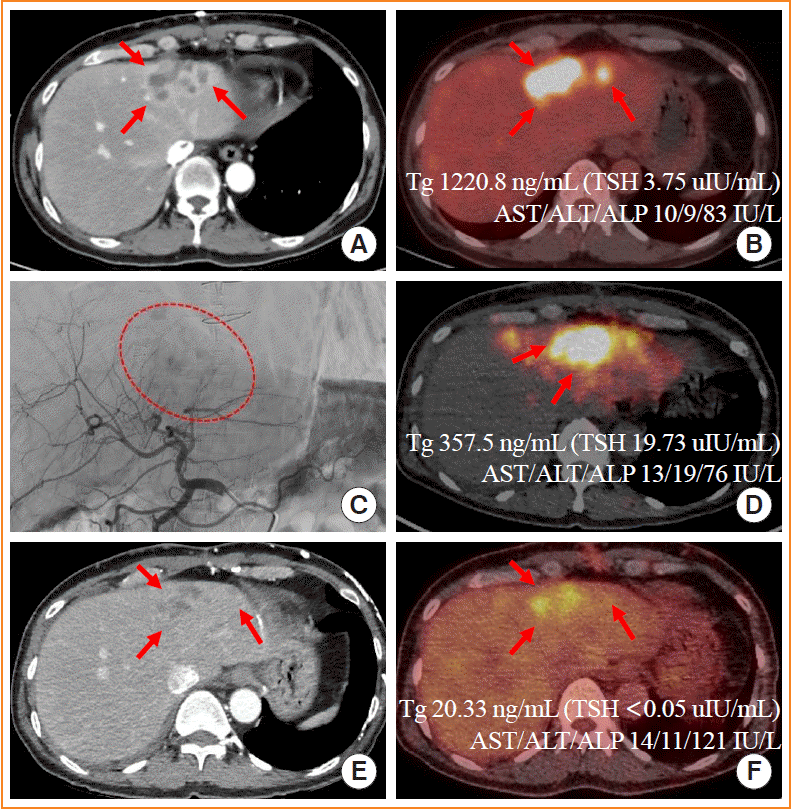
A 55-year-old man was referred for a huge neck mass and was suspected of having metastatic thyroid cancer. The primary thyroid mass in the right lobe invaded the trachea, and very large ipsilateral metastatic lymph nodes encased the right common carotid artery. A bone-destructive mass in the anterior mediastinum extended to the upper mediastinum and compressed the left innominate vein. Other hypermetabolic masses were found in the right acetabulum, both lungs, and the left lobe of the liver. The liver masses showed heterogeneous enhancement on arterial-phase dynamic computed tomography (CT) and intense hypermetabolism on 18F-fluorodeoxyglucose (FDG) positron emission tomography (PET)/CT (Fig. 1A, B).
Total thyroidectomy and neck dissection, innominate vein resection and reconstruction, en bloc resection of the sternum, and wedge resection of the lung were performed. Pathologic diagnosis concluded that the tumor was a PDCA originating from the tall cell variant of papillary thyroid cancer. BRAFV600E and TERTC228T mutations were identified. Planning angiography and a lung shunt scan showed minimal vascularity of the liver metastases (Fig. 1C) and negligible extrahepatic activity, respectively.
For hepatic metastasis, loco-regional treatments such as surgical resection, radiofrequency ablation therapy (RFA), transarterial chemoembolization (TACE), or external radiation therapy (EBRT) and/or systemic 131I therapy are traditionally considered [2]. However, the applications of loco-regional treatment options to large masses with weak arterial-phase hyperenhancement (APHE) are limited. In this particular case, surgical treatment was not suitable because of the patient’s previous major surgery. The tumor (5.2 cm) was too large to be treated by RFA. 131I was expected to be relatively ineffective as the tumor was PDCA and showed high FDG uptake. TACE was also not suitable because of weak APHE. We thoroughly discussed the possible treatment options, including EBRT and TARE, with the patient; finally, TARE was chosen.
Radioactive 90Y-labeled resin microspheres (total 1.2 GBq) were injected into the segment IV artery and the segment II/III artery, and the mean target absorbed dose was 110 Gy (Fig. 1D). The patient neither suffered from post-embolization symptoms, such as fever and right upper abdominal pain, nor showed any signs of hepatic failure. One month after the procedure, contrast-enhanced dynamic CT and 18F-FDG PET/CT demonstrated markedly reduced arterial enhancement and metabolism, respectively (Fig. 1E, F).
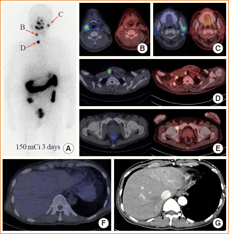
After TARE treatment, the patient received EBRT to the neck for locoregional disease control, followed by 131I therapy with 150 mCi for systemic disease control. A whole-body scan performed 3 days after 131I therapy identified radioactive iodine (RAI)-avid masses in the neck (Fig. 2A-D) and RAI-refractory masses in the liver and right acetabulum (Fig. 2E, F). Three months after TARE, dynamic CT showed progressive reduction of the metastatic mass in the liver (Fig. 2G). We then planned lenvatinib treatment for the RAI-refractory metastatic masses.
It has been reported that TARE led to a lower frequency of post-embolization syndrome, a longer time to progression, and a comparable overall survival rate relative to TACE in unresectable hepatocellular carcinoma [3,4]. One interesting case of liver metastasis of papillary thyroid cancer managed with TARE was reported. The metastatic mass in the liver was RAI-refractory and unsuitable for TACE because of its large size (20 cm). Primary surgical resection was attempted, but failed due to a large amount of blood loss from multiple enlarged feeding veins extending into the tumor. After TARE treatment, the tumor’s size was reduced to 15 cm. The metastatic mass, which had changed to become dense and fibrotic, could be surgically removed [5]. Likewise, TARE was successfully applied for a large, hypervascular metastatic mass in the liver from thyroid cancer as a bridging therapy to surgical resection. Both in this case and our case, the expected effects were not influenced by the tumor differentiation status, which is the main limitation of 131I therapy. Therefore, TARE could be considered alternatively as an effective locoregional treatment option not only for metastatic masses unsuitable for surgery or TACE, but also for large poorly differentiated tumors with weak APHE.
ACKNOWLEDGMENTS
We thank Injae Wang for proofreading the manuscript. There is no funding information to declare.
REFERENCES
1. Salem R, Lewandowski RJ, Sato KT, Atassi B, Ryu RK, Ibrahim S, et al. Technical aspects of radioembolization with 90Y microspheres. Tech Vasc Interv Radiol. 2007; 10:12–29.
2. Haugen BR, Alexander EK, Bible KC, Doherty GM, Mandel SJ, Nikiforov YE, et al. 2015 American Thyroid Association management guidelines for adult patients with thyroid nodules and differentiated thyroid cancer: the American Thyroid Association Guidelines task force on thyroid nodules and differentiated thyroid cancer. Thyroid. 2016; 26:1–133.

3. Salem R, Lewandowski RJ, Kulik L, Wang E, Riaz A, Ryu RK, et al. Radioembolization results in longer time-to-progression and reduced toxicity compared with chemoembolization in patients with hepatocellular carcinoma. Gastroenterology. 2011; 140:497–507.e2.

Fig. 1.
(A) A dynamic computed tomography (CT) scan of the hepatic arterial phase performed before transarterial radioembolization (TARE) shows heterogeneous enhancement (low in the central area and high in peripheral area) of liver metastases (red arrows). (B) An 18F-fluorodeoxyglucose (18F-FDG) positron emission tomography (PET)/CT scan at the same level shows metastatic masses with hypermetabolism (red arrows). (C) A common hepatic angiogram shows minimal hypervascularity of liver metastases (red circle with dotted line). (D) PET/CT immediately after TARE shows high radioactivity within the tumor (red arrows). (E) A CT scan of the hepatic arterial phase performed 1 month after TARE shows reduced arterial enhancement (red arrows). (F) An 18F-FDG PET/CT scan at the same level as (B) shows reduced metabolism (red arrows). Tg, thyroglobulin; TSH, thyroid-stimulating hormone; AST, aspartate aminotransferase; ALT, alanine aminotransferase; ALP, alkaline phosphatase.

Fig. 2.
(A) Whole-body scan performed 3 days after 131I therapy. Red arrows indicate radioactive iodine (RAI)-avid masses in the neck. (B, C, D, E) RAI-avid masses in the neck (B, C, D) and RAI-refractory masses in the right acetabulum (E). The left panels show 131I single-photon emission computed tomography/computed tomography (SPECT/CT) scans, and the right panels show 18F-fluorodeoxyglucose (18F-FDG) PET/CT scans at the same level. (F) A RAI-refractory mass in the liver. (G) A dynamic CT scan 3 months after transarterial radioembolization.


 PDF
PDF Citation
 Print
Print


 XML Download