Abstract
Background
Immunoglobulin G4 (IgG4)-related disease is an entity that can involve the thyroid gland. The spectrum of IgG4-related thyroid disease (IgG4-RTD) includes Hashimoto thyroiditis (HT) and its fibrotic variant, Riedel thyroiditis, as well as Graves’ disease. The early diagnosis of IgG4-RTD is important because it is a medically treatable disease, and a delay in the diagnosis might result in unnecessary surgery. We present a case series of IgG4-RTD with a review of the literature.
Methods
We retrospectively reviewed the clinical presentation and the radiological and pathological findings of patients diagnosed with IgG4-RTD between 2017 and 2021 at a tertiary medical center in Korea. We also conducted a literature review of IgG4-RTD.
Results
Five patients were diagnosed with IgG4-RTD during the study period. The patients’ age ranged from 31 to 76 years, and three patients were men. Most patients visited the clinic for a neck mass, and hypoechogenic nodular lesions were observed on neck ultrasonography. Three patients had IgG4 HT, and two patients had IgG4 Riedel thyroiditis. All patients developed hypothyroidism that necessitated L-thyroxine replacement. The diagnosis of IgG4-RTD was confirmed after a pathological examination of the surgical specimen in the first two cases. However, the early diagnosis was possible after a core needle biopsy in three clinically suspected patients.
Conclusion
The diagnosis of IgG4-RTD requires clinical suspicion combined with serology and histological analyses using IgG4 immunostaining. The early diagnosis of IgG4-RTD is difficult; thus, biopsy with IgG4 immunostaining and serum IgG4 measurements will help diagnose patients suspected of having IgG4-RTD.
Immunoglobulin G4 (IgG4)-related disease, a disease entity involving multiple organs, is characterized by dense lymphoplasmacytic infiltration of IgG4-positive plasma cells in various involved tissues [1-3]. Since autoimmune pancreatitis was reported in 2001, similar fibro-inflammatory diseases such as Mikulicz syndrome, retroperitoneal fibrosis, Kűttner tumor, and Riedel thyroiditis (RT) have been unified under the unique spectrum of IgG4-related disease [1,3-7]. It is typically characterized by elevated serum IgG4 levels and a good response to steroid therapy [2,8]. Thyroid gland involvement in IgG4-related disease was suggested based on the frequent observation of hypothyroidism and thyroid autoantibodies in patients with autoimmune pancreatitis [9,10].
A unique subgroup of Hashimoto thyroiditis (HT) with increased IgG4-positive plasma cells in the thyroid tissue was first reported in 2009 [11]. Since then, the identification of other subtypes of IgG4-rich thyroid conditions has been reported. This expanding spectrum of IgG4-related thyroid disease (IgG4-RTD) now includes HT and its fibrotic variant (FVHT), RT, and Graves’ disease (GD) [8]. The pathogenesis of IgG4-RTD involves genetic factors, antigen-antibody reactions, and allergic phenomena, but remains poorly understood [2,12]. Due to insufficient awareness of this clinical entity, the prevalence of IgG4-RTD is likely to be underestimated. Most studies regarding IgG4 HT were reported in Japan, with the remainder in China and Germany [13-16].
IgG4-RTD is a medically treatable disease in the vast majority of cases. Nonetheless, rapid progression of the disease and a delayed diagnosis might result in unnecessary surgery. Despite its clinical importance, there have been only a few case reports of IgG4-RTD, including RT, in Korea [17-19]. IgG4 immunostaining data were insufficient in these reports, and the diagnosis was confirmed after total thyroidectomy. In some recent cases, we have diagnosed IgG4-RTD by core needle biopsy (CNB) before surgery. Therefore, we report a case series of IgG4-RTD from a single institution and present a literature review of IgG4-RTD focusing on IgG4-related HT.
We retrospectively reviewed the medical records of five patients diagnosed with IgG4-related thyroiditis between 2017 and 2021 at a tertiary medical center in Korea. For each case, clinical presentation, radiology, pathology, treatment, and clinical outcomes were described in detail. This study was approved by the Institutional Review Board of Asan Medical Center (No. 2021-0867). The requirement of informed consent from the patients was waived due to the retrospective study design.
The reference ranges of thyroid-stimulating hormone (TSH) and free thyroxine (fT4) were 0.4 to 4.5 mIU/L and 0.80 to 1.90 mg/dL, respectively. The anti-thyroid peroxidase antibody (TPOAb) level was determined by radioimmunoassay (BRAHMS anti-TPOn RIA, Thermo Fisher, Waltham, MA, USA), and a value of ≥60 U/mL was considered positive. The anti-thyroglobulin antibody (TgAb) level was also measured by radioimmunoassay (BRAHMS anti-Tgn RIA), and a value of ≥60 U/mL was considered positive [20]. Serum IgG was measured using nephelometry (nephelometer, Dade Behring, Siemens, Munich, Germany) and IgG4 using a single radial immunodiffusion method (The Binding Site, Birmingham, UK) [21]. A serum IgG4 level ≥135 mg/dL was considered to indicate positivity [2,22]. An experienced endocrine pathologist (D.E.S.) reviewed all the specimens and confirmed the diagnosis of IgG4-RTD. The histological criteria for diagnosis of IgG4-thyroiditis were as follows: ≥20 IgG4-positive plasma cells/high power field (HPF) and an IgG4/IgG ratio >30% [13]. The diagnosis of IgG4-RTD was categorized as definite, probable, or possible based on the recently updated criteria of IgG4-RTD [22].
We performed a literature review using the keywords “IgG4,” “thyroid,” “Hashimoto,” “Riedel’s thyroiditis,” and “Graves’ disease” in the PubMed database. We also found related studies by referring to several recent reviews [8,23-25]. We identified nine studies on IgG4-related HT, one study on FVHT, 10 studies on IgG4-related RT, and eight studies on IgG4-related GD. Each spectrum of disease was briefly summarized.
We report five cases of IgG4-RTD. Their baseline characteristics and clinical courses are summarized in Table 1. Three patients had IgG4-related HT and two patients had IgG4-related RT. IgG4-RTD was diagnosed after total thyroidectomy in two cases and CNB with IgG4 immunostaining in three cases. All the patients developed hypothyroidism that necessitated L-thyroxine replacement; two of them experienced transient thyrotoxicosis. Hypoechoic nodular lesions were revealed on neck ultrasonography (US), and increased serum IgG4 levels were found in two patients. The detailed case descriptions follow.
A 31-year-old man visited the clinic with progressive enlargement of a large neck mass. Physical examination revealed a large diffuse goiter without tenderness. The serum TSH and fT4 levels were 28.4 mIU/L and 0.91 ng/dL, respectively. Both TPOAb and TgAb were positive.
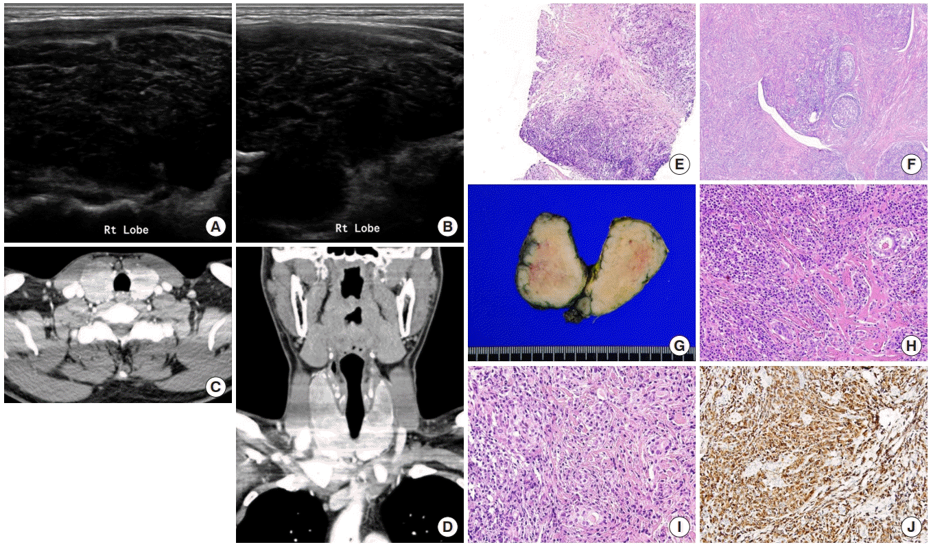
He was treated for subclinical hypothyroidism with L-thyroxine for 6 months. During follow-up, the size of the goiter progressively increased, and the initial CNB-based diagnosis was granulomatous thyroiditis with dense stromal fibrosis (Fig. 1E, F). IgG and IgG4 staining were not performed on the sample from this CNB. He underwent total thyroidectomy due to a progressive increase of goiter and a thyroid weight of up to 135 g (Fig. 1G). Pathological examination revealed a dense lymphoplasmacytic infiltrate, interlobular and interfollicular fibrosis, and increased IgG4-positive plasma cells (up to 152 cells/HPF) with an IgG4/IgG ratio of 55%, confirming the diagnosis of IgG4-related HT (Fig. 1H-J).
A 41-year-old man visited the clinic with a neck mass and dysphagia. A firm fixed large neck mass was palpable on physical examination. He was treated with L-thyroxine due to overt hypothyroidism (TSH, 76.8 mIU/L; fT4, 0.19 ng/dL). Both TPOAb and TgAb were positive.
He underwent total thyroidectomy after CNB, which indicated suspicious papillary carcinoma (Bethesda category V) with marked stromal fibrosis. Pathology revealed extensive stromal fibrosis with patchy lymphoplasmacytic infiltration, indicating IgG4-related RT. IgG4-positive plasma cells had increased up to 93 cells/HPF, and the IgG4/IgG ratio was 42%. There was no evidence of malignancy in surgical pathology.
A 76-year-old woman visited our clinic with a palpable neck mass without pain. Her serum TSH and fT4 levels were 0.09 mIU/L and 2.0 ng/dL, respectively. TPOAb and TgAb were negative.
The CNB pathology revealed lymphocytic thyroiditis with focal granulomatous thyroiditis features, interfollicular fibrosis, and increased IgG4-positive plasma cells (up to 63 cells/HPF) with an IgG4/IgG ratio of 35%, suggesting IgG4-related HT (Fig. 2C-E). The serum IgG4 level had increased to 107 mg/dL.
To evaluate the possible presence of systemic disease, we performed neck, chest, and abdominal-pelvic CT; there was no evidence of other organ involvement. She received L-thyroxine replacement therapy due to hypothyroidism during follow-up. Steroid or surgical treatments were considered should the thyroid nodule become larger.
A 72-year-old man was referred to our clinic with a thyroid nodule identified during a routine health examination. His serum TSH and fT4 levels were 0.04 mIU/L and 3.73 ng/dL, respectively, indicating thyrotoxicosis.
There was no evidence of other organ involvement in a systemic evaluation. Four months after the thyrotoxicosis phase, we started L-thyroxine therapy for the newly developed hypothyroidism. Currently, the patient is being followed up with thyroid function tests and US without steroids or surgery.
A 66-year-old woman visited our clinic with general weakness. Her serum TSH and fT4 levels were 12.5 mIU/L and 1.1 ng/dL, respectively, indicating subclinical hypothyroidism.
The serum IgG4 level had increased to 1,070 mg/dL, with multiple enlarged mesenteric lymph nodes on abdominal-pelvic CT, necessitating follow-up. She is being treated with L-thyroxine and followed with neck US, and is under consideration for steroid treatment in case of deterioration.
We presented three cases (cases 1, 3, and 5) of IgG4-related HT in this study. Autoantibodies and serum IgG4 had increased in two patients (cases 1 and 5). One patient underwent total thyroidectomy for progressive goiter, and two patients were being treated with L-thyroxine and closely followed up. We found nine studies in the literature on IgG4-related HT from several countries (Table 2) [11,13-16,26-29]. Most of the early research began in Japan, and the authors proposed the new term “IgG4 thyroiditis” [11,13,26]. Based on the immunohistochemical staining pattern for IgG4 on surgical pathology, IgG4 thyroiditis can be sub-classified as distinct from HT, with a more severe lymphoplasmacytic infiltration, dense fibrosis, and marked follicular cell degeneration [11]. In 2012, the same group performed a larger study including 105 patients with HT; 28 had IgG4 thyroiditis and 77 had non-IgG4 thyroiditis [13]. They extended the previous histopathological findings by describing the pattern of stromal fibrosis. IgG4 thyroiditis was significantly associated with the presence of predominant interfollicular fibrosis, whereas non-IgG4 thyroiditis was characterized by predominant interlobular fibrosis [13]. The prevalence of IgG4 thyroiditis in all HT patients ranged from 12.6% to 38.5% in several studies using surgical specimen databases. The discrepancies between Europe and Asia might be explained by genetic, dietary, and geographic differences [23]. Furthermore, one reason might be the histological criteria for the diagnosis of IgG4 thyroiditis [8,15]. In contrast to these studies, which used surgical specimens as the data source, two studies that screened serum total IgG levels in patients with HT diagnosed with IgG4 thyroiditis based on the serum criterion (IgG4 >135 mg/dL) showed lower prevalence (4.0% to 5.3%) [27,28,30]. In these two studies using serological databases, five and six patients were diagnosed with IgG4 thyroiditis, one and two of whom were confirmed by biopsy, respectively [27,28].
Since IgG4 thyroiditis began to emerge as a new clinicopathological entity, subsequent studies were reported to investigate the further meaning of this sub-classification, including the serum IgG4 level. Unlike the earlier study by Li et al. [26], Zhang et al. [16], and Raess et al. [29] reported that there were no significant differences in serum total IgG and IgG4 levels between the IgG4 thyroiditis and non-IgG4 thyroiditis groups. Furthermore, other studies reported that only 4.0% to 5.3% of patients with HT were diagnosed with IgG4 thyroiditis based on the serum criterion [2,27,28]. These findings suggest that serum IgG and IgG4 levels were neither necessary nor adequate for the diagnosis of IgG4 thyroiditis. If additional criteria are met, “probable” IgG4-RTD can be diagnosed without serum IgG4 elevation according to the latest diagnostic criteria [22]. This is also supported by our study, which showed that serum IgG4 levels were elevated in two of three patients. However, the levels of IgG4 binding to specific thyroid antigens, such as TPOAb and TgAb, were significantly higher in the IgG4 thyroiditis group [16]. Thus, these markers, including TPOAb IgG4 and TgAb IgG4, might be helpful for differentiating the diagnosis.
In addition to histopathological findings, Li et al. [26] reported distinct clinical and ultrasonographic features between IgG4 thyroiditis and non-IgG4 thyroiditis. Compared with patients with non-IgG4 thyroiditis, patients with IgG4 thyroiditis were more likely to be younger and male, and showed more rapid progress, more subclinical hypothyroidism than euthyroidism, more diffuse low echogenicity, and higher levels of circulating antibodies [26]. These clinical features had some degree of discrepancy in other studies. In terms of age, Zhang et al. [16] and Jokisch et al. [15] also reported that patients with IgG4 thyroiditis were significantly younger than patients with non-IgG4 thyroiditis. However, this was not confirmed in the study by Takeshima et al. [28]. This may have resulted from the low prevalence of disease and regional differences. In general, according to previous studies, the mean age of patients with IgG4 thyroiditis was 42 to 74 years, with middle-aged men being typically affected (Table 2). Although a lower female-to-male ratio is a well-established finding in IgG4-related HT compared to non-IgG4 thyroiditis, 50% to 100% of patients diagnosed with IgG4 thyroiditis were women (Table 2). Despite its small numbers, our study revealed a comparable patient profile.
FVHT is a rare form of HT, accounting for 10% of patients with HT [31]. The main histopathological feature of FVHT is marked fibrous replacement of more than one-third of the thyroid parenchyma [31]. Thus, it is characterized by severe constrictive symptoms and a very firm thyroid gland with rapid enlargement. Deshpande et al. [32] first proposed that FVHT belongs to the spectrum of IgG4-RTD in 2012. These authors evaluated 28 cases of HT and nine cases of FVHT and found that compared to typical HT, FVHT showed a significantly higher IgG4-positive cell count, IgG4/IgG ratio, and hypothyroidism. The median age of the nine patients was 50 years, and one of them was male. Due to the rarity of the disease and the small numbers of the current study, we were unable to present an FVHT case in this study. More research is needed in the future.
RT is a rare form of chronic thyroiditis that characteristically extends through surrounding tissues with a prevalence of 1.06 cases per 100,000 patients [33]. Although the precise etiology of this disease remains unknown, in some cases it has been considered as the thyroid manifestation of multifocal fibrosis in combination with other fibrotic disorders. In 2010, Dahlgren et al. [7] reported surplus IgG4 plasma cells in three cases of RT surgical specimens, and they suggested that RT was a part of the IgG4-related systemic disease spectrum. A few case studies have evaluated RT in the literature, including a recent systematic review, as briefly presented in Table 3 [7,24,34-40]. The mean age of the 24 patients was 42 years (range, 21 to 81) and most (78.9%) were women (Table 3). Typically, hypothyroidism is not described in RT, and only 33% of the patients had hypothyroidism. We presented two cases (case 2 and 4) of IgG4-related RT in this study. Both of these patients were men and developed hypothyroidism. As RT is a rare disease, there have been neither treatment guidelines nor large prospective studies. Based on these studies, multiple treatment approaches for RT, including systemic steroids, tamoxifen, and thyroidectomy have been used [39]. Glucocorticoids are still the mainstay of therapy, and surgical management is administered when a histopathological diagnostic confirmation is needed or when acute compressive symptoms exist. In the current case series, one patient underwent total thyroidectomy to rule out malignancy; however, the other one is being followed up with thyroid function tests and US without steroid treatment or surgery. Additionally, IgG4-related RT was linked with other fibrosclerotic diseases; thus, a systemic work-up for other organs is important.
Both FVHT and RT are characterized by destruction of the thyroid architecture by stromal fibrosis, which blurs the differential diagnosis. Most importantly, FVHT is typically limited to the thyroid without extracapsular extension, unlike RT [8,41]. RT is also associated with lower thyroid antibody titers, the more frequent presence of autoimmune disease, and diminished Doppler flow on US compared to FVHT [2,41].
Since the first case of a patient with GD whose histological and serological findings indicated IgG4-RTD, studies on the relationship between GD and IgG4 have been actively conducted [42]. A subset of patients with GD have elevated serum IgG4 levels, which have been reported to be associated with thyroid eye disease (TED). As summarized in Table 4, the prevalence of elevated serum IgG4 ranges from 6.4% to 23.1% [25,43-50]. These patients were older at diagnosis and responded excellently to anti-thyroid drug therapy [23,43,46]. These studies reported that the prevalence of TED in patients with IgG4-related GD was as high as 37.5% to 100%. Furthermore, Yu et al. [48] reported that serum IgG4 levels were associated with the severity of TED. However, these studies focused on the relationship between serum IgG4 and GD, rather than histological confirmation, and the criteria for serum IgG4 elevation were varied. Therefore, the role of IgG4 in GD needs to be validated in the future. As with FVHT, we were unable to include IgG4-related GD patients in this study.
IgG4-RTD includes a heterogeneous spectrum of disease features; therefore, the approach to treating this disease may be complex. The recently proposed criteria by the Japan Endocrine Society comprise five items: (1) enlargement of the thyroid, (2) hypoechoic lesions in the thyroid evidenced by US, (3) elevated serum IgG4 levels, (4) histopathological findings in the thyroid lesion, and (5) the involvement of other organs [22]. The diagnosis is categorized as “definitive,” “probable,” or “possible” based on the degree to which the above five items are present.
Because histological features are the mainstay of the diagnosis, physicians’ decision to perform a biopsy with additional IgG4 immunostaining is most important for the early diagnosis of IgG4-RTD. Based on our experience and the literature review, enlargement of the thyroid lobe with hypoechoic lesions on US and combined thyroid dysfunction might be important factors. In our study, we did not diagnose the first two patients with IgG4-RTD by CNB because we did not perform IgG4 immunostaining. However, we performed CNB with immunostaining of IgG4 in the three more recent clinically suspected cases. Because IgG4-RTD is a medically treatable disease, an early diagnosis might reduce unnecessary surgery and subsequent complications.
Another issue is the technique of performing CNB in IgG4-RTD. The lesion in IgG4-RTD is particularly hard; thus, inserting the core needle into the lesion is challenging and needs caution. When the stylet of the core needle and the cutting cannula are fired, the needle recoils. The recoil must be minimized to collect an accurate sample and to minimize damage to the surrounding structures.
It is currently impossible to assess the prevalence of IgG4-RTD in Korea, because only a few cases have recently begun to be diagnosed. In addition, the treatment of patients (three out of five) in this study was not substantially different from the existing treatment of non-IgG4-RTD. However, it cannot be assumed that the findings are of little clinical significance for these reasons. Due to a lack of awareness and suspicion of this disease, IgG4-RTD might be underdiagnosed. In this study, two patients underwent surgery before the diagnosis of IgG4-RTD, and three cases were diagnosed in a single year thereafter. Therefore, we expect awareness and suspicion of this disease entity will lead to more diagnoses and an improved understanding of the disease. Furthermore, the prognosis and treatment will be evaluated accurately through longer follow-up.
This study describes the first case series of IgG4-RTD in Korea and reviews the current knowledge of IgG4-RTD. The early diagnosis of IgG4-RTD requires clinical features combined with serology and histological features with IgG4 immunostaining. We expect further research that will lead to a deeper understanding of IgG4-RTD in Korea.
ACKNOWLEDGMENTS
This study was supported by a grant (2021IL0039) from the Asan Institute for Life Sciences, Asan Medical Center, Seoul, Korea.
A part of this study was presented as poster at the Ninth Seoul International Congress of Endocrinology and Metabolism, 2021.
REFERENCES
1. Stone JH, Khosroshahi A, Deshpande V, Chan JK, Heathcote JG, Aalberse R, et al. Recommendations for the nomenclature of IgG4-related disease and its individual organ system manifestations. Arthritis Rheum. 2012; 64:3061–7.
2. Kottahachchi D, Topliss DJ. Immunoglobulin G4-related thyroid diseases. Eur Thyroid J. 2016; 5:231–9.
3. Hamano H, Kawa S, Horiuchi A, Unno H, Furuya N, Akamatsu T, et al. High serum IgG4 concentrations in patients with sclerosing pancreatitis. N Engl J Med. 2001; 344:732–8.
4. Yamamoto M, Takahashi H, Ohara M, Suzuki C, Naishiro Y, Yamamoto H, et al. A new conceptualization for Mikulicz’s disease as an IgG4-related plasmacytic disease. Mod Rheumatol. 2006; 16:335–40.
5. Fujimori N, Ito T, Igarashi H, Oono T, Nakamura T, Niina Y, et al. Retroperitoneal fibrosis associated with immunoglobulin G4-related disease. World J Gastroenterol. 2013; 19:35–41.
6. Geyer JT, Ferry JA, Harris NL, Stone JH, Zukerberg LR, Lauwers GY, et al. Chronic sclerosing sialadenitis (Kuttner tumor) is an IgG4-associated disease. Am J Surg Pathol. 2010; 34:202–10.
7. Dahlgren M, Khosroshahi A, Nielsen GP, Deshpande V, Stone JH. Riedel’s thyroiditis and multifocal fibrosclerosis are part of the IgG4-related systemic disease spectrum. Arthritis Care Res (Hoboken). 2010; 62:1312–8.
8. Rotondi M, Carbone A, Coperchini F, Fonte R, Chiovato L. Diagnosis of endocrine disease: IgG4-related thyroid autoimmune disease. Eur J Endocrinol. 2019; 180:R175–83.
9. Komatsu K, Hamano H, Ochi Y, Takayama M, Muraki T, Yoshizawa K, et al. High prevalence of hypothyroidism in patients with autoimmune pancreatitis. Dig Dis Sci. 2005; 50:1052–7.
10. Sah RP, Chari ST. Clinical hypothyroidism in autoimmune pancreatitis. Pancreas. 2010; 39:1114–6.
11. Li Y, Bai Y, Liu Z, Ozaki T, Taniguchi E, Mori I, et al. Immunohistochemistry of IgG4 can help subclassify Hashimoto’s autoimmune thyroiditis. Pathol Int. 2009; 59:636–41.
12. Topliss DJ. Clinical update in aspects of the management of autoimmune thyroid diseases. Endocrinol Metab (Seoul). 2016; 31:493–9.
13. Li Y, Zhou G, Ozaki T, Nishihara E, Matsuzuka F, Bai Y, et al. Distinct histopathological features of Hashimoto’s thyroiditis with respect to IgG4-related disease. Mod Pathol. 2012; 25:1086–97.
14. Zhao Z, Lee YJ, Zheng S, Khor LY, Lim KH. IgG4-related disease of the thyroid gland requiring emergent total thyroidectomy: a case report. Head Neck Pathol. 2019; 13:523–7.
15. Jokisch F, Kleinlein I, Haller B, Seehaus T, Fuerst H, Kremer M. A small subgroup of Hashimoto’s thyroiditis is associated with IgG4-related disease. Virchows Arch. 2016; 468:321–7.
16. Zhang J, Zhao L, Gao Y, Liu M, Li T, Huang Y, et al. A classification of Hashimoto’s thyroiditis based on immunohistochemistry for IgG4 and IgG. Thyroid. 2014; 24:364–70.
17. Lee IS, Lee JU, Lee KJ, Jang YS, Lee JM, Kim HS. Painful immunoglobulin G4-related thyroiditis treated by total thyroidectomy. Korean J Intern Med. 2016; 31:399–402.
18. Kim CS, Lee SJ, Park JS, Nam JY, Kim DM, Ahn CW, et al. A case of Riedel’s thyroiditis in a patient with a history of subacute thyroiditis. J Korean Soc Endocrinol. 2003; 18:414–9.
19. Lee DY, Moon JS, Kim GE, Kim HK, Kang HC. Riedel thyroiditis in a patient with graves disease. Endocrinol Metab (Seoul). 2013; 28:138–43.
20. Song E, Oh HS, Jeon MJ, Chung KW, Hong SJ, Ryu JS, et al. The value of preoperative antithyroidperoxidase antibody as a novel predictor of recurrence in papillary thyroid carcinoma. Int J Cancer. 2019; 144:1414–20.
21. Choi EK, Kim MH, Lee TY, Kwon S, Oh HC, Hwang CY, et al. The sensitivity and specificity of serum immunoglobulin G and immunoglobulin G4 levels in the diagnosis of autoimmune chronic pancreatitis: Korean experience. Pancreas. 2007; 35:156–61.
22. Takeshima K, Li Y, Kakudo K, Hirokawa M, Nishihara E, Shimatsu A, et al. Proposal of diagnostic criteria for IgG4-related thyroid disease. Endocr J. 2021; 68:1–6.
23. Li Y, Inomata K, Nishihara E, Kakudo K. IgG4 thyroiditis in the Asian population. Gland Surg. 2020; 9:1838–46.
24. Yu Y, Liu J, Yu N, Zhang Y, Zhang S, Li T, et al. IgG4 immunohistochemistry in Riedel’s thyroiditis and the recommended criteria for diagnosis: a case series and literature review. Clin Endocrinol (Oxf). 2021; 94:851–7.
25. Olejarz M, Szczepanek-Parulska E, Dadej D, Sawicka-Gutaj N, Domin R, Ruchala M. IgG4 as a biomarker in Graves’ orbitopathy. Mediators Inflamm. 2021; 2021:5590471.
26. Li Y, Nishihara E, Hirokawa M, Taniguchi E, Miyauchi A, Kakudo K. Distinct clinical, serological, and sonographic characteristics of Hashimoto’s thyroiditis based with and without IgG4-positive plasma cells. J Clin Endocrinol Metab. 2010; 95:1309–17.
27. Kawashima ST, Tagami T, Nakao K, Nanba K, Tamanaha T, Usui T, et al. Serum levels of IgG and IgG4 in Hashimoto thyroiditis. Endocrine. 2014; 45:236–43.
28. Takeshima K, Ariyasu H, Inaba H, Inagaki Y, Yamaoka H, Furukawa Y, et al. Distribution of serum immunoglobulin G4 levels in Hashimoto’s thyroiditis and clinical features of Hashimoto’s thyroiditis with elevated serum immunoglobulin G4 levels. Endocr J. 2015; 62:711–7.
29. Raess PW, Habashi A, El Rassi E, Milas M, Sauer DA, Troxell ML. Overlapping morphologic and immunohistochemical features of Hashimoto thyroiditis and IgG4-related thyroid disease. Endocr Pathol. 2015; 26:170–7.
30. Umehara H, Okazaki K, Masaki Y, Kawano M, Yamamoto M, Saeki T, et al. Comprehensive diagnostic criteria for IgG4-related disease (IgG4-RD), 2011. Mod Rheumatol. 2012; 22:21–30.
31. Katz SM, Vickery AL Jr. The fibrous variant of Hashimoto’s thyroiditis. Hum Pathol. 1974; 5:161–70.
32. Deshpande V, Huck A, Ooi E, Stone JH, Faquin WC, Nielsen GP. Fibrosing variant of Hashimoto thyroiditis is an IgG4 related disease. J Clin Pathol. 2012; 65:725–8.
34. Pusztaszeri M, Triponez F, Pache JC, Bongiovanni M. Riedel’s thyroiditis with increased IgG4 plasma cells: evidence for an underlying IgG4-related sclerosing disease? Thyroid. 2012; 22:964–8.
35. Cameselle-Teijeiro J, Ladra MJ, Abdulkader I, Eloy C, Soares P, Barreiro F, et al. Increased lymphangiogenesis in Riedel thyroiditis (immunoglobulin G4-related thyroid disease). Virchows Arch. 2014; 465:359–64.
36. Takeshima K, Inaba H, Ariyasu H, Furukawa Y, Doi A, Nishi M, et al. Clinicopathological features of Riedel’s thyroiditis associated with IgG4-related disease in Japan. Endocr J. 2015; 62:725–31.
37. Stan MN, Sonawane V, Sebo TJ, Thapa P, Bahn RS. Riedel’s thyroiditis association with IgG4-related disease. Clin Endocrinol (Oxf). 2017; 86:425–30.
38. Simoes CA, Tavares MR, Andrade NM, Uehara TM, Dedivitis RA, Cernea CR. Does the intensity of IGG4 immunostaining have a correlation with the clinical presentation of Riedel’s thyroiditis? Case Rep Endocrinol. 2018; 2018:4101323.
39. Falhammar H, Juhlin CC, Barner C, Catrina SB, Karefylakis C, Calissendorff J. Riedel’s thyroiditis: clinical presentation, treatment and outcomes. Endocrine. 2018; 60:185–92.
40. Blanco VM, Paez CA, Victoria AM, Arango LG, Arrunategui AM, Escobar J, et al. Riedel’s thyroiditis: report of two cases and literature review. Case Rep Endocrinol. 2019; 2019:5130106.
41. Hennessey JV. Clinical review: Riedel’s thyroiditis: a clinical review. J Clin Endocrinol Metab. 2011; 96:3031–41.
42. Nishihara E, Hirokawa M, Takamura Y, Ito M, Nakamura H, Amino N, et al. Immunoglobulin G4 thyroiditis in a Graves’ disease patient with a large goiter developing hypothyroidism. Thyroid. 2013; 23:1496–7.
43. Takeshima K, Inaba H, Furukawa Y, Nishi M, Yamaoka H, Miyamoto W, et al. Elevated serum immunoglobulin G4 levels in patients with Graves’ disease and their clinical implications. Thyroid. 2014; 24:736–43.
44. Bozkirli E, Bakiner OS, Ersozlu Bozkirli ED, Eksi Haydardedeoglu F, Sizmaz S, Torun AI, et al. Serum immunoglobulin G4 levels are elevated in patients with Graves’ ophthalmopathy. Clin Endocrinol (Oxf). 2015; 83:962–7.
45. Sy A, Silkiss RZ. Serum total IgG and IgG4 levels in thyroid eye disease. Int Med Case Rep J. 2016; 9:325–8.
46. Torimoto K, Okada Y, Kurozumi A, Narisawa M, Arao T, Tanaka Y. Clinical features of patients with Basedow’s disease and high serum IgG4 levels. Intern Med. 2017; 56:1009–13.
47. Martin CS, Sirbu AE, Betivoiu MA, Florea S, Barbu CG, Fica SV. Serum immunoglobulin G4 levels and Graves’ disease phenotype. Endocrine. 2017; 55:478–84.
48. Yu SH, Kang JG, Kim CS, Ihm SH, Choi MG, Yoo HJ, et al. Clinical implications of immunoglobulin G4 to Graves’ ophthalmopathy. Thyroid. 2017; 27:1185–93.
49. Hiratsuka I, Yamada H, Itoh M, Shibata M, Takayanagi T, Makino M, et al. Changes in serum immunoglobulin G4 levels in patients with newly diagnosed Graves’ disease. Exp Clin Endocrinol Diabetes. 2020; 128:119–24.
50. Luo B, Yuan X, Wang W, Zhang J, Liu R, Hu W, et al. Ocular manifestations and clinical implications of serum immunoglobulin G4 levels in Graves’ ophthalmopathy patients. Ocul Immunol Inflamm. 2020; Oct. 15. [Epub]. https://doi.org/10.1080/09273948.2020.1826537.
Table 1.
Table 2.
Study | Year | Country | Study design | Data source | Prevalence (IgG4 thyroiditis/total HT) | Sex, F/M (F proportion) | Age, yr |
---|---|---|---|---|---|---|---|
Li et al. [11] | 2009 | Japan | Retrospective | Surgical specimens | 5/13 (38.5) | 4/1 (80) | 56±19 |
Li et al. [26] | 2010 | Japan | Retrospective | Surgical specimens | 19/70 (27.1) | 14/5 (73.7) | 53±10 |
Li et al. [13] | 2012 | Japan | Retrospective | Surgical specimens | 28/105 (27.7) | 21/7 (75) | 52±10 |
Zhang et al. [16] | 2014 | China | Retrospective | Surgical specimens | 12/53 (22.6) | 11/1 (91.7) | 43±19 |
Kawashima et al. [27] | 2014 | Japan | Prospective | Serological data | 5/94 (5.3) | 5/0 (100) | 58±15 |
Takeshima et al. [28] | 2015 | Japan | Prospective | Serological data | 6/149 (4.0) | 3/3 (50) | 74±7 |
Raess et al. [29] | 2015 | USA | Retrospective | Surgical specimens | 8/23 (34.8) | NA | NA |
Jokisch et al. [15] | 2016 | Germany | Retrospective | Surgical specimens | 24/191 (12.6) | 11/1 (91.7) | 42±15 |
Zhao et al. [14] | 2018 | Singapore | Case report | Surgical specimens | - | 1/0 (100) | 47 |
Table 3.
Study | Year | Country | No. of cases | Sex, F/M (F proportion) | Age, yr | Hypothyroidism | Glucocorticoid/thyroidectomy |
---|---|---|---|---|---|---|---|
Dahlgren et al. [7] | 2010 | USA | 3 | 3/0 (100) | 39, 43, 37 | 2 | 1/2 |
Pusztaszeri et al. [34] | 2012 | Switzerland | 1 | 1/0 (100) | 57 | 0 | 0/1 |
Camerselle-Teijeiro et al. [35] | 2014 | Spain | 1 | 1/0 (100) | 39 | 0 | 0/1 |
Takeshima et al. [36] | 2015 | Japan | 2 | 1/1 (50) | 31, 27 | 0 | 1/1 |
Lee et al. [17] | 2016 | Korea | 1 | 1/0 (100) | 35 | 1 | 1/1a |
Stan et al. [37] | 2017 | USA | 5b | NA | NA | NA | 0/5 |
Simoes et al. [38] | 2018 | Brazil | 1 | 1/0 (100) | 40 | 1 | 1/0 |
Falhammar et al. [39] | 2018 | Sweden | 3c | 2/1 (66.7) | 81, 35, 25 | 2 | 2/1a |
Blanco et al. [40] | 2019 | Colombia | 2 | 1/1 (50) | 38, 56 | 1 | 0/2 |
Yu et al. [24] | 2021 | China | 5 | 4/1 (80) | 33, 56, 34, 45, 54 | 0 | 3/2 |
Total | 24 | 15/4 (78.9) | 42±13 | 7 (36.8) | 9/16 |
Table 4.
Study | Year | Country | Study design | Prevalence of elevated IgG4 in GD | Sex, F/M (F proportion) | Age, yr | Prevalence of TED in the GD group with elevated IgG4 |
---|---|---|---|---|---|---|---|
Takeshima et al. [43] | 2014 | Japan | Prospective | 7/109 (6.4) | 6/1 | 55±6 | 3/7 (42.9) |
Bozkirli et al. [44] | 2015 | Turkey | Cross-sectional | 15/65 (23.1) | NA | NA | 12/15 (80) |
Sy et al. [45]a | 2016 | USA | Case series | 2/24 (8.3)b | 2/0 (100) | 44±16 | 24/24 (100) |
Torimoto et al. [46] | 2017 | Japan | Cross-sectional | 5/72 (6.9) | 1/4 (20) | 43±19 | NA |
Martin et al. [47] | 2017 | Romania | Prospective | 8/80 (10) | 5/3 (62.5) | 39±15 | 3/8 (37.5) |
Yu et al. [48] | 2017 | Korea | Case-control | 6/64 (9.4) | 4/2 (66.7) | 32±10 | 6/6 (100) |
Hiratsuka et al. [49]a | 2018 | Japan | Longitudinal | 2/28 (7.1)b | 2/0 (100) | 66, 70 | 1/2 (50) |
Luo et al. [50] | 2020 | China | Case-control | 8/57 (14.0)a | 6/1 (85.7) | 51±13 | 57/57 (100) |