INTRODUCTION
METHODS
Patients
FNA and CNB procedures
Categorical diagnosis of FNA and CNB
Statistical analysis
RESULTS
Table 1
| Characteristic | CNB (n=1,381) | FNA (n=2,223) | P value |
|---|---|---|---|
| Age, yr | 47.1±13.7 | 48.9±16.2 | 0.534 |
|
|
|||
| Sex | 0.477 | ||
| Female | 1,045 (75.7) | 1,658 (74.6) | |
| Male | 336 (24.3) | 565 (25.4) | |
|
|
|||
| Diagnostic category | |||
| I | 39 (2.8) | 250 (11.2) | <0.001 |
| II | 841 (60.9) | 1,121 (50.4) | <0.001 |
| III | 17 (1.2) | 138 (6.2) | <0.001 |
| IV | 241 (17.5) | 45 (2.0) | <0.001 |
| V | 7 (0.5) | 68 (3.1) | <0.001 |
| VI | 236 (17.1) | 601 (27.0) | <0.001 |
|
|
|||
| Histologically confirmed cases | 429 (31.1) | 666 (30.0) | 0.557 |
|
|
|||
| Histologic diagnosis | |||
| FA/HA | 70 (16.3) | 27 (4.1) | <0.001 |
| Other benign lesions | 32 (7.5) | 76 (11.4) | 0.042 |
| NIFTP | 15 (3.5) | 7 (1.1) | 0.005 |
| PTC | 244 (56.9) | 539 (80.9) | <0.001 |
| FTC/HCC | 32 (7.5) | 9 (1.4) | <0.001 |
| PDTC/ATC | 9 (2.1) | 2 (0.3) | 0.004 |
| MTC | 3 (0.7) | 3 (0.5) | 0.587 |
| Lymphoma | 18 (4.2) | 0 | <0.001 |
| Other malignancy | 6 (1.4) | 3 (0.5) | 0.091 |
|
|
|||
| Nodule size of histologically confirmed cases, cma | |||
| Mean | 2.0±1.5 | 1.1±0.8 | <0.001 |
| ≤1.0 | 111 (25.9) | 428 (64.3) | <0.001 |
| 1.1–2.0 | 143 (33.3) | 177 (26.6) | 0.045 |
| 2.1–3.0 | 101 (23.5) | 38 (5.7) | <0.001 |
| >3.0 | 74 (17.3) | 23 (3.4) | <0.001 |
CNB, core needle biopsy; FNA, fine needle aspiration; FA, follicular adenoma; HA, Hürthle cell adenoma; NIFTP, noninvasive follicular thyroid neoplasm with papillary-like nuclear feature; PTC, papillary thyroid carcinoma; FTC, follicular thyroid carcinoma; HCC, Hürthle cell carcinoma; PDTC, poorly differentiated thyroid carcinoma; ATC, anaplastic thyroid carcinoma; MTC, medullary thyroid carcinoma.
Malignancy risk for each category in CNB
Table 2
Values are expressed as number (%). The upper bound of the ROM was calculated by having all resected cases as denominator. The malignant cases (category VI) diagnosed by CNB were considered as histologically confirmed cases. The lower bound of ROM was calculated by having the total number of CNB cases as denominator.
Malignancy risk of each category in FNA
Table 3
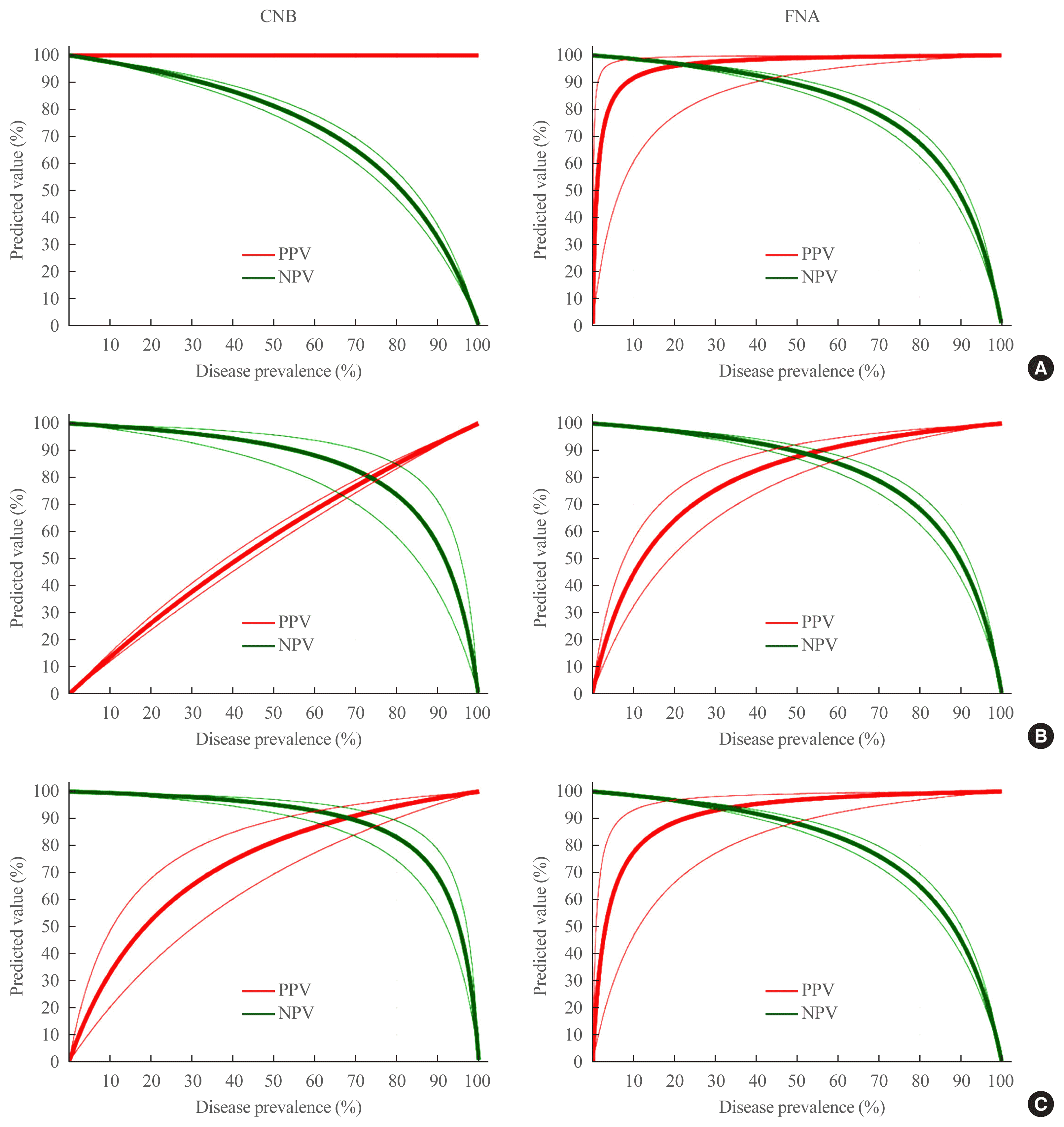
Diagnostic performance of CNB and FNA
Table 4
| Outcomes | CNB (n=429) | FNA (n=666) | P value | ||
|---|---|---|---|---|---|
|
|
|
|
|||
| Analysis 1a | Malignant | Benign+NIFTP | Malignant | Benign+NIFTP | |
| Category V–VI | 240 | 0 | 489 | 1 | |
| Category I–IV | 72 | 117 | 67 | 109 | |
| Diagnostic performance | Incidence | 95% CI | Incidence | 95% CI | |
| Disease prevalence | 55.9% | 49.1–63.5 | 73.4% | 67.1–80.0 | <0.001 |
| Sensitivity | 76.9% | 71.8–81.5 | 88.0% | 85.0–90.5 | 0.089 |
| Specificity | 100% | 96.9–100.0 | 99.1% | 95.0–100.0 | 0.945 |
| PPV | 100% | 100.0 | 99.8% | 98.6–100.0 | 0.979 |
| NPV | 61.9% | 57.0–66.6 | 61.9% | 56.5–67.1 | 0.997 |
| Accuracy | 83.2% | 79.3–86.6 | 89.8% | 87.2–92.0 | 0.256 |
|
|
|||||
| Analysis 2b | Malignant+NIFTP | Benign | Malignant+NIFTP | Benign | P value |
|
|
|||||
| Category IV–VI | 318 | 70 | 506 | 13 | |
| Category I–III | 9 | 32 | 57 | 90 | |
| Diagnostic performance | Incidence | 95% CI | Incidence | 95% CI | |
| Disease prevalence | 76.2% | 68.2–85.0 | 84.5% | 77.7–91.8 | 0.137 |
| Sensitivity | 97.2% | 94.8–98.7 | 89.9% | 87.1–92.2 | 0.271 |
| Specificity | 31.4% | 22.5–41.3 | 87.4% | 79.4–93.1 | <0.001 |
| PPV | 82.0% | 79.9–80.2 | 97.5% | 95.9–98.5 | 0.015 |
| NPV | 78.0% | 63.7–87.8 | 61.2 | 55.0–67.1 | 0.237 |
| Accuracy | 81.6% | 77.6–85.1 | 89.5% | 86.9–91.7 | 0.170 |
|
|
|||||
| Analysis 3c | Neoplastic (surgical disease) | Non-neoplastic | Neoplastic (surgical disease) | Non-neoplastic | P value |
|
|
|||||
| Category IV–VI | 381 | 7 | 517 | 2 | |
| Category I–III | 16 | 25 | 78 | 69 | |
| Diagnostic performance | Incidence | 95% CI | Incidence | 95% CI | |
| Disease prevalence | 88.8% | 80.1–98.2 | 77.6% | 71.1–84.6 | 0.046 |
| Sensitivity | 96.0% | 93.5–97.7 | 86.9% | 83.9–89.5 | 0.141 |
| Specificity | 78.1% | 60.0–90.7 | 97.2% | 90.2–99.7 | 0.349 |
| PPV | 98.2% | 96.6–99.1 | 99.6% | 98.5–99.9 | 0.832 |
| NPV | 61.0% | 48.3–72.3 | 46.9% | 41.7–52.2 | 0.261 |
| Accuracy | 94.6% | 92.1–96.6 | 88.0% | 85.3–90.4 | 0.259 |
NIFTP, noninvasive follicular thyroid neoplasm with papillary-like nuclear feature; CNB, core needle biopsy; FNA, fine needle biopsy; CI, confidence interval; PPV, positive predictive value; NPV, negative predictive value.
a Categories V and VI were considered diagnostic-positive results for malignancy. Diagnostic performance was evaluated for the differentiating malignancy from nonmalignant lesions including NIFTP;
Fig. 1


 Citation
 Print
Print


 XML Download