

 PDF
PDF Citation
 Print
Print


INTRODUCTION
C. auris 균주의 세계적 출현
국내 C. auris 분리 및 감염 현황
균종 동정법
Table 1
| Identification method | Instrument | Database/software | Specific method | No. of isolates | Correct ID | Incomplete ID | No ID | Incorrect ID | [Ref] |
|---|---|---|---|---|---|---|---|---|---|
| MALDI-TOF MS | Bruker Biotyper | Library v4.0 | In-tube FA/ACN | 73 | 100 | 0 | 0 | 0 | [41] |
| On-plate FA | 73 | 83.6 | 0 | 16.4 | 0 | [41] | |||
| RUO library v3.3.1.0 | In-tube FA/ACN | 61 | 75.4 | 13.1 | 11.5 | 0 | [26] | ||
| CMdb alone | In-tube FA/ACN | 33 | 100 | 0 | 0 | 0 | [43] | ||
| RUO | In-tube FA/ACN | 33 | 39.4 | NA | NA | NA | [43] | ||
| EPdbs | In-tube FA/ACN | 33 | 100 | 0 | 0 | 0 | [43] | ||
| MBT Compass Library, Revision E MBT 7854 MSP library | Plate agar (SDA) | 50 | 94.0 | 6.0 | 0 | 0 | [44] | ||
| Positive blood culture | 50 | 92.0 | 8.0 | 0 | 0 | [44] | |||
| ASTA | CoreDB v1.27.02* | On-plate FA | 73 | 100 | 0 | 0 | 0 | [41] | |
| VITEK MS | RUO library v4.14 | On-plate FA | 61 | 93.4 | 6.6 | 0 | 0 | [26] | |
| IVD library v3.2 | On-plate FA | 61 | 96.7 | 0 | 3.3 | 0 | [26] | ||
| Biochemical | Vitek 2 | v8.01† | 35 | 52.4 | 26.7 | 0 | 21.0‡ | [45] |
*MALDI-TOF MS system in this study (ASTA) can differentiate clade II C. auris isolates from non-clade II.
![]()
항진균제 내성 현황
Table 2
| Region | Year | No. of isolates | Sample (No. of isolates) | Method | Resistance for antifungal agents (%)* | ||||||||
|---|---|---|---|---|---|---|---|---|---|---|---|---|---|
| FLU | VRC | AMB | CAS | MFG | AFG | 5FC | MDR | [Ref] | |||||
| UK | 2016 | 50 | Blood, sputum, environmental and body swap† | SYO | 100 | NA | NA | 0‡ | 0 | NA | [6] | ||
| Pakistan, India, South Africa, Venezuela | 2017 | 54 | Blood (27), urine (10), soft tissue (5), wound (4), BAL (3), CVC tip (2), other sites (3) | CLSI BMD | 92.6 | 53.7 | 35.2 | 3.7 | 3.7 | 3.7 | 5.6 | 41 | [11] |
| India | 2018 | 350 | Blood (267), urine (28), tissue (25), sputum (12), skin swap (9), pus (6), other sites (3) | CLSI BMD | 90.3 | 14.9 | 7.7 | NA§ | 2.0 | 2.0 | NA | 25.1 | [7] |
| US | 2018 | 51 | Blood (31), bile (3), urine (4), respiratory specimens (4), wounds (3), catheter tips (2), other sites (3) | Custom TREK frozen BMD panels (Etest for AMB, 5FC) | 98.0 | NA | 29.4 | 0 | 0 | 0 | NA | NA | [14] |
| US | 2018 | 99 | NA | CLSI BMD (Etest for AMB) | 88.9 | NA | 33.3 | 6.1‡ | NA | 39.4 | [36] | ||
| UK | 2018 | 80 | NA∥ | SYO | 100 | 97.5 | 17.7 | NA | 0 | NA | 0 | NA | [48] |
| Kuwait | 2018 | 56 | Blood (13), urine (27), tracheal aspirate (21), catheter tip (5), sputum (6), vaginal swab (4), other sites (12) | Etest | 100 | 73.2 | 23.2 | 1.8 | 1.8 | NA | NA | 19.6 | [49] |
| Colombia | 2018 | 93 | NA | CLSI BMD (Etest for AMB) | 30.1 | NA | 21.5 | NA | NA | 1.1 | NA | 1.1 | [50] |
| Korea | 2019 | 61 | Blood (4), ear (57) | CLSI | 62.3 | 9.8 | 0 | 0 | 0 | NA | NA | 0 | [26] |
| Pakistan | 2019 | 63 | NA | SYO, Etest | 100 | 28.6 | 7.9 | 0 | 0 | 0 | NA | 4.8 | [31] |
| Japan | 2019 | 13 | Otorrhea (12), eustachian tube (1) | NA | 15.4 | 7.7 | 0 | 0 | 0 | NA | 0 | 0 | [54] |
| Kuwait | 2020 | 314 | Blood (58), urine (124), respiratory (98), other sites (34)¶ | Etest | 100 | 41.1 | 27.1 | NA | 1.7 | NA | 0 | NA | [33] |
| Kuwait | 2020 | 62 | Blood (16)/colonization (46) | MICRONAUT-AM AST/Etest | 93.8/ 87.0 | 93.8/ 23.8 | 0/0 | NA§ | 0/4.3 | 0/4.3 | NA | NA | [34] |
| South Africa | 2020 | 85 | Urine (22), blood (20), CVC tips (19), irrigation fluid (5), tissue (4), respiratory tract specimen (3), miscellaneous sites (12) | BMD panels containing alamar blue | 96.5 | 7.1 | 0 | NA§ | 8.2 | 1.2 | 0 | 8.2 | [35] |
| US | 2020 | 277/ 116** | Blood (140), Urine (64), wound (37), lung (23), Bile (4), corneal, eye (2), ear (1), bone (1), stool (1), unspecified (4)/NA | CLSI (Etest for AMB, 5FC) | 99.6/ 100 | 80.9/ 82.8 | 61.4/ 57.8 | 0/2.6 | 0/3.4 | 0/2.6 | 0.7/5.1 | NA | [37] |
| South Africa | 2021 | 77 | Blood (77) | CLSI | 89.6 | NA | 29.9 | 2.6 | 2.6 | 0 | NA | 9.1 | [38] |
*Results of resistance were analyzed using the tentative MIC breakpoints for C. auris published by the CDC, but resistance for voriconazole, flucytosine were determined by using MIC breakpoints 2 µg/mL (except 1 µg/mL for [48,49] , 4 µg/mL for [31]), 128 µg/mL, respectively.
§Caspofungin MICs were not interpreted because caspofungin is an unreliable indicator of echinocandin resistance.
Abbreviations: FLU, fluconazole; VRC, voriconazole; AMB, amphotericin B; CAS, caspofungin; MFG, micafungin; AFG, anidulafungin; 5FC, flucytosine; MDR, multidrug resistance; SYO, Sensititre YeastOne; NA, not available; BAL, bronchoalveolar lavage; CVC, central venous catheter; CLSI, Clinical Laboratory Standard Institute; BMD, broth microdilution; AST, antifungal susceptibility test; MIC, minimum inhibitory concentration; CDC, Centers for Disease Control and Prevention.
![]()
감염 관리 및 전파 예방에 대한 전략
1. 임상특성
2. C. auris의 감염 예방 및 관리
Table 3
| Infection prevention and control measures | Recommendation | [Ref] | |
|---|---|---|---|
| Patients control | Single room | Isolate patients in a single room | [22-25,59] |
| Priority assignment of single rooms to patients at high risk of pathogen transmission | [22] | ||
| Ideally with negative pressure, and preferably with an ante-room and en-suite bathroom/toilet | [24,25,59] | ||
| Shared room | Isolate Patients with the C. auris in the same room when single rooms are not available | [22-25,59] | |
| Recommended practices to reduce transmission in shared rooms;Maintain at least 3 feet distance between roommatesUse privacy curtainsClean and disinfect any shared reusable equipment and environmental surfacesChange PPE and perform hand hygiene when moving between patients | [22] | ||
| Cohorting | Consider cohorting patients with C. auris together in one unit if multiple cases have occurred | [22-24,59] | |
| Consider cohorting healthcare personnel (e.g., dedicated nursing staff) | [22,23] | ||
| Patients movement | If a patient needs to be taken out of the isolation room (e.g., for imaging, dialysis, rehabilitation);Scheduled them last on the list for the dayClean and disinfect the environment after they have been used | [22,24,25,59] | |
| Transmission-based precaution | Hand hygiene | Follow standard hand hygiene practices;Adherence to the five moments of hand hygieneUse a 70% alcohol-based hand sanitizerWash with soap and water If hands are visibly soiled | [22-25,59] |
| Increase hand hygiene audits; Re-educate healthcare personnel on hand hygiene, if audits demonstrate low adherence to recommended hand hygiene practices | [22-24] | ||
| Contact precautions |
Consider using single-patient items (e.g., blood pressure cuffs, pillows) Clean and disinfect shared equipment |
[22,24,25,59] | |
| Use PPE when in contact with patients | [22,24,59] | ||
| Continue setting appropriate TBP for the entire duration of the patient’s stay in the facility | [22,59] | ||
| Prevention of invasive infections | Appropriate care of invasive medical devices |
Strict adherence to central and peripheral catheter care bundles, urinary catheter care bundle and care of the tracheostomy site Continuously assess the need for invasive devices |
[22,24] |
| Environmental disinfection and cleaning | Cleaning and disinfection | Perform thorough daily and terminal cleaning and disinfection of patients’ rooms;High-touch surfaces (bedside tables and bedrails)General environmental surfaces (e.g., floor, walls, windowsills)Mobile equipment that is shared between patients | [22-25,59] |
| Disinfectant | CDC recommends use of an EPA–registered hospital-grade disinfectant effective against C. auris; List P | [22,61] | |
| Use of an EPA-registered hospital-grade disinfectant effective against Clostridium difficile spores (List K) is possible | [22,62] | ||
| Follow all manufacturers’ directions for use of surface disinfectants and applying the product for the correct contact time | [22-25,61] |
![]()
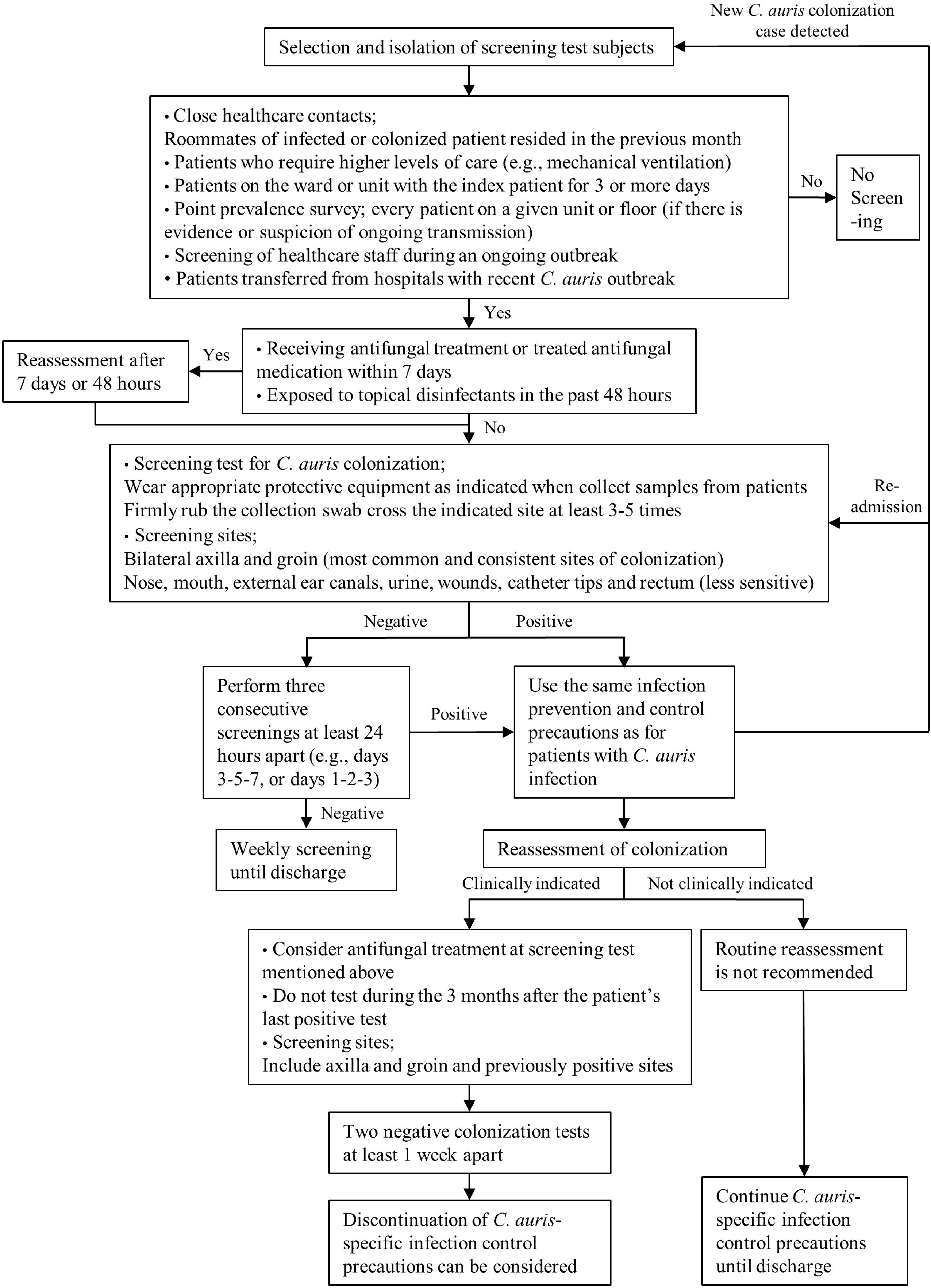
3. C. auris 선별검사
4. 검사실 및 시스템적 중재 방안
Table 4
| Infection prevention and control measures | Recommendation | [Ref] | |
|---|---|---|---|
| Laboratory consideration | Retrospective investigation | Check whether there was an increase in Candida species in the same unit for 4 weeks before diagnosis of C. auris | [22] |
| Prospective surveillance | Speciate all Candida isolates from the same unit to the species level for the subsequent four weeks | [22-24] | |
| Safety concern |
Use personal protective equipment, at least lab coat and gloves Use a biological safety cabinet (in at least BSL-2) Decontaminate the biological safety cabinet with 10% bleach (or another product on List P) and perform hand hygiene after work with C. auris |
[22] | |
| Use reliable identification method | Notify laboratory/microbiologist that C. auris is being investigated; in order that correct methods can be applied to diagnostic samples | [25] | |
| Systemic considerations | Communication within facility | Prompt notification of C. auris to the clinician, laboratory, and infection control teams | [23,25] |
| Education/training/monitoring | Educate all healthcare workers, including cleaning staff, patients, and visitors about C. auris and appropriate infection prevention and control precautions | [22-25] | |
|
Monitor adherence to infection control practices including hand hygiene Cleaning and disinfecting should be monitored and audited |
[22,25] | ||
| Electronic flagging system | Label C. auris patients with an infection control flag on electronic system;clinicians are immediately alerted when the patient is readmitted | [22,24,59] | |
| Antimicrobial stewardship |
An environment with a high level of broad-spectrum antibacterial and antifungal use will favor the emergence of multidrug-resistant yeasts Mitigate the risks of C. auris acquisition and transmission Essential component of strategies to reduce antimicrobial resistance in general The need for antifungal prophylaxis should be reviewed |
[22,23,25] | |
| Communication to other facilities |
Inform the receiving health care facility of the patient's C. auris infection status Consider the ability of the accepting facility to provide care for patient |
[22,23,59] | |
| National guidelines |
Consider prepare national guidelines for laboratory testing and infection control measures for C. auris Designate national mycology reference laboratory Consider updating list of surveillance systems for healthcare-associated infections to include C. auris |
[23] |
![]()
 XML Download