Abstract
Facial degloving injuries are due to separation between the skin and subcutaneous tissues from the underlying muscles, bones, and fascia. These injuries often create a reconstructive challenge for surgeons especially when there are associated complications like wound infection or necrosis of the avulsed flap. This case report presents management of a case of facial degloving injury with full thickness necrosis of the avulsed flap. The authors concluded that treatment of such complex wounds requires a multi-disciplinary approach along with proper planning and staging of the surgical procedures for optimum aesthetic and functional outcomes.
Reconstruction of complex facial wounds is challenging for surgeons for a multitude of reasons. The surgeons have to balance between achievement of an acceptable cosmetic result and attainment of optimal functional outcomes.
Degloving lacerations are severe soft tissue injuries characterized by avulsions or detachment of the skin and subcutaneous tissues from the underlying muscles, bones, and fascia1. These injuries usually occur due to a sudden shearing force applied to the skin surface2.
Facial degloving injuries are rare, and there are no pre-established protocols for treatment3. Delayed treatment of these injuries can lead to infection and necrotizing fasciitis of the avulsed flap4. Moreover, full-thickness necrosis of the flap can also occur due to traumatic damage to its vascular supply5.
The authors present a case report of one such degloving facial injury with full-thickness loss of the avulsed skin flap and discuss the management of such a complex wound.
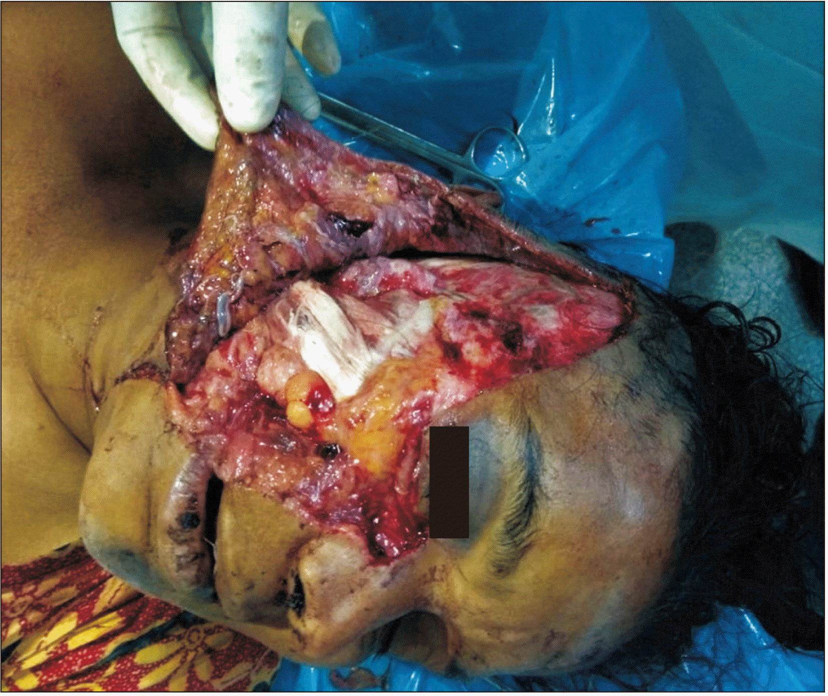
A 42-year-old female presented in the emergency department with history of a traffic accident. There was a degloving injury over the right side of her face involving the cheek, oral commissure, lower eyelid, and temporal scalp.(Fig. 1) The external auditory canal involving the fibro-cartilaginous area was also severed distally.
The patient had a patent airway and was breathing well. She was immediately administered fluids for maintenance of circulation, and control of bleeding was achieved. Although the patient was alert, oriented, and ambulatory during examination, she reported a history of transient loss of consciousness after the accident. There was no sign of brain injury on computed tomography, but a fracture was noted in the ramus on the right mandible. There was no sign of globe injury, and vision was intact along with pupillary reflexes.
Thorough debridement of the wound was performed with normal saline under local anesthesia, and all devitalized tissues were excised from the wound. The degloved flap was repositioned and sutured to relocate the avulsed structures to their respective original positions. The avulsed external auditory canal was sutured to its proximal part using continuous suturing. The lower eyelid was repositioned and sutured in layers. Postoperative dressing was applied to prevent hematoma. The patient was transferred to the general ward for observation and was kept on antibiotic therapy to prevent wound infection. Since the patient had experienced a fractured mandible, she was kept on a soft diet. The patient was planned for open reduction and internal fixation of the mandibular fracture under general anesthesia.
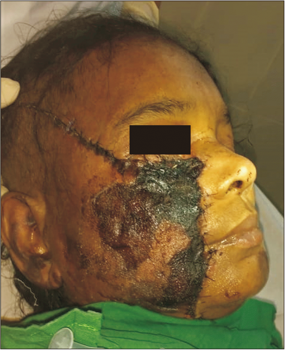
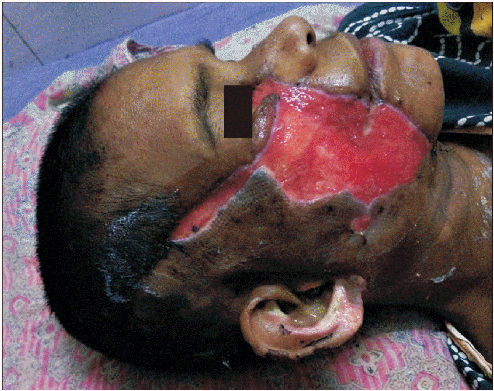
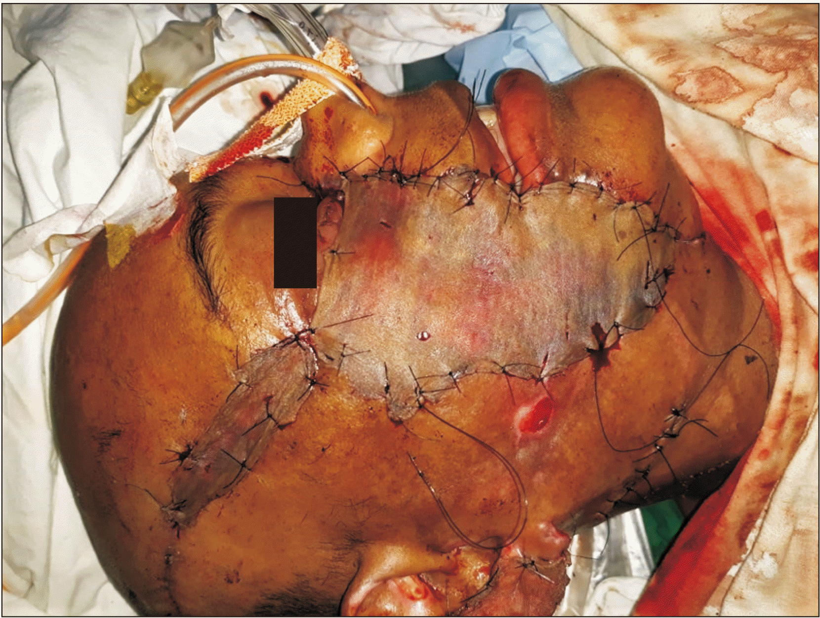
Unfortunately, the patient showed signs of necrosis of the avulsed flap on the following day.(Fig. 2) This led to full thickness loss of the distal half of the avulsed flap (Fig. 3) and loss of a small portion of the right ear pinna due to perichondritis. The patient subsequently underwent serial debridement until healthy granulation tissue was formed in the wound bed.
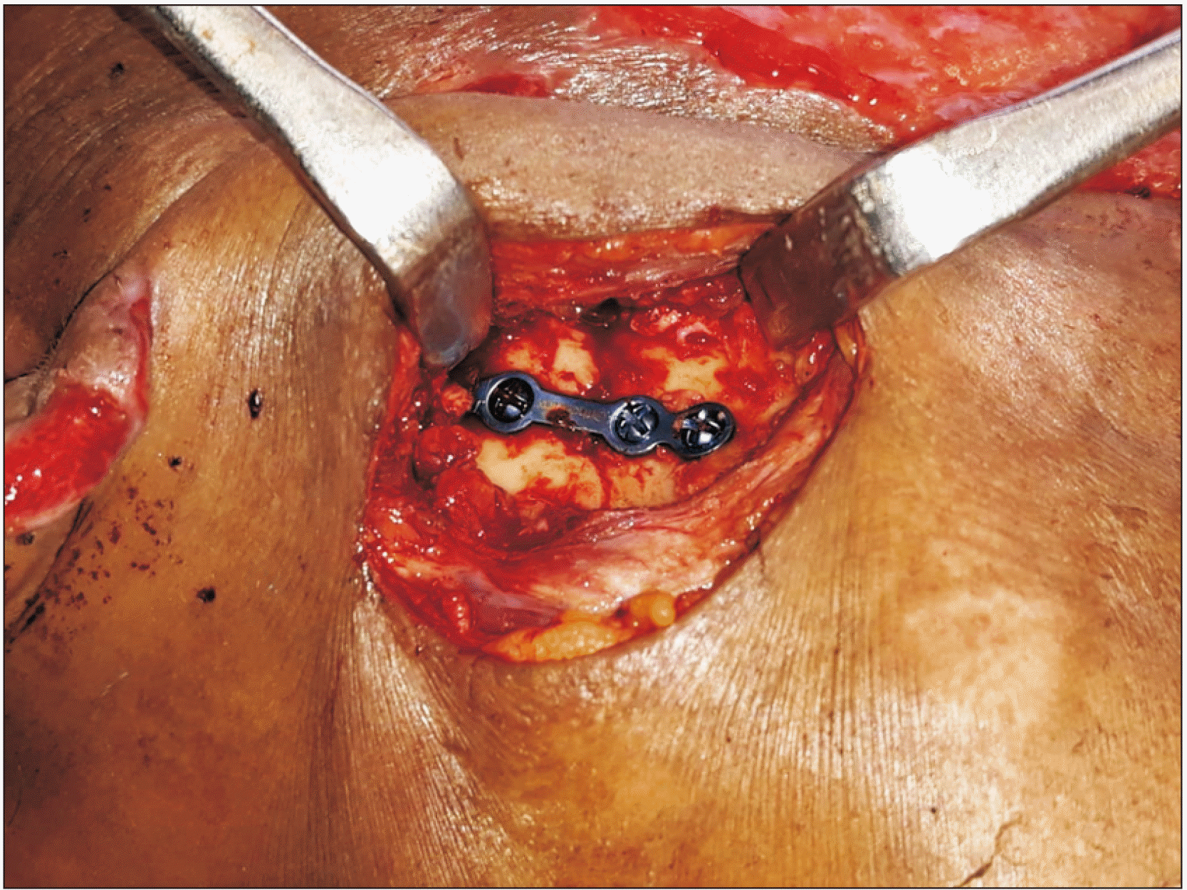
Finally, the patient underwent definitive surgery after one week. Under general anesthesia, the fracture site on the right ramus of the mandible was exposed using a submandibular skin incision. The fracture was reduced and fixed with a 2.0-mm titanium miniplate system.(Fig. 4) Satisfactory dental occlusion was achieved after fracture fixation, and the incision was closed in layers. The soft tissue defect over the right side of the face was reconstructed using a split-thickness skin graft (Fig. 5) harvested from the left thigh region of the patient.
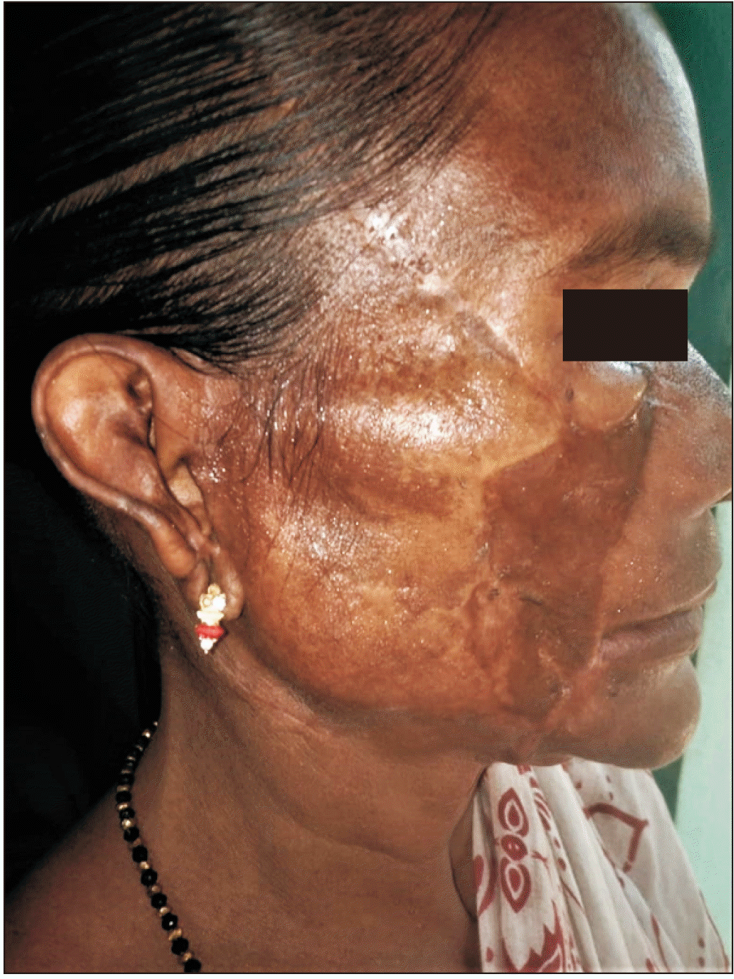
The postoperative healing was uneventful.(Fig. 6) There were no deficits in hearing or oral competency at one year after surgery, but the patient had epiphora from the right eye. There was a mismatch of skin color between the grafted area and the normal facial skin. The patient was given options for staged removal of the skin graft for cosmetic improvement and was recommended surgical treatment of the epiphora. However, the patient was satisfied and refused additional cosmetic surgeries. The patient was planned for recanalization of the lacrimal drainage apparatus on a later date. Healing of the skin graft donor site was satisfactory.
Degloving soft tissue injury comprises 4% of all traumatic injuries. These injuries show a marked predilection in males and young patients. Motor vehicle accidents are the most common cause of degloving injuries of the head and neck region6.
Degloving soft tissue injuries can be classified as either open or closed lesions4. Open degloving injury generally manifests as soft tissue avulsion, whereas closed degloving injury presents as a cavity filled with hematoma and liquefied fat7. Open degloving lacerations are more common in the head and neck region, whereas closed lesions are more frequent in the trunk and extremities.
Treatment of open degloving injuries ranges from simple debridement with primary skin closure to complex reconstructions with skin grafts, local flaps, microvascular free flaps, replantation, or revascularization. The choice of treatment depends on the site, extent, and severity of the wound.
Vacuum-assisted wound closure is often used for open degloving wounds to promote faster formation of granulation tissue prior to skin grafting8. However, presence of multiple contours and of hair hinders achievement of an air-tight seal in the head and neck region9.
The primary management of facial degloving injuries focuses on definitive primary skin cover and early return of function. However, complications can occur even after meticulous wound debridement and closure.
Complications such as infection and skin necrosis can be caused by delay in the treatment of degloving injuries10. This was not the cause of complication for the presented case as the patient was immediately treated with wound debridement and primary closure. The authors believe that the cause of loss of the avulsed flap is due to damage to the blood supply of the cutaneous tissues and to the extent of the injury.
The main limitation of this case report is that the postoperative epiphora could have been prevented by performing recanalization of the lacrimal drainage apparatus during the definitive surgery after confirming the blockade using the Jones test. Although the patient was satisfied with her appearance, the aesthetics could be improved further using facial cosmetic surgeries.
Treatment of this type of open degloving soft tissue injury is a challenge since there is no established evidence-based approach for management of this type of wound. A multidisciplinary approach is required for successful management of these types of injuries. Proper planning and staging of the surgical procedures are required for achievement of optimal aesthetic and functional outcomes in these types of complex facial injuries.
Acknowledgements
The authors thank the entire faculty and theatre staff of Department of Oral and Maxillofacial Surgery, SCB Dental College and Hospital, Cuttack, India.
Notes
Authors’ Contributions
D.D. participated in the data collection and wrote the manuscript. D.F.S. participated in the case report design and coordination and helped to draft the manuscript. All authors read and approved the final manuscript.
References
1. Morris M, Schreiber MA, Ham B. 2009; Novel management of closed degloving injuries. J Trauma. 67:E121–3. https://doi.org/10.1097/TA.0b013e31803420be. DOI: 10.1097/TA.0b013e31803420be. PMID: 19820564.

2. Nicolai JP, van der Zwan J, van der Veen JA, Dokter BJ, van Oort RP. 1980; The vertical avulsion flap. Int J Oral Surg. 9:99–102. https://doi.org/10.1016/s0300-9785(80)80045-7. DOI: 10.1016/S0300-9785(80)80045-7. PMID: 6773902.

3. Jadhav NC, Ramdas S, Lingam PP, Sateesh S. 2016; Complex traumatic facial degloving injury. J Craniofac Surg. 27:1051–2. https://doi.org/10.1097/SCS.0000000000002613. DOI: 10.1097/SCS.0000000000002613. PMID: 27171962.

4. Latifi R, El-Hennawy H, El-Menyar A, Peralta R, Asim M, Consunji R, et al. 2014; The therapeutic challenges of degloving soft-tissue injuries. J Emerg Trauma Shock. 7:228–32. https://doi.org/10.4103/0974-2700.136870. DOI: 10.4103/0974-2700.136870. PMID: 25114435. PMCID: PMC4126125.

5. Yan H, Gao W, Li Z, Wang C, Liu S, Zhang F, et al. 2013; The management of degloving injury of lower extremities: technical refinement and classification. J Trauma Acute Care Surg. 74:604–10. https://doi.org/10.1097/TA.0b013e31827d5e00. DOI: 10.1097/TA.0b013e31827d5e00. PMID: 23354258.

6. Hakim S, Ahmed K, El-Menyar A, Jabbour G, Peralta R, Nabir S, et al. 2016; Patterns and management of degloving injuries: a single national level 1 trauma center experience. World J Emerg Surg. 11:35. https://doi.org/10.1186/s13017-016-0093-2. DOI: 10.1186/s13017-016-0093-2. PMID: 27468300. PMCID: PMC4962500.

7. Gummalla KM, George M, Dutta R. 2014; Morel-Lavallee lesion: case report of a rare extensive degloving soft tissue injury. Ulus Travma Acil Cerrahi Derg. 20:63–5. https://doi.org/10.5505/tjtes.2014.88403. DOI: 10.5505/tjtes.2014.88403. PMID: 24639319.

8. Lee SJ, Kwon J, Lim KM, Kim HJ, Cha IH, Nam W. 2010; Postoperative orocutaneous fistula closure using a vacuum-assisted closure system: a case report. J Korean Assoc Oral Maxillofac Surg. 36:413–6. https://doi.org/10.5125/jkaoms.2010.36.5.413. DOI: 10.5125/jkaoms.2010.36.5.413.

9. Andrews BT, Smith RB, Goldstein DP, Funk GF. 2006; Management of complicated head and neck wounds with vacuum-assisted closure system. Head Neck. 28:974–81. https://doi.org/10.1002/hed.20496. DOI: 10.1002/hed.20496. PMID: 16983694.

10. Faria PE, de Souza Carvalho AC, Masalskas B, Chihara L, Sant'Ana E, Filho OM. 2016; Surgical reconstruction of lower face degloving. J Craniofac Surg. 27:e683–5. https://doi.org/10.1097/SCS.0000000000003067. DOI: 10.1097/SCS.0000000000003067. PMID: 27763946.


 PDF
PDF Citation
 Print
Print


 XML Download