Journal List > J Korean Assoc Oral Maxillofac Surg > v.48(3) > 1516078823

Parmentier, Nys, Verstraete, and Politis: A systematic review of treatment and outcomes in patients with mandibular coronoid process hyperplasia

Abstract

Treatment of mandibular coronoid process hyperplasia (MCPH) has been described and explored in the literature. This systematic review aims to provide a comprehensive overview of the surgical and non-surgical treatment options for MCPH in pediatric and adult populations. Three databases were searched for treatment of MCPH patients (MEDLINE, Embase, and Web of Science). Two reviewers selected case reports and case series based on titles and abstracts. Finally, 55 studies reporting a total of 127 cases were included for qualitative synthesis and data extraction. The mean age at symptom onset was 15.6 years, while the mean age at diagnosis was 23.5 years. Of the included cases, 83.7% were male, and the condition was bilateral in more than 81% of the cases. Coronoidectomy was performed in 82.7% of the included cases, while coronoidotomy was performed in 3.9% of the cases. In 85.0% of the surgically treated cases, the approach was intraoral. The mean maximal intraoperative mouth opening was 38.1 mm compared with 16.5 mm at diagnosis. The mean maximal postoperative mouth opening was 35.3 mm, and the mean follow-up period was 16.3 months. Maximum mouth opening was achieved intraoperatively, and non-surgical treatment after surgery aims to reduce the risk of relapse. Additional research with a higher level of evidence is necessary to confirm these findings.

I. Introduction

The coronoid process is an anatomical part of the mandible that serves as the attachment for the temporalis muscle, buccinator muscle, and the anterior part of the masseter muscle. In classic anatomy, it is described as a sharp triangular-shaped structure in extension of the anterior border of the mandibular ramus. In reality, this structure shows great morphological variety, including hook-shaped type, triangular type, or rounded type. In the study of Lalitha and Sridevi1, the majority (73.9%) of mandibles had the same type of coronoid process on both sides, while 26.1% of the cases showed different types on the two sides. Allometric variation can be established in mandibular shape in humans, with taller individuals having superoinferiorly taller rami with more anteriorly-oriented and higher coronoid processes and a corresponding deeper sigmoid notch2.
Mandibular coronoid process hyperplasia (MCPH) is an uncommon congenital or developmental condition that is characterized by a macroscopic increase in the size of the coronoid process with a normal histologic structure of the bone. MCPH can exist as a uni- or bilateral condition and causes a slow and progressive reduction of mouth opening. Restricted mouth opening results from impingement of the coronoid process on the medial surface of the zygomatic arch3-5. Unilateral MCPH can involve facial asymmetry with deviation toward the affected side5. Langenbeck was the first to report MCPH in 1853, and the first case of restricted mouth opening due to coronoid process enlargement was reported in 1899 by Jacob3. Jacob’s disease refers to the condition where the coronoid process creates a new joint with the zygomatic process.
The pathogenesis of MCPH remains unclear. Several factors might be associated with development of MCPH6,7. History of facial trauma and, in particular, zygomatic arch trauma is thought to be a contributing factor in some cases8-11. Hall et al.12 hypothesized that MCPH results from a developmental bone defect in neoplastic or cartilaginous growth centers of the coronoid processes, causing continued growth and eventual hyperplasia. Endocrine stimulation also can play a role in the pathogenesis of MCPH. Chung et al.13 reported that two years of growth hormone therapy for children with short stature led to increase of posterior facial height and growth of the mandible. Only one case was reported on the relationship between growth hormone and growth of the coronoid process of the mandible14. However, hypotheses involving increased activity of the temporal muscles have received the most support. Temporomandibular joint (TMJ) disorders with limited mandibular movement due to disc displacement or TMJ ankylosis were considered to be related to MCPH due to compensatory hyperactivity of the temporal muscles15-19. Such findings also were reported in a study by Isberg et al.20 with experimentally induced mandibular hypomobility in rhesus monkeys. Other findings supporting these theories are the shortened muscle tendon units, including fibrous and hypertrophic masticatory muscle tendons and hypertrophic or hypertonic temporalis muscles, in some syndromes5. The most common disorder associated with congenital bilateral MCPH is trismus pseudocamptodactyly syndrome (TPS)21. Genetic analysis of patients with TPS reveals a defective MYH8 gene, which presumably interferes with myosin activity and, possibly, the binding of myosin to actin. This genetic defect leads to multiple muscle contractures and arthrogryposis. Other syndromes with a common finding of hypotonia can be associated with MCPH. Moebius syndrome is characterized by uni- or bilateral paralysis of the facial mimetic muscles and the lateral rectus muscle of the eye and has been associated with bilateral MCPH22,23. Kabuki syndrome and Pena-Shokeir syndrome are also both associated with hypotonia of the mandibular depressors, and it was hypothesized that the absence of mandibular movements and deglutition can result in relative hyperactivity of the temporalis muscle, resulting in reactive enlargement of the coronoid process24. Genetic inheritance is another factor that can contribute to the development of MCPH. An example is mucopolysaccharidoses, a lysosomal storage disease, where generalized skeletal changes in animal models are caused by the effects of glycosaminoglycan deposition with induced modification of morphology and function of osteoclasts. Possible craniofacial characteristics are macrocephaly, thickened calvarium, underdevelopment of the mandibular condyle, and MCPH25.
Males are more commonly affected by MCPH than females, with a reported ratio of 5:17. MCPH is alternatively termed “coronoid impingement syndrome” (CIS)7. CIS is described in literature as an acquired condition after extended craniofacial and orthognathic surgery, where the coronoid process interferes with the zygomatic arch, and contraction of the temporal muscle can prevent the osteotomized mandible from further setback26,27.
In addition to progressive mouth opening limitation, patients suspected of MCPH can have a hard end feel and pain in the zygomatic area with maximum mouth opening5. However, only a small percentage of these patients actually has symptoms. Pain or crepitation with mouth opening are present in 7% or 8% of patients, respectively28. In the neonatal population, limited mouth opening can result in important sequelae, such as feeding difficulties, risk of malnutrition, and a compromised airway with apnea22.
If treated conservatively, no improvement of these symptoms can be expected, and further diagnostic modalities are indicated. Before diagnosis of this rare condition, patients might be misdiagnosed with another and more common temporomandibular disorder as listed in the Diagnostic Criteria for Temporomandibular Disorders (DC/TMD) by Schiffman et al.29. The final diagnosis can be made by a thorough clinical (re)examination, combined with additional imaging. In the literature, panoramic radiograph (orthopantomogram [OPG]), cone-beam computed tomography (CBCT), computed tomography (CT), and bone scintigraphy have been used to diagnose MCPH15.
Treatment of MCPH has been described and explored substantially in the literature. Common treatment modalities for patients with MCPH include both surgical and non-surgical procedures4,7,8,28,30,31. The aim of this study is to perform a systematic review on treatment modalities and outcomes for patients in pediatric and adult populations with MCPH.

II. Methods

1. Protocol design

This protocol was drafted according to the Cochrane Guidelines for review protocols (http://training.cochrane.org/) and was submitted in Prospero, international prospective register of systematic reviews (http://www.crd.york.ac.uk/PROSPERO), registration number CRD42021267132. This systematic review aims to comply with the PRISMA (Preferred Reporting Items for Systematic reviews and Meta-Analyses) statement.

2. Selection of studies

1) Criteria for considering studies for this review (PICOS)

(1) Types of participants (P)
Study populations including living infants, adolescents, and adults were considered. No restrictions were made based on race or sex. Studies on animals or study models were excluded.
(2) Types of interventions (I)
Studies describing treatment of unilateral or bilateral MCPH were included.
We did not include treatment of osteochondroma, osteoma, trauma, or oncological treatment of the coronoid process. We also did not include treatment of zygomatic malformation, hypoplasia of the masticatory muscle tendon aponeurosis, and trismus of an unknown cause.
(3) Types of controls (C)
The control for treatment of MCPH and its postoperative maximal mouth opening is preoperative maximal mouth opening.
(4) Types of outcome measures (O)
The included studies were required to provide outcome measures of postoperative mouth opening, surgical approach, and any alternative or additional treatment.
(5) Types of studies (S)
Case reports, case series, cross-sectional observational studies, and cohort observational studies were included. No restrictions were made based on the country or date of publication. Studies in English and French were included. Review articles were excluded.

3. Search methods

According to the inclusion and exclusion criteria, studies were identified in searches in MEDLINE (via the PubMed interface), Embase (via the Embase.com interface), and Web of Science (via the Web of Science interface). The specific search string for each database is displayed in Table 1.

4. Review and selection of studies

Two authors (G.I.L.P. and M.N.) independently screened the titles and abstracts of the obtained search results. References were managed using Covidence software (Covidence systematic review software; Veritas Health Innovation, Melbourne, Australia; https://www.covidence.org/) for study selection and deduplication, and Zotero software was used for deduplication of search results. After a first selection of articles, the authors considered and compared their selections to achieve consensus. Of the retained abstracts, the full text paper was assessed independently for eligibility. Discrepancies between reviewers were identified and resolved by discussion to reach consensus. The reasons for excluding studies at each step (either title and abstract or full text) are shown as follows.
1) Wrong patient population (P)
2) Wrong intervention (I)
3) Wrong outcome measures (O)
4) Wrong study design (S): review
5) Full text not available
6) Wrong route of administration
7) Wrong language

5. Data extraction and management

Study characteristics and outcome data were extracted by one reviewer (G.I.L.P.). The data extraction table summarizes data on study population characteristics such as age of symptom onset, age of administration, age of diagnosis, diagnostic tools, preoperative maximal mouth opening, treatment, treatment results, and follow-up period.

6. Quality assessment of studies

The quality of the selected articles was assessed by the reviewer using the items and questions in Table 2. The used tool was based on the Quality Assessment Tool for Case Series Studies by NHLBI (National Heart, Lung, and Blood Institute; https://www.nhlbi.nih.gov/health-topics/study-quality-assessment-tools).

7. Statistical analysis for study characteristics

Data were collected using Microsoft Excel 2010 (Microsoft, Redmond, WA, USA) and analyzed using IBM SPSS Statistics for Windows (ver. 26; IBM, Armonk, NY, USA). Descriptive statistics were calculated.

III. Results

1. Selection of studies

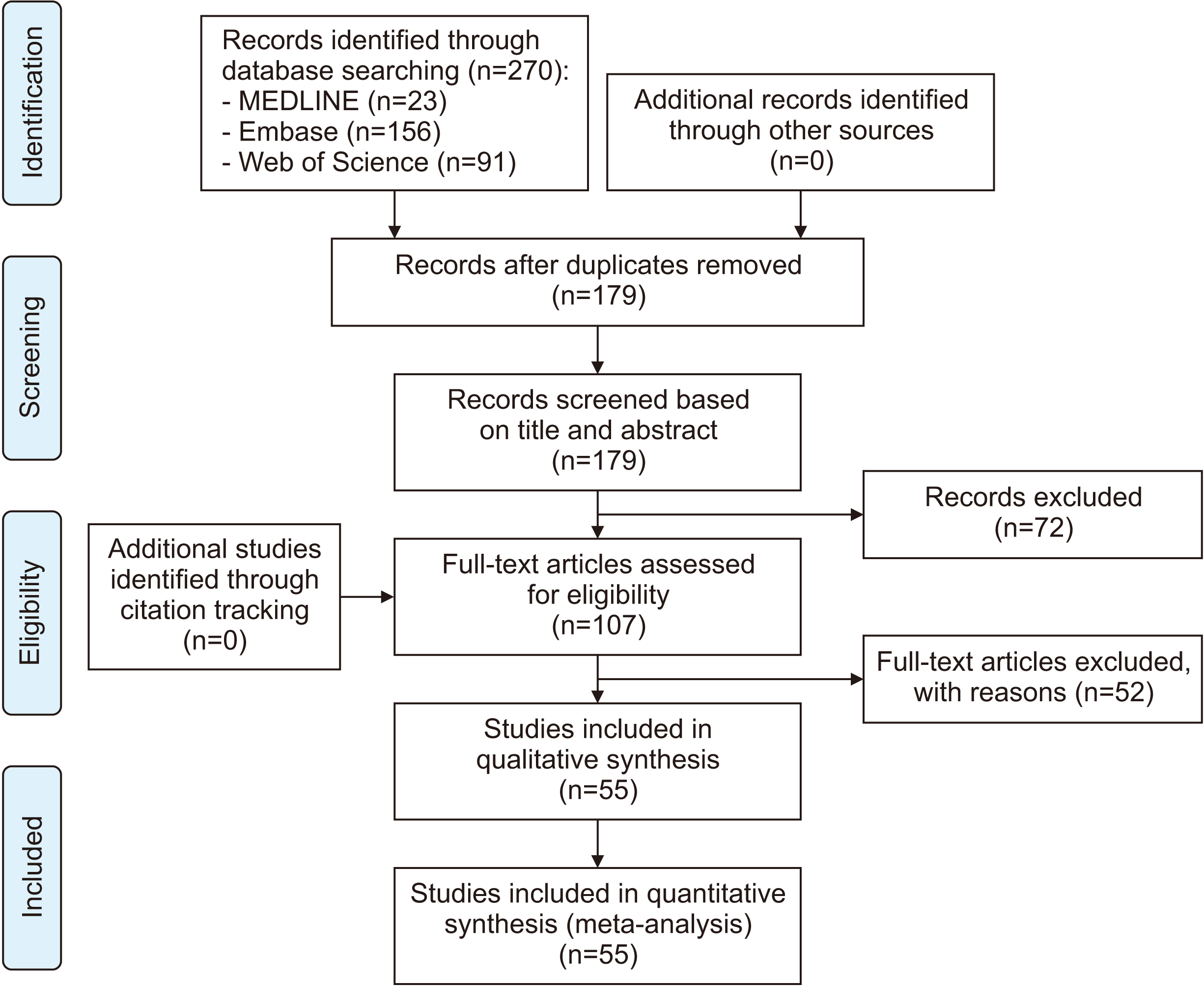
The database search rendered 270 records: 23 records in MEDLINE, 156 records in Embase, and 91 records in Web of Science. After deduplication, 179 records were screened based on title and abstract by two reviewers. After achieving consensus regarding selection of articles, 107 full text papers were evaluated independently for eligibility. Finally, 55 studies reporting 127 cases were included for qualitative synthesis and data extraction.(Fig. 1)

2. Quality and characteristics of the included studies

1) Quality assessment of the included studies

A quality assessment was performed for each included study. As shown in Table 33,6-10,14-17,22-25,30-70, all case reports or case series were verified and assessed for the provided information regarding the study objective. All studies were checked and scored if a case or several cases were fully described or defined with sufficient details to allow the practitioner to make inferences related to their own practice. The description of the treatment of MCPH, the outcome measures, and the follow-up were verified and scored for every case. In general, all studies had a clear objective, although the case definition was not mentioned in 3 studies, and the intervention was not clearly described in 9 studies. Four studies lacked follow-up reports, 10 studies did not describe the cases with sufficient details, and the outcome measures were not sufficiently reported in 11 studies. No studies were excluded due to quality issues. All case reports and case series were included to provide as much information as possible concerning this condition and its treatment.

2) Study characteristics

Results on case reports and case series are displayed alphabetically in Table 43,6-10,14-17,22-25,30-70.
Case populations were considered and described. The age of onset (when the first symptoms were noticed), age at diagnosis, and sex were collected. In addition, the used imaging techniques, maximal mouth opening at diagnosis, possible causes, and the side of coronoid hyperplasia were recorded. The mean and standard deviation age of onset was 15.6±10.3 years, while the mean age at diagnosis was 23.5±14.0 years. In 33.86% of the cases, the age of onset for restricted mouth opening was not clearly specified, while the age at diagnosis was not mentioned in 4 cases. Approximately 81.1% of the patients with MCPH were male, while 15.7% were female; in four cases, the sex was not mentioned. The mean maximal mouth opening was 16.5±7.16 mm at diagnosis. In 10 cases, the maximal mouth opening at diagnosis was not reported. MCPH was bilateral in 81.1% of the cases and unilateral in 18.9% of the cases.
The type of surgical procedure and the surgical approach were recorded, as was maximal intraoperative mouth opening. Coronoidectomy was performed in 82.7% (n=105), coronoidotomy in 3.9% (n=5) and no surgery was performed in 9.4% (n=12) of the cases. In five cases (3.9%), the surgical intervention was not described. In the cases where surgical intervention was executed and reported, surgery was performed by an intraoral approach in 82.7% (n=72), extraoral approach in 9.2% (n=8), and combined intraoral and extraoral approach in 8% (n=7) of the cases. The mean maximal intraoperative mouth opening was 38.1±9.24 mm compared with 16.5±7.16 mm at diagnosis.
The type of postoperative supportive intervention and follow-up period were recorded, and postoperative mouth opening and maximum mouth opening were reported. Postoperative supportive physiotherapy was applied in 91.8% (n=101) of the cases.
In 16 cases (12.5%), the type of postoperative supportive therapy was not described. The Therabite appliance (TheraBite Jaw Motion Rehabilitation System; Atos Medical AB, Hörby, Sweden) was used in 23.4% (n=26) of the cases that reported postoperative physiotherapy. Although Therabite was preferred in a substantial amount of cases to maintain and improve the postoperative result, no study could confirm the superiority of one specific type of postoperative physiotherapy4. The mean maximal postoperative mouth opening was 35.0±9.6 mm after postoperative supportive therapy. The mean postoperative follow-up period was 16.1 months, with a range between 1.0 and 72.0 months.

IV. Discussion

Progressive limitation of maximal mouth opening is the hallmark of MCPH. Restricted mouth opening, a hard end feel when opening the mouth, pain or slight asymmetry in the zygomatic area, and no improvement of symptoms despite repeated conservative treatment are clinical signs suspicious for MCPH15. Patients with mouth opening limitation who have no symptoms related to a temporomandibular or masticatory muscle disorder after initial clinical and radiographic examinations should undergo CT for a more accurate diagnosis.
A considerable difference between the age of onset of the first symptoms and the age of diagnosis is noted in the literature4,5. We confirm these findings with a mean difference of 7.92 years in our study. As a rare condition with often painless progressive restricted mouth opening, MCPH is prone to underreporting, misdiagnosis, and delayed diagnosis59. In patients with limited mouth opening, temporomandibular disorders typically are considered first32.
In 1963, Rowe57 was the first to classify MCPH as uni- or bilateral. Izumi et al.71 classified MCPH into three categories (high, middle, low) by radiologically evaluating the tip of the coronoid process in relation to the zygomatic arch.
A coronoid/condyle ratio of <1.0 has been proposed as the definition of normal anatomy60. A CT scan can provide detailed imaging of the coronoid process and its anatomical relation with the zygomatic bone. A scan performed in the maximal mouth opening position can be useful to locate the exact location of interference and can provide added value in surgical planning32,58.
Evaluation of the coronoid process is commonly performed based on CT images. Due to development in radiographic imaging, lower-dose CBCT is more frequently performed and can also be used for this assessment60.
Osteochondromas or osteomas can cause unilateral coronoid hyperplasia and might present with similar symptoms, but their radiological and histological characteristics reveal difference from true MCPH. These neoplasms of the coronoid process should not be termed unilateral coronoid hyperplasia since there are no histological features of neoplasia in MCPH, but they show histologically normal bone with abnormal bony elongation4,28,72. In most patients diagnosed with unilateral MCPH, some abnormality was also detected on the contralateral coronoid process8.
The aim of treatment of MCPH is restoration of mouth opening and maintenance of a long-term and stable result30. Ineffective long-term conservative treatment from misdiagnosis of MCPH can lead to unsatisfied patients and patient discomfort with loss of quality of life but also to important health risks15. Therefore, an early diagnosis of this entity is important. A point of interest is that TMD and MCPH can co-exist in the same patient and lead to undiagnosed MCPH17. The treatment of coronoid process hyperplasia is primarily surgical, since the problem is mechanical due to interference by the zygomatic arch16,56.
Coronoidectomy was the treatment of choice in 95.6% of the included surgically treated cases in this study. This procedure involves removal of the osteotomized coronoid process and stripping of the insertion of the temporalis muscle. Some authors preferred coronoidotomy with the osteotomized coronoid segment remaining in situ. This technique shows a good outcome, but a limited number of cases have been reported. Coronoidotomy requires less tissue manipulation, reducing tissue damage and scarring and is considered less invasive4,65. To avoid reattachment of the osteotomized coronoid process, Chen et al.73 modified their surgical technique by performing a gap coronoidotomy.
The intraoral surgical approach is preferred in most cases. It has the advantages of providing sufficient access in most cases without producing an extraoral scar and reduces the risk of damage to the branches of the facial nerve. The complications are postoperative hematoma formation and postoperative fibrosis8,15. An endoscopically-assisted approach can reduce the complications associated with the intraoral approach74.
The extraoral coronal approach allows the ability to resect the inferior muscle belly of the temporalis muscle more thoroughly, since aberrant activity of the temporalis muscle can contribute to development of MCPH41.
It can be difficult to determine the best timing of surgery for MCPH in infants or children. If surgery is performed at an early age, clinical improvement of trismus will be achieved, although postoperative recurrence and dysgnathia are possible52. However, most authors agree that, except in patients with very severe limitation of mouth opening, it is best to perform the operation once growth has finished to avoid recurrence, deformity, or restricted mobility of the mandible75,76. Thus, in mild cases, surgical intervention should be delayed until skeletal maturity69. Relapse can be induced by persistent underlying causes of coronoid hypertrophy, postoperative fibrosis, or an inadequate physiotherapy program59. Change in muscular activity due to detachment of the temporalis muscle and postoperative fibrosis can lead to displacement of the mandible, malocclusion, and an anterior open bite42,62,65.
After surgical intervention, continuous and active postoperative physiotherapy is key for successful treatment with a long-term result15,59. Obtaining a satisfactory outcome depends largely on proper postoperative rehabilitation. Regular and long-term follow-up is important to assure patient compliance and postoperative physiotherapy. Regrowth of the previously resected coronoid process, hematoma formation, or fibrosis can lead to an unsatisfactory prognosis15,34,41. It is recommended to start physiotherapy between three days and seven days postoperatively56,77. Active physiotherapy should be continued for at least 6 months for satisfactory results. Postoperative cases with limited mouth opening caused by fibrosis secondary to incorrect reorganization of a hematoma at the site of operation and even recurrence in the growth of the coronoid process have been described. Regrowth of a hyperplastic coronoid process can be explained by fibrosis of a postoperative hematoma showing pathological calcification10,75. Poor compliance for postoperative physiotherapy is associated with increased risk of relapse. Compliance for physiotherapy has a bimodal distribution; patients younger than 2 years old and older than 16 years had better postoperative results22. Lefaivre and Aitchison78 noted that long-term physiotherapy might be the most important variable in long-term postoperative results and advised postponing intervention in children until optimal cooperation is possible.
Therabite and other appliances have been used to improve limited mouth opening and also to stretch fibrotic tissue around the jaws and prevent further trismus31. Importantly, the use of a Therabite in treatment of uni- or bilateral MCPH is only useful as a supportive treatment after surgical intervention, since the responsible mechanical obstruction needs to be removed to obtain acceptable maximal mouth opening. Postoperatively, patients must be educated about the importance of physiotherapy and the use of the Therabite device since postoperative tissue scarring can compromise the obtained maximal mouth opening5,30,66.
In the literature, the results after coronoidectomy were generally disappointing28. However, one must consider the definition of a disappointing result. The DC/TMD protocol considers an unassisted maximal mouth opening of 35 mm or greater or an assisted maximal mouth opening of 40 mm or greater to be a normal interincisal distance29. Others considered a mouth opening of 30 mm or greater to be a successful result. However, the postoperative result in studies varies in a range of 30-35 mm and is not consistent to calculate an overall success rate. More important is the clinical result and subjective patient satisfaction and quality of life, which are not always proportionate to the objective findings. Notably, in most studies, it is not mentioned if the reported maximal mouth opening is assisted or unassisted. A more uniform system to report these measures would be helpful to investigate these findings more thoroughly.
In this review, we included only case reports and case studies, which might contribute to reporting bias. Additional research including more cases with a higher level of evidence is necessary to confirm these findings.

Notes

Authors’ Contributions

G.I.L.P. and C.P. participated in data collection and G.I.L.P. wrote the manuscript. G.I.L.P. and L.V. participated in the study design and performed the statistical analysis. G.I.L.P. and M.N. participated in the study design and coordination and helped to draft the manuscript. All authors read and approved the final manuscript.

Conflict of Interest

No potential conflict of interest relevant to this article was reported.

References

1. Lalitha B, Sridevi NS. 2016; Variations in the shape of coronoid process of Indian adult dry human mandibles. Int J Sci Stud. 4:22–5.
2. Nicholson E, Harvati K. 2006; Quantitative analysis of human mandibular shape using three-dimensional geometric morphometrics. Am J Phys Anthropol. 131:368–83. https://doi.org/10.1002/ajpa.20425. DOI: 10.1002/ajpa.20425. PMID: 16617436.
crossref
3. Starch-Jensen T, Kjellerup AD. 2017; Bilateral elongated mandibular coronoid process and restricted mouth opening: a case report. Open Dent J. 11:670–8. https://doi.org/10.2174/1874210601711010670. DOI: 10.2174/1874210601711010670. PMID: 29387283. PMCID: PMC5750776.
crossref
4. Goh YC, Tan CC, Lim D. 2020; Coronoid hyperplasia: a review. J Stomatol Oral Maxillofac Surg. 121:397–403. https://doi.org/10.1016/j.jormas.2019.12.019. DOI: 10.1016/j.jormas.2019.12.019. PMID: 31904534.
crossref
5. Mulder CH, Kalaykova SI, Gortzak RA. 2012; Coronoid process hyperplasia: a systematic review of the literature from 1995. Int J Oral Maxillofac Surg. 41:1483–9. https://doi.org/10.1016/j.ijom.2012.03.029. DOI: 10.1016/j.ijom.2012.03.029. PMID: 22608198.
crossref
6. Ghazizadeh M, Sheikhi M, Salehi MM, Khaleghi A. 2017; Bilateral coronoid hyperplasia causing painless limitation of mandibular movement. Radiol Case Rep. 13:112–7. https://doi.org/10.1016/j.radcr.2017.11.001. DOI: 10.1016/j.radcr.2017.11.001. PMID: 29487645. PMCID: PMC5826470.
crossref
7. Acharya P, Stewart A, Naini FB. 2017; Coronoid impingement syndrome: literature review and clinical management. Maxillofac Plast Reconstr Surg. 39:11. https://doi.org/10.1186/s40902-017-0111-7. DOI: 10.1186/s40902-017-0111-7. PMID: 28529934. PMCID: PMC5418167.
crossref
8. McLoughlin PM, Hopper C, Bowley NB. 1995; Hyperplasia of the mandibular coronoid process: an analysis of 31 cases and a review of the literature. J Oral Maxillofac Surg. 53:250–5. https://doi.org/10.1016/0278-2391(95)90219-8. DOI: 10.1016/0278-2391(95)90219-8. PMID: 7861274.
crossref
9. Bayar GR, Akcam T, Gulses A, Sencimen M, Gunhan O. 2012; An excessive coronoid hyperplasia with suspected traumatic etiology resulting in mandibular hypomobility. Cranio. 30:144–9. https://doi.org/10.1179/crn.2012.021. DOI: 10.1179/crn.2012.021. PMID: 22606859.
crossref
10. Smyth AG, Wake MJ. 1994; Recurrent bilateral coronoid hyperplasia: an unusual case. Br J Oral Maxillofac Surg. 32:100–4. https://doi.org/10.1016/0266-4356(94)90138-4. DOI: 10.1016/0266-4356(94)90138-4. PMID: 8199139.
crossref
11. Praal FR. 1984; Limitation of mandibular movement due to bilateral mandibular coronoid process enlargement. J Oral Maxillofac Surg. 42:534–6. https://doi.org/10.1016/0278-2391(84)90013-2. DOI: 10.1016/0278-2391(84)90013-2. PMID: 6588171.
crossref
12. Hall RE, Orbach S, Landesberg R. 1989; Bilateral hyperplasia of the mandibular coronoid processes: a report of two cases. Oral Surg Oral Med Oral Pathol. 67:141–5. https://doi.org/10.1016/0030-4220(89)90318-6. DOI: 10.1016/0030-4220(89)90318-6. PMID: 2919057.
crossref
13. Chung SH, Kim JW, Park YH, Hwang CJ, Lee HK. 2010; The effect of growth hormone treatment on craniofacial growth in short stature children. Korean J Orthod. 40:227–38. https://doi.org/10.4041/kjod.2010.40.4.227. DOI: 10.4041/kjod.2010.40.4.227.
crossref
14. Lee ST, Chung IK. 2012; Severe trismus due to bilateral coronoid process hyperplasia in growth hormone therapy patient: a case report. J Korean Assoc Oral Maxillofac Surg. 38:249–54. https://doi.org/10.5125/jkaoms.2012.38.4.249. DOI: 10.5125/jkaoms.2012.38.4.249.
crossref
15. Kim SM, Lee JH, Kim HJ, Huh JK. 2014; Mouth opening limitation caused by coronoid hyperplasia: a report of four cases. J Korean Assoc Oral Maxillofac Surg. 40:301–7. https://doi.org/10.5125/jkaoms.2014.40.6.301. DOI: 10.5125/jkaoms.2014.40.6.301. PMID: 25551096. PMCID: PMC4279969.
crossref
16. Costa YM, Porporatti AL, Stuginski-Barbosa J, Cassano DS, Bonjardim LR, Conti PC. 2012; Coronoid process hyperplasia: an unusual cause of mandibular hypomobility. Braz Dent J. 23:252–5. https://doi.org/10.1590/s0103-64402012000300012. DOI: 10.1590/S0103-64402012000300012. PMID: 22814695.
crossref
17. Zhong SC, Xu ZJ, Zhang ZG, Zheng YH, Li TX, Su K. 2009; Bilateral coronoid hyperplasia (Jacob disease on right and elongation on left): report of a case and literature review. Oral Surg Oral Med Oral Pathol Oral Radiol Endod. 107:e64–7. https://doi.org/10.1016/j.tripleo.2008.10.017. DOI: 10.1016/j.tripleo.2008.10.017. PMID: 19157918.
crossref
18. Farrar WB, McCarty WL Jr. 1979; Inferior joint space arthrography and characteristics of condylar paths in internal derangements of the TMJ. J Prosthet Dent. 41:548–55. https://doi.org/10.1016/0022-3913(79)90092-1. DOI: 10.1016/0022-3913(79)90092-1. PMID: 286048.
crossref
19. Isberg A, Eliasson S. 1990; A cephalometric analysis of patients with coronoid process enlargement and locking. Am J Orthod Dentofacial Orthop. 97:35–40. https://doi.org/10.1016/S0889-5406(05)81706-8. DOI: 10.1016/S0889-5406(05)81706-8. PMID: 2296941.
crossref
20. Isberg AM, McNamara JA Jr, Carlson DS, Isacsson G. 1990; Coronoid process elongation in rhesus monkeys (Macaca mulatta) after experimentally induced mandibular hypomobility. A cephalometric and histologic study. Oral Surg Oral Med Oral Pathol. 70:704–10. https://doi.org/10.1016/0030-4220(90)90004-c. DOI: 10.1016/0030-4220(90)90004-C. PMID: 2263326.
crossref
21. Jaskolka MS, Eppley BL, van Aalst JA. 2007; Mandibular coronoid hyperplasia in pediatric patients. J Craniofac Surg. 18:849–54. https://doi.org/10.1097/scs.0b013e3180a772ba. DOI: 10.1097/scs.0b013e3180a772ba. PMID: 17667676.
crossref
22. Wallender A, Ahson I, Steinberg B. 2015; Neonatal coronoid hyperplasia: a report of a case and concepts to promote early diagnosis and treatment. J Oral Maxillofac Surg. 73:1615.e1–7. https://doi.org/10.1016/j.joms.2015.03.014. DOI: 10.1016/j.joms.2015.03.014. PMID: 25865712.
crossref
23. Turk AE, McCarthy JG, Nichter LS, Thorne CH. 1999; Moebius syndrome: the new finding of hypertrophy of the coronoid process. J Craniofac Surg. 10:93–6. DOI: 10.1097/00001665-199901000-00018. PMID: 10388433.
24. Puche M, Guijarro-Martínez R, Pérez-Herrezuelo G, Miragall L, Iglesias ME, Martínez-Costa C. 2012; The hypothetical role of congenital hypotonia in the development of early coronoid hyperplasia. J Craniomaxillofac Surg. 40:e155–8. https://doi.org/10.1016/j.jcms.2011.08.005. DOI: 10.1016/j.jcms.2011.08.005. PMID: 21880499.
crossref
25. Khadembaschi D, Arvier J, Beech N, Dolan D, McGill J. 2020; Successful management of severe coronoid process hyperplasia in a patient with mucopolysaccharidosis VI: a case report. J Surg Case Rep. 2020:rjz388. https://doi.org/10.1093/jscr/rjz388. DOI: 10.1093/jscr/rjz388. PMID: 31976064. PMCID: PMC6963165.
crossref
26. Greives MR, Figueroa AA, Reid RR. 2016; Successful treatment of postoperative mouth opening limitation following le fort III distraction with bilateral coronoidectomies. J Maxillofac Oral Surg. 15:127–30. https://doi.org/10.1007/s12663-015-0798-x. DOI: 10.1007/s12663-015-0798-x. PMID: 26929565. PMCID: PMC4759030.
crossref
27. Hwang K, Kim DH. 2010; Oblique osteotomy and coronoidectomy in extreme prognathism of Apert syndrome. J Craniofac Surg. 21:555–6. https://doi.org/10.1097/SCS.0b013e3181d02558. DOI: 10.1097/SCS.0b013e3181d02558. PMID: 20216439.
crossref
28. Wenghoefer M, Martini M, Anwander T, Götz W, Reich R, Bergé SJ. 2006; [Hyperplasia of the coronoid process: diagnosis and treatment]. Mund Kiefer Gesichtschir. 10:409–14. German. https://doi.org/10.1007/s10006-006-0028-2. DOI: 10.1007/s10006-006-0028-2. PMID: 17028843.
crossref
29. Schiffman E, Ohrbach R, Truelove E, Look J, Anderson G, Goulet JP, et al. 2014; Diagnostic criteria for temporomandibular disorders (DC/TMD) for clinical and research applications: recommendations of the International RDC/TMD Consortium Network and Orofacial Pain Special Interest Group. J Oral Facial Pain Headache. 28:6–27. https://doi.org/10.11607/jop.1151. DOI: 10.11607/jop.1151. PMID: 24482784. PMCID: PMC4478082.
crossref
30. Fernández Ferro M, Fernández Sanromán J, Sandoval Gutierrez J, Costas López A, Etayo Pérez A. López de Sánchez A. 2008; Treatment of bilateral hyperplasia of the coronoid process of the mandible. Presentation of a case and review of the literature. Med Oral Patol Oral Cir Bucal. 13:E595–8. PMID: 18758406.
31. Gibbons AJ, Abulhoul S. 2007; Use of a Therabite appliance in the management of bilateral mandibular coronoid hyperplasia. Br J Oral Maxillofac Surg. 45:505–6. https://doi.org/10.1016/j.bjoms.2006.05.005. DOI: 10.1016/j.bjoms.2006.05.005. PMID: 16842894.
crossref
32. Akan H, Mehreliyeva N. 2006; The value of three-dimensional computed tomography in diagnosis and management of Jacob's disease. Dentomaxillofac Radiol. 35:55–9. https://doi.org/10.1259/dmfr/52275596. DOI: 10.1259/dmfr/52275596. PMID: 16421267.
crossref
33. Baraldi CE, Martins GL, Puricelli E. 2010; Pseudoankylosis of the temporomandibular joint caused by zygomatic malformation. Int J Oral Maxillofac Surg. 39:729–32. https://doi.org/10.1016/j.ijom.2010.02.013. DOI: 10.1016/j.ijom.2010.02.013. PMID: 20227851.
crossref
34. Bronstein SL, Osborne JJ. 1984; Mandibular limitation due to bilateral coronoid enlargement: management by surgery and physical therapy. Cranio. 3:58–62. https://doi.org/10.1080/08869634.1984.11678086. DOI: 10.1080/08869634.1984.11678086. PMID: 6594401.
crossref
35. Capote A, Rodríguez FJ, Blasco A, Muñoz MF. 2005; Jacob's disease associated with temporomandibular joint dysfunction: a case report. Med. Oral Patol Oral Cir Bucal. 10:210–4.
36. Choi JG, Kim SY, Perez-Atayde AR, Padwa BL. 2013; Bilateral coronoid process hyperplasia with pseudocartilaginous joint formation: Jacob disease. J Oral Maxillofac Surg. 71:316–21. https://doi.org/10.1016/j.joms.2012.05.029. DOI: 10.1016/j.joms.2012.05.029. PMID: 22871308.
crossref
37. Colquhoun A, Cathro I, Kumara R, Ferguson MM, Doyle TC. 2002; Bilateral coronoid hyperplasia in two brothers. Dentomaxillofac Radiol. 31:142–6. https://doi.org/10.1038/sj.dmfr.4600672. DOI: 10.1038/sj.dmfr.4600672. PMID: 12076056.
crossref
38. Vert Klok E, Mommaerts MY. de la Torre ÒE i. Roig AM i. Ayats JP i. 2001; Jacob's disease: report of two cases and review of the literature. J Craniomaxillofac Surg. 29:372–6. https://doi.org/10.1054/jcms.2001.0247. DOI: 10.1054/jcms.2001.0247. PMID: 11777357.
crossref
39. Duffy BL. 1980; Case report--bilateral mandibular coronoid process hyperplasia. Anaesth Intensive Care. 8:484–6. https://doi.org/10.1177/0310057X8000800418. DOI: 10.1177/0310057X8000800418. PMID: 7457822.
crossref
40. Fitzpatrick BN. 1970; Bilateral hyperplasia of the mandibular coronoid process. Oral Surg Oral Med Oral Pathol. 29:184–90. https://doi.org/10.1016/0030-4220(70)90079-4. DOI: 10.1016/0030-4220(70)90079-4. PMID: 2919057.
crossref
41. Galiè M, Consorti G, Tieghi R, Denes SA, Fainardi E, Schmid JL, et al. 2010; Early surgical treatment in unilateral coronoid hyperplasia and facial asymmetry. J Craniofac Surg. 21:129–33. https://doi.org/10.1097/SCS.0b013e3181c46a30. DOI: 10.1097/SCS.0b013e3181c46a30. PMID: 20072021.
crossref
42. Gerbino G, Bianchi SD, Bernardi M, Berrone S. 1997; Hyperplasia of the mandibular coronoid process: long-term follow-up after coronoidotomy. J Craniomaxillofac Surg. 25:169–73. https://doi.org/10.1016/s1010-5182(97)80010-8. DOI: 10.1016/S1010-5182(97)80010-8. PMID: 9234098.
crossref
43. Giacomuzzi D. 1986; Bilateral enlargement of the mandibular coronoid processes: review of the literature and report of case. J Oral Maxillofac Surg. 44:728–31. DOI: 10.1016/0278-2391(86)90044-3. PMID: 3462364.
crossref
44. Hayter JP, Robertson JM. 1989; Surgical access to bilateral coronoid hyperplasia using the bicoronal flap. Br J Oral Maxillofac Surg. 27:487–93. https://doi.org/10.1016/s0266-4356(89)80007-5. DOI: 10.1016/S0266-4356(89)80007-5. PMID: 2597660.
crossref
45. Huang W, Akashi M, Nishio T, Negi N, Kimoto A, Hasegawa T. 2020; Can four-dimensional computed tomography support diagnosis and treatment planning?: a case report before and after coronoidectomy. Oral Maxillofac Surg. 24:515–20. https://doi.org/10.1007/s10006-020-00876-1. DOI: 10.1007/s10006-020-00876-1. PMID: 32621034.
crossref
46. Inoue K, Nakano H, Yonenaga T, Ogura A, Omori M, Yamamoto N, et al. 2020; A case of coronoidectomy simulated using threedimensional models and surgical guide for severe trismus caused by bilateral coronoid hyperplasia. J Oral Maxillofac Surg Med Pathol. 32:450–3. https://doi.org/10.1016/j.ajoms.2020.06.002. DOI: 10.1016/j.ajoms.2020.06.002.
crossref
47. Kraut RA. 1985; Bilateral coronoid hyperplasia: report of two cases. J Oral Maxillofac Surg. 43:612–4. https://doi.org/10.1016/0278-2391(85)90130-2. DOI: 10.1016/0278-2391(85)90130-2. PMID: 3859611.
crossref
48. Kreutz RW, Sanders B. 1985; Bilateral coronoid hyperplasia resulting in severe limitation of mandibular movement. Report of a case. Oral Surg Oral Med Oral Pathol. 60:482–4. https://doi.org/10.1016/0030-4220(85)90233-6. DOI: 10.1016/0030-4220(85)90233-6. PMID: 3864110.
crossref
49. Leovic D, Djanic D, Zubcic V. 2006; Mandibular locking due to bilateral coronoid process hyperplasia. Wien Klin Wochenschr. 118:594. https://doi.org/10.1007/s00508-006-0663-5. DOI: 10.1007/s00508-006-0663-5. PMID: 17136333.
crossref
50. Loh HS, Ling SY, Lian CB, Shanmuhasuntharam P. 1997; Bilateral coronoid hyperplasia--a report with a view on its management. J Oral Rehabil. 24:782–7. https://doi.org/10.1046/j.1365-2842.1997.00561.x. DOI: 10.1046/j.1365-2842.1997.00561.x. PMID: 9372470.
crossref
51. Lucaya J, Herrera M, Vera J. 1982; Unilateral hyperplasia of the coronoid process in a child: a cause of restricted opening of the mouth. Radiology. 144:528. https://doi.org/10.1148/radiology.144.3.7100466. DOI: 10.1148/radiology.144.3.7100466. PMID: 7100466.
crossref
52. Mano T, Ueyama Y, Koyama T, Nishiyama A, Matsumura T. 2005; Trismus due to bilateral coronoid hyperplasia in a child: case report. J Oral Maxillofac Surg. 63:399–401. https://doi.org/10.1016/j.joms.2004.07.018. DOI: 10.1016/j.joms.2004.07.018. PMID: 15742295.
crossref
53. Monevska DP, Benedetti A, Popovski V, Naumovski S, Grcev A, Bozovic S, et al. 2016; Mandibular movement restoration in a child with bilateral coronoid hyperplasia: a case report. Open Access Maced J Med Sci. 4:293–5. https://doi.org/10.3889/oamjms.2016.049. DOI: 10.3889/oamjms.2016.049. PMID: 27335604. PMCID: PMC4908749.
crossref
54. Monks FT. 1978; Bilateral hyperplasia of the mandibular coronoid processes: a case report. Br J Oral Surg. 16:31–7. https://doi.org/10.1016/s0007-117x(78)80053-5. DOI: 10.1016/S0007-117X(78)80053-5. PMID: 280368.
crossref
55. Ramalho-Ferreira G, Faverani LP, Fabris AL, Pastori CM, Magro-Filho O, Ponzoni D, et al. 2011; Mandibular movement restoration through bilateral coronoidectomy by intraoral approach. J Craniofac Surg. 22:988–91. https://doi.org/10.1097/SCS.0b013e3182101674. DOI: 10.1097/SCS.0b013e3182101674. PMID: 21558896.
crossref
56. Romano M, Porcellini G, Rossi D, Bolzoni A, Giannì AB, Silvestre FJ, et al. 2017; Bilateral coronoid hyperplasia: a report of six cases. J Biol Regul Homeost Agents. 31(2 Suppl 1):139–45. PMID: 28691465.
57. Rowe NL. 1963; Bilateral developmental hyperplasia of the mandibular coronoid process. A report of two cases. Br J Oral Surg. 1:90–104. https://doi.org/10.1016/s0007-117x(63)80056-6. DOI: 10.1016/S0007-117X(63)80056-6. PMID: 14089492.
crossref
58. Satoh K, Ohno S, Aizawa T, Imamura M, Mizutani H. 2006; Bilateral coronoid hyperplasia in an adolescent: report of a case and review of the literature. J Oral Maxillofac Surg. 64:334–8. https://doi.org/10.1016/j.joms.2005.10.032. DOI: 10.1016/j.joms.2005.10.032. PMID: 16413908.
crossref
59. Schneble EJ, Moore RD, Pettersson DR, Pollock JM, Barajas RF Jr. 2019; Coronoid process hyperplasia: a rare disorder masquerading as temporomandibular joint disease. Clin Neuroradiol. 29:787–9. https://doi.org/10.1007/s00062-019-00778-0. DOI: 10.1007/s00062-019-00778-0. PMID: 30989247.
crossref
60. Tavassol F, Spalthoff S, Essig H, Bredt M, Gellrich NC, Kokemüller H. 2012; Elongated coronoid process: CT-based quantitative analysis of the coronoid process and review of literature. Int J Oral Maxillofac Surg. 41:331–8. https://doi.org/10.1016/j.ijom.2011.10.033. DOI: 10.1016/j.ijom.2011.10.033. PMID: 22192388.
crossref
61. Tucker MR, Guilford WB, Howard CW. 1984; Coronoid process hyperplasia causing restricted opening and facial asymmetry. Oral Surg Oral Med Oral Pathol. 58:130–2. https://doi.org/10.1016/0030-4220(84)90124-5. DOI: 10.1016/0030-4220(84)90124-5. PMID: 6592504.
crossref
62. Wenghoefer M, Martini M, Allam JP, Novak N, Reich R, Bergé SJ. 2008; Hyperplasia of the coronoid process in patients with ankylosing spondylitis (Bechterew disease). J Craniofac Surg. 19:1114–8. https://doi.org/10.1097/SCS.0b013e318176ac3b. DOI: 10.1097/SCS.0b013e318176ac3b. PMID: 18650743.
crossref
63. Yamaguchi T, Komatsu K, Yura S, Totsuka Y, Nagao Y, Inoue N. 1998; Electromyographic activity of the jaw-closing muscles before and after unilateral coronoidectomy performed on a patient with coronoid hyperplasia: a case study. Cranio. 16:275–82. https://doi.org/10.1080/08869634.1998.11746068. DOI: 10.1080/08869634.1998.11746068. PMID: 10029756.
crossref
64. Yoshida H, Sako J, Tsuji K, Nakagawa A, Inoue A, Yamada K, et al. 2008; Securing the coronoid process during a coronoidotomy. Int J Oral Maxillofac Surg. 37:181–2. https://doi.org/10.1016/j.ijom.2007.07.021. DOI: 10.1016/j.ijom.2007.07.021. PMID: 17825527.
crossref
65. Yura S, Ohga N, Ooi K, Izumiyama Y. 2009; Mandibular coronoid hyperplasia: a case report. Cranio. 27:275–9. https://doi.org/10.1179/crn.2009.039. DOI: 10.1179/crn.2009.039. PMID: 19891262.
crossref
66. Gibbons AJ, Byrne AJ, Key SJ. 2001; Trismus due to bilateral mandibular coronoid hyperplasia. J R Army Med Corps. 147:311–3. https://doi.org/10.1136/jramc-147-03-11. DOI: 10.1136/jramc-147-03-11. PMID: 11766215.
crossref
67. Shurman J. 1975; Bilateral hypertrophy of the coronoid processes. Anesthesiology. 42:491–2. https://doi.org/10.1097/00000542-197504000-00022. DOI: 10.1097/00000542-197504000-00022. PMID: 1119717.
crossref
68. Totsuka Y, Fukuda H. 1991; Bilateral coronoid hyperplasia. Report of two cases and review of the literature. J Craniomaxillofac Surg. 19:172–7. https://doi.org/10.1016/s1010-5182(05)80308-7. DOI: 10.1016/S1010-5182(05)80308-7. PMID: 1880211.
crossref
69. Utsman RA, Klasser GD, Padilla M. 2013; Coronoid hyperplasia in a pediatric patient: case report and review of the literature. J Calif Dent Assoc. 41:766–70. PMID: 24340429.
70. Tieghi R, Galiè M, Piersanti L, Clauser L. 2005; Bilateral hyperplasia of the coronoid processes: clinical report. J Craniofac Surg. 16:723–6. https://doi.org/10.1097/01.scs.0000157202.81438.ea. DOI: 10.1097/01.SCS.0000157202.81438.EA. PMID: 16077327.
crossref
71. Izumi M, Isobe M, Toyama M, Ariji Y, Gotoh M, Naitoh M, et al. 2005; Computed tomographic features of bilateral coronoid process hyperplasia with special emphasis on patients without interference between the process and the zygomatic bone. Oral Surg Oral Med Oral Pathol Oral Radiol Endod. 99:93–100. https://doi.org/10.1016/j.tripleo.2004.04.013. DOI: 10.1016/j.tripleo.2004.04.013. PMID: 15599354.
crossref
72. Kubota Y, Takenoshita Y, Takamori K, Kanamoto M, Shirasuna K. 1999; Levandoski panographic analysis in the diagnosis of hyperplasia of the coronoid process. Br J Oral Maxillofac Surg. 37:409–11. https://doi.org/10.1054/bjom.1999.0159. DOI: 10.1054/bjom.1999.0159. PMID: 10577758.
crossref
73. Chen CM, Chen CM, Ho CM, Huang IY. 2011; Gap coronoidotomy for management of coronoid process hyperplasia of the mandible. Oral Surg Oral Med Oral Pathol Oral Radiol Endod. 112:e1–4. https://doi.org/10.1016/j.tripleo.2011.03.040. DOI: 10.1016/j.tripleo.2011.03.040. PMID: 21705243.
crossref
74. Robiony M, Casadei M, Costa F. 2012; Minimally invasive surgery for coronoid hyperplasia: endoscopically assisted intraoral coronoidectomy. J Craniofac Surg. 23:1838–40. https://doi.org/10.1097/SCS.0b013e3182645ebf. DOI: 10.1097/SCS.0b013e3182645ebf. PMID: 23147302.
crossref
75. Newaskar V, Idrees F, Patel P. 2012; Unilateral coronoid hyperplasia treated by coronoidectomy using a transzygomatic approach. Natl J Maxillofac Surg. 3:195–8. https://doi.org/10.4103/0975-5950.111380. DOI: 10.4103/0975-5950.111380. PMID: 23833497. PMCID: PMC3700156.
crossref
76. Rivas PH. 1979; Reduction of mouth opening due to mandibular coronoid progess enlargement. Report of a case. Oral Surg Oral Med Oral Pathol. 47:131–4. https://doi.org/10.1016/0030-4220(79)90166-x. DOI: 10.1016/0030-4220(79)90166-X. PMID: 284276.
crossref
77. van Hoof RF, Besling WF. 1973; Coronoid process enlargement. Br J Oral Surg. 10:339–48. https://doi.org/10.1016/s0007-117x(72)80065-9. DOI: 10.1016/S0007-117X(72)80065-9. PMID: 4516118.
crossref
78. Lefaivre JF, Aitchison MJ. 2003; Surgical correction of trismus in a child with Hecht syndrome. Ann Plast Surg. 50:310–4. https://doi.org/10.1097/01.sap.0000046787.17899.83. DOI: 10.1097/01.SAP.0000046787.17899.83. PMID: 12800911.
crossref

Fig. 1
PRISMA (Preferred Reporting Items for Systematic reviews and Meta-Analyses) flowchart of the literature search and study selection process.
jkaoms-48-3-133-f1.tif
Table 1
Database-specific search strings for MEDLINE, Embase, and Web of Science
Database Database-specific search string
MEDLINE (((“coronoid”[tiab] OR “coronoid process”[tiab]) AND (“therapy”[tiab] OR “therapeutics” [MeSH Terms] “surgery”[tiab] OR “surgical procedures, operative”[MeSH Terms] OR “general surgery”[MeSH Terms] “physiotherapy”[tiab] OR “physical therapy modalities”[MeSH Terms] “coronoidectomy”[tiab] OR “coronoidotomy”[tiab])) AND (“trismus”[MeSH Terms] OR “coronoid hyperplasia”[tiab] OR “hyperplasia” [MeSH Terms] OR “enlargement” [tiab] OR “elongation” [tiab] OR “limited mouth opening”[tiab]))
Embase ‘coronoid’/exp OR ‘coronoid’:ti,ab,kw OR ‘coronoid process’/exp OR ‘coronoid process’:ti,ab,kw OR ‘mandible’/exp OR ‘mandible’:ti,ab,kw AND ‘trismus’/exp OR ‘trismus’:ti,ab,kw OR ‘coronoid hyperplasia’/exp OR ‘coronoid hyperplasia’:ti,ab,kw OR
‘hyperplasia’/exp OR ‘hyperplasia’:ti,ab,kw OR
‘elongation’/exp OR ‘elongation’:ti,ab,kw
AND ‘therapy’/exp OR ‘therapy’:ti,ab,kw OR ‘surgery’/exp OR ‘surgery’:ti,ab,kw OR ‘physiotherapy’/exp OR ‘physiotherapy’:ti,ab,kw OR ‘coronoidectomy’/exp OR ‘coronoidectomy’:ti,ab,kw OR ‘surgical technique’/exp OR ‘surgical technique’:ti,ab,kw OR ‘general surgery’/exp OR ‘general surgery’:ti,ab,kw
Web of Science TS=(“coronoid” OR “coronoid process”) AND TS=(“trismus” OR “coronoid hyperplasia” OR “hyperplasia” OR “elongation” OR “enlargement” OR “limited mouth opening”) AND TS=(“therapy” OR “coronoidectomy” OR “surgery” OR “physiotherapy” OR “surgical technique” OR “general surgery” OR “coronoidotomy”)
Table 2
Quality assessment tool for case studies
Question + ?
1. Was the study question or objective clearly stated?
2. Was the study case clearly and fully described, including a case definition?
3. Was the intervention clearly described?
4. Was follow-up long enough for outcomes to occur?
5. Is the case(s) described with sufficient details to allow other investigators to make inferences related to their own practice?
6. Were the outcome measures clearly defined, valid, and reliable?

(+: yes, ?: unclear, –: no)

Table 3
Quality assessment for every included case report or case series
No. Study 1 2 3 4 5 6
1 Acharya et al.7 (2017) + ? + + + +
2 Akan and Mehreliyeva32 (2006) + ? + ? ? +
3 Baraldi et al.33 (2010) + + + + + +
4 Bayar et al.9 (2012) + + + ? ?
5 Bronstein and Osborne34 (1984) + + + ? +
6 Capote et al.35 (2005) + + + +
7 Choi et al.36 (2013) + + ? + ? +
8 Colquhoun et al.37 (2002) + ? ? - -
9 Costa et al.16 (2012) + + + + + +
10 de la Torre et al.38 (2001) + ? + ? ?
11 Duffy39 (1980) + ? + +
12 Fernández Ferro et al.30 (2008) + ? + + + +
13 Fitzpatrick40 (1970) + ? + + + +
14 Galiè et al.41 (2010) + ? + + + +
15 Gerbino et al.42 (1997) + ? + + + +
16 Ghazizadeh et al.6 (2017) + ? + +
17 Giacomuzzi et al.43 (1986) + + + + + +
18 Gibbons et al.66 (2001) + ? + ? ? ?
19 Gibbons and Abulhoul31 (2007) + ? + + + ?
20 Hayter and Robertson44 (1989) + + + + +
21 Huang et al.45 (2020) + ? + +
22 Inoue et al.46 (2020) + ? + + ? +
23 Khadembaschi et al.25 (2020) ? + + + + +
24 Kim et al.15 (2014) ? + + v +
25 Kraut47 (1985) + + + + + +
26 Kreutz and Sanders48 (1985) + ? + + + +
27 Lee and Chung14 (2012) + + + +
28 Leovic et al.49 (2006) + + ? ? ? ?
29 Loh et al.50 (1997) + ? + ? ? +
30 Lucaya et al.51 (1982) + ? ? ? ?
31 Mano et al.52 (2005) + + + + + +
32 McLoughlin et al.8 (1995) + ? + ? ?
33 Monevska et al.53 (2016) + + + ? ? +
34 Monks54 (1978) + + + + + +
35 Puche et al.24 (2012) + + + + ? +
36 Ramalho-Ferreira et al.55 (2011) + + + + + +
37 Romano et al.56 (2017) ? + +
38 Rowe57 (1963) ? + + +
39 Satoh et al.58 (2006) ? + + + + +
40 Schneble et al.59 (2019) + + + ? +
41 Shurman67 (1975) + + ? ?
42 Smyth and Wake10 (1994) ? + + + + +
43 Starch-Jensen and Kjellerup3 (2017) + + + + + +
44 Tavassol et al.60 (2012) + + ? ?
45 Tieghi et al.70 (2005) + + + + + +
46 Totsuka and Fukuda68 (1991) + + + + + +
47 Tucker et al.61 (1984) ? + + + + +
48 Turk et al.23 (1999) + ? +
49 Utsman et al.69 (2013) + + ? ? ?
50 Wallender et al.22 (2015) + + + + +
51 Wenghoefer et al.62 (2008) + + + ?
52 Yamaguchi et al.63 (1998) + + + + + +
53 Yoshida et al.64 (2008) ? + + + ? +
54 Yura et al.65 (2009) + + + + + +
55 Zhong et al.17 (2009) ? + + + + +

Refer to Table 2 for the content of questions 1 to 6.

Table 4
Overview of included case reports and case series
Study Possible cause Age of onset (yr) Age at diagnosis (yr) Sex Diagnostic imaging Mouth opening diagnosis (mm) Coronoid hyperplasia Surgical intervention Surgical approach Intraoperative mouth opening (mm) Supportive postoperative intervention Postoperative mouth opening (mm) Follop-up (mo)
Acharya et al.7 (2017) NA 13 16 M OPG, MRI, CT 15 Bilateral Coronoidectomy Intraoral NA Therabite 27 3
Akan and Mehreliyeva32 (2006) NA 20 24 M MRI, CT 15 Bilateral Coronoidectomy Intraoral NA Physiotherapy 30 NA
Baraldi et al.33 (2010) Fetal trauma (twin) Since adolescence 21 F MRI, CT 12 Right Coronoidectomy Intraoral 30 Physiotherapy 35 7
Bayar et al.9 (2012) Trauma 19 yr 19 21 M OPG, CT 23 Right Coronoidectomy Extraoral (preaurcular)+ intraoral NA Physiotherapy 38 NA
Bronstein and Osborne34 (1984) Trauma 2 yr NA 20 M OPG, tomo, fluoro 20 Bilateral Coronoidectomy NA 30 Physiotherapy, ultrasound, pulsed galvanis stimulation 39 3
Capote et al.35 (2005) Trauma 2 yr 17 23 F OPG, CT 30 Unilateral Coronoidectomy+ arthroscopy Intraoral NA Physiotherapy NA 12
Choi et al.36 (2013) No history 12 13 M CT, MRI 15 Bilateral Coronoidectomy Intraoral 45 NA 40 4
Choi et al.36 (2013) NA 13 13 M OPG, MRI, CT 20 Bilateral Coronoidectomy Intraoral 50 NA 45 6
Choi et al.36 (2013) NA 16 16 OPG 15 Bilateral Redo coronoidectomy Intraoral 45 NA 42 12
Colquhoun et al.37 (2002) Trauma 29 yr 29 32 M OPG, CT 21 Bilateral Right coronoidectomy Intraoral NA NA NA 18
Colquhoun et al.37 (2002) NA 23 26 M OPG, CT 22 Bilateral Coronoidectomy Intraoral 35 NA 22 30
Costa et al.16 (2012) No history NA 18 M OPG, CBCT, MRI 20 Bilateral Coronoidectomy Intraoral NA Physiotherapy 50 5
de la Torre et al.38 (2001) NA 17 19 M Tomo, RX 7 Left Coronoidectomy Intraoral NA Physiotherapy NA NA
de la Torre et al.38 (2001) NA 14 16 NA OPG, CT 14 Bilateral Coronoidectomy Intraoral 55 NA 22 4
de la Torre et al.38 (2001) NA 16 NA Unknown 25 Bilateral Coronoidectomy+ debridement and stretching Intraoral 52 Physiotherapy 49 12
Duffy39 (1980) NA NA 15 M Unknown 8 Bilateral Coronoidectomy Intraoral 30 Physiotherapy NA 30
Fernández Ferro et al.30 (2008) NA NA 28 M OPG, CT 13 Bilateral Coronoidectomy Intraoral NA Therabite 40 12
Fitzpatrick40 (1970) NA 18 27 M Tomo 5 Bilateral Coronoidectomy Intraoral 22 Physiotherapy 40 24
Galiè et al.41 (2010) NA A few months old 3 F CT 5 Unilateral left Coronoidectomy+ fasciotomy and myotomy Extraoral (coronal) NA Physiotherapy 30 18
Gerbino et al.42 (1997) NA 14 15 M OPG, Water's view, CT 15 Unilateral right Right coronoidotomy Intraoral 46 Physiotherapy 41 12
Gerbino et al.42 (1997) NA 15 32 M OPG, Water's view, CT 20 Bilateral Coronoidotomy Intraoral 42 Physiotherapy 38 60
Gerbino et al.42 (1997) NA 12 14 M OPG, Water's view, CT 12 Unilateral right Right coronoidotomy Intraoral 46 Physiotherapy 48 60
Gerbino et al.42 (1997) NA 10 13 M OPG, Water's view, CT 18 Bilateral Coronoidotomy Intraoral 40 Physiotherapy 38 15
Gerbino et al.42 (1997) NA 14 16 M OPG, Water's view, CT 20 Bilateral Coronoidotomy Intraoral 38 Physiotherapy 45 60
Ghazizadeh et al.6 (2017) NA Childhood 30 M OPG, CT 21 Bilateral Coronoidectomy Intraoral NA Physiotherapy NA 3
Giacomuzzi43 (1986) No history 11 19 M OPG, tomo 11 Bilateral No surgery (refused by patient) No surgery No surgery NA No surgery No surgery
Gibbons et al.66 (2001) NA 16 28 M OPG, CT 15 Bilateral Coronoidectomy Right extraoral, left intraoral 30 Interdental screw appliance 40 NA
Gibbons and Abulhoul31 (2007) NA 16 36 M OPG, CT 20 Bilateral Coronoidectomy Intraoral 30 Therabite 38 12
Hayter and Robertson44 (1989) No history 15 16 M RX 22 Bilateral Coronoidectomy Bicoronal NA Physiotherapy NA 1
Huang et al.45 (2020) NA When she was young 59 F OPG, CT, MRI 25 Bilateral Coronoidectomy NA 41 NA 30 15
Inoue et al.46 (2020) NA Junior high school In his 50s M OPG, CT 18 Bilateral Coronoidectomy Intraoral NA Physiotherapy 30 6
Khadembaschi et al.25 (2020) Mucopolysaccharidosis 12 14 M CT 8 Bilateral Coronoidectomy, removal of zygomatic arch exostoses Intraoral+ bicoronal 40 Therabite 45 18
Kim et al.15 (2014) NA 13 43 M OPG, MRI, CT 25 Bilateral No surgery (refused by patient) No surgery No surgery NA No surgery No surgery
Kim et al.15 (2014) NA NA 21 M MRI, CT 28 Right Coronoidectomy NA 43 Physiotherapy 63 15
Kim et al.15 (2014) NA 15 19 M MRI, CT 32 Bilateral Coronoidectomy NA 35 Physiotherapy 65 18
Kim et al.15 (2014) NA 18 18 M CT 12 Right Coronoidectomy NA NA Physiotherapy 54 15
Kraut47 (1985) NA NA 38 F OPG 14 Bilateral Coronoidectomy Intraoral 43 Physiotherapy 27 11
Kraut47 (1985) NA 17 17 M OPG, CT 19 Bilateral Coronoidectomy Intraoral 40 Physiotherapy 40 2
Kreutz and Sanders48 (1985) NA 44 54 F OPG, Water's view 16 Bilateral Coronoidectomy Submandibular 50 Physiotherapy 45 24
Lee and Chung14 (2012) Somatropin 12 17 M OPG, CBCT 10 Bilateral Coronoidectomy left submandibular, right intraoral 53 NA 40 1
Leovic et al.49 (2006) NA 17 35 M CT 15 Bilateral Coronoidectomy NA NA Physiotherapy 35 NA
Loh et al.50 (1997) NA 12 14 M RX 13 Bilateral Coronoidectomy Intraoral 22 Physiotherapy 37 3
Loh et al.50 (1997) NA NA 41 M NA 15 Bilateral Coronoidectomy Intraoral 25 Physiotherapy 25 NA
Loh et al.50 (1997) NA 18 22 M NA 5 Bilateral Coronoidectomy Extraoral NA Physiotherapy 20 NA
Loh et al.50 (1997) NA NA 25 F NA 16 Bilateral Coronoidectomy Intraoral NA Physiotherapy 30 NA
Lucaya et al.51 (1982) NA 9 9 F RX 22 Right Coronoidectomy NA NA NA Normal NA
Mano et al.52 (2005) No history 5 6 M OPG, tomo, CT 17 Bilateral Coronoidectomy Intraoral NA Physiotherapy 40 72
McLoughlin et al.8 (1995) NA 1 7 M NA 12 Bilateral Coronoidectomy Intraoral NA Physiotherapy 17 NA
McLoughlin et al.8 (1995) NA 7 8 F NA 15 Bilateral Coronoidectomy Intraoral NA Physiotherapy 39 NA
McLoughlin et al.8 (1995) NA 12 13 M NA 22 Bilateral No surgery No surgery NA Physiotherapy NA NA
McLoughlin et al.8 (1995) NA 8 13 M NA NA Bilateral Coronoidectomy Intraoral NA Physiotherapy NA NA
McLoughlin et al.8 (1995) NA 11 13 M NA 14 Bilateral Coronoidectomy Intraoral NA Physiotherapy 29 NA
McLoughlin et al.8 (1995) NA 17 17 M NA 13 Bilateral Coronoidectomy Intraoral NA Physiotherapy 38 NA
McLoughlin et al.8 (1995) NA NA 18 M NA NA Bilateral Coronoidectomy Intraoral NA Physiotherapy NA NA
McLoughlin et al.8 (1995) NA 13 18 M NA 19 Bilateral Coronoidectomy Intraoral NA Physiotherapy 35 NA
McLoughlin et al.8 (1995) NA 13 18 M NA 21 Bilateral Coronoidectomy Intraoral NA Physiotherapy 33 NA
McLoughlin et al.8 (1995) NA 14 19 M NA NA Bilateral No surgery No surgery NA Physiotherapy NA NA
McLoughlin et al.8 (1995) NA 1 21 M NA 10 Bilateral Coronoidectomy Intraoral NA Physiotherapy 21 NA
McLoughlin et al.8 (1995) NA 21 25 M NA NA Bilateral Coronoidectomy Intraoral NA Physiotherapy NA NA
McLoughlin et al.8 (1995) NA 31 31 M NA 29 Bilateral No surgery No surgery NA Physiotherapy NA NA
McLoughlin et al.8 (1995) NA 24 34 M NA 22 Bilateral Coronoidectomy Intraoral NA Physiotherapy 29 NA
McLoughlin et al.8 (1995) NA 22 33 F NA 4 Bilateral Coronoidectomy Intraoral NA Physiotherapy 35 NA
McLoughlin et al.8 (1995) NA NA 33 M NA 17 Bilateral No surgery No surgery NA Physiotherapy NA NA
McLoughlin et al.8 (1995) NA 34 34 M NA 22 Bilateral No surgery No surgery NA Physiotherapy 40 NA
McLoughlin et al.8 (1995) NA 26 36 M NA 13 Bilateral Coronoidectomy Intraoral NA Physiotherapy 28 NA
McLoughlin et al.8 (1995) NA 16 36 M NA 48 Bilateral No surgery No surgery NA Physiotherapy NA NA
McLoughlin et al.8 (1995) NA 23 43 M NA 18 Bilateral Coronoidectomy Intraoral NA Physiotherapy NA NA
McLoughlin et al.8 (1995) NA 36 44 M NA NA Bilateral NA NA NA Physiotherapy NA NA
McLoughlin et al.8 (1995) NA NA 47 F NA 21 Bilateral No surgery No surgery NA Physiotherapy NA NA
McLoughlin et al.8 (1995) NA 58 78 M NA 13 Bilateral Coronoidectomy Intraoral NA Physiotherapy 29 NA
McLoughlin et al.8 (1995) NA 2 3 M NA 13 Unilateral Coronoidectomy Intraoral NA Physiotherapy 14 NA
McLoughlin et al.8 (1995) NA 13 15 M NA 22 Unilateral Coronoidectomy Intraoral NA Physiotherapy 40 NA
McLoughlin et al.8 (1995) NA 14 18 M NA 20 Unilateral Coronoidectomy Intraoral NA Physiotherapy 25 NA
McLoughlin et al.8 (1995) NA 7 19 M NA 19 Unilateral Coronoidectomy Extraoral NA Physiotherapy 24 NA
McLoughlin et al.8 (1995) NA NA 29 M NA NA Unilateral NA NA NA Physiotherapy NA NA
McLoughlin et al.8 (1995) NA 16 31 M NA 25 Unilateral Coronoidectomy Intraoral NA Physiotherapy 38 NA
McLoughlin et al.8 (1995) NA 20 32 M NA 17 Unilateral Coronoidectomy Intraoral NA Physiotherapy 25 NA
McLoughlin et al.8 (1995) NA 40 42 F NA 18 Unilateral Coronoidectomy Intraoral NA Physiotherapy NA NA
Monevska et al.53 (2016) NA NA 3 F OPG, CT 2 Bilateral Coronoidectomy Intraoral 30 Therabite 25 NA
Monks54 (1978) Difficult extraction 11 50 M RX 8 Bilateral Coronoidectomy Intraoral right, extraoral left 20 Physiotherapy 20 24
Puche et al.24 (2012) Kabuki syndrome (hypotonia) Congenital 0.67 M CT 9 Bilateral Coronoidectomy Intraoral 22 Physiotherapy 24 27
Puche et al.24 (2012) Pena-Shokeir syndrome (hypotonia) Congenital 0.83 M CT 11 Bilateral Coronoidectomy Intraoral 24 Physiotherapy 24 25
Ramalho-Ferreira et al.55 (2011) No history 23 26 M OPG, CT 19 Bilateral Coronoidectomy Intraoral 29 Physiotherapy 31 30
Romano et al.56 (2017) NA NA 37 M OPG, MRI, CT 16 Bilateral Coronoidectomy Intraoral 39 Physiotherapy+ Therabite 39 12
Romano et al.56 (2017) NA NA 25 M OPG, MRI, CT 20 Bilateral Coronoidectomy Intraoral 38 Physiotherapy+ Therabite 40 12
Romano et al.56 (2017) NA NA 43 M OPG, MRI, CT 13 Bilateral Coronoidectomy Intraoral 41 Physiotherapy+ Therabite 39 12
Romano et al.56 (2017) NA NA 27 M OPG, MRI, CT 19 Bilateral Coronoidectomy Extraoral (preaurcular) 47 Physiotherapy+ Therabite 45 12
Romano et al.56 (2017) NA NA 29 M OPG, MRI, CT 13 Bilateral Coronoidectomy Extraoral (preaurcular) 38 Physiotherapy+ Therabite 43 12
Romano et al.56 (2017) NA NA 23 M OPG, MRI, CT 17 Bilateral Coronoidectomy Extraoral (preaurcular) 39 Physiotherapy+ Therabite 40 12
Rowe et al.57 (1963) No history 12 15 M RX 15 Bilateral Coronoidectomy Intraoral NA Mechanical exerciser 35 14
Rowe et al.57 (1963) No history 13 15 M RX 10 Bilateral No surgery (refused by patient) No surgery No surgery NA No surgery 36
Satoh et al.58 (2006) No history 12 13 M OPG, CBCT 27 Bilateral Coronoidectomy Intraoral 40 Physiotherapy 45 8
Schneble et al.59 (2019) No history 0 4 M CT 15 Bilateral Coronoidectomy NA 25 None 15 60
Shurman67 (1975) Facial injury 2 yr prior 42 44 M RX 22 Bilateral Coronoidectomy NA 22 Physiotherapy 40-50 Next few weeks
Smyth and Wake10 (1994) Adenoidectomy 8 15 M RX, CT 4 Bilateral Coronoidectomy Intraoral+ bicoronal 50 Physiotherapy 23 12
Smyth and Wake10 (1994) Adenoidectomy 8 23 M OPG, CT 11 Bilateral Redo coronoidectomy Intraoral+ bicoronal 40 Physiotherapy 50 12
Starch-Jensen and Kjellerup3 (2017) No history NA 18 M OPG, CBCT 22 Bilateral Coronoidectomy Intraoral 42 Physiotherapy+ Therabite (not motivated) 32 3
Tavassol et al.60 (2012) NA 11 13 M CT, MRI 10 Bilateral Coronoidectomy Intraoral 48 NA Still normal NA
Tieghi et al.70 (2005) Trauma, age 4 yr 13 15 M CT 25 Bilateral Coronoidectomy Intraoral 40 Bite block, physiotherapy (butterfly jaw exerciser) 46 6
Tieghi et al.70 (2005) No history NA 17 F OPG, CT 25 Bilateral Coronoidectomy Intraoral 40 Bite block, physiotherapy (butterfly jaw exerciser) 40 20
Totsuka and Fukuda68 (1991) No history 14 17 M RX,OPG, CT 17 Bilateral Coronoidectomy Intraoral 45 Physiotherapy+ mechanical extenser 52 10
Totsuka and Fukuda68 (1991) No history 10 13 M RX, CT 29 Bilateral Coronoidectomy Intraoral 48 Physiotherapy 45 3
Tucker et al.61 (1984) No history 14 16 M OPG, tomo, CT 22 Unilateral right Coronoidectomy Intraoral 40 Physiotherapy 44 3
Turk et al.23 (1999) Moebius syndrome At birth 1.5 M CT 8 Bilateral Coronoidectomy Intraoral NA NA 25 4
Turk et al.23 (1999) Moebius syndrome At birth NA NA CT 10 Bilateral Coronoidectomy Intraoral 30 NA 40 NA
Utsman et al.69 (2013) NA 9 12 M CBCT 15 Bilateral (right>left) NA NA NA Daily exercises NA 2
Wallender et al.22 (2015) Prematurity, duplication of chromosome 7P21.1 0 0.16 M CT 4 Bilateral Coronoidectomy Intraoral 25 Jaw physiotherapy 25 1 wk
Wenghoefer et al.62 (2008) NA NA 2 F NA 10 Bilateral Coronoidectomy NA NA Physiotherapy+ Therabite 25 NA
Wenghoefer et al.62 (2008) AS NA 45 M NA 10 Bilateral Coronoidectomy NA NA Physiotherapy+ Therabite 31 NA
Wenghoefer et al.62 (2008) NA NA 16 M NA 14 Bilateral Coronoidectomy NA NA Physiotherapy+ Therabite 40 NA
Wenghoefer et al.62 (2008) NA NA 18 M NA 16 Bilateral Coronoidectomy NA NA Physiotherapy+ Therabite >30 NA
Wenghoefer et al.62 (2008) NA NA 24 M NA 18 Bilateral Coronoidectomy NA NA Physiotherapy+ Therabite 40 NA
Wenghoefer et al.62 (2008) NA NA 5 F NA 4 Bilateral Coronoidectomy NA NA Physiotherapy+ Therabite 32 NA
Wenghoefer et al.62 (2008) CNS disorder, hypertonic muscles NA 35 M NA 5 Bilateral Coronoidectomy NA NA Physiotherapy+ Therabite 31 NA
Wenghoefer et al.62 (2008) NA NA 23 M NA 10 Bilateral Coronoidectomy NA NA Physiotherapy+ Therabite 23 NA
Wenghoefer et al.62 (2008) AS NA 38 M NA 7 Right Coronoidectomy NA NA Physiotherapy+ Therabite 35 NA
Wenghoefer et al.62 (2008) NA NA 53 F NA NA Left Coronoidectomy NA NA Physiotherapy+ Therabite >30 NA
Wenghoefer et al.62 (2008) NA NA 4 M NA NA Bilateral Coronoidectomy NA NA Physiotherapy+ Therabite >30 NA
Wenghoefer et al.62 (2008) NA NA 14 M NA 15 Bilateral Coronoidectomy NA NA Physiotherapy+ Therabite 33 NA
Wenghoefer et al.62 (2008) CNS disorder, hypertonic muscles NA 28 M NA NA Bilateral Coronoidectomy NA NA Physiotherapy+ Therabite >30 NA
Wenghoefer et al.62 (2008) NA NA 18 F NA NA Left Coronoidectomy NA NA Physiotherapy+ Therabite >30 NA
Wenghoefer et al.62 (2008) AS NA 52 M NA 22 Left NA NA NA NA NA NA
Wenghoefer et al.62 (2008) AS NA 56 M NA 25 Bilateral NA NA NA NA NA NA
Yamaguchi et al.63 (1998) NA 17 25 M CT 24 Left Coronoidectomy Intraoral NA Physiotherapy 43 8
Yoshida et al.64 (2008) Bilateral hypertrophy of masseter muscles 28 34 F RX, CT, MRI 18 Bilateral Redo coronoidectomy Intraoral 44 NA 38 6
Yura et al.65 (2009) No history 15 28 M OPG, CT 30 Left Coronoidectomy Intraoral 50 Physiotherapy+ mouth opening exerciser 43 15
Zhong et al.17 (2009) No history 26 39 F OPG, CT 8 Bilateral Coronoidectomy Intraoral 40 Physiotherapy 31 9

(NA: not available or not correctly reported, M: male, F: female, OPG: orthopantomography, MRI: magnetic resonance imaging, CT: computed tomography, tomo: tomography, fluoro: fluoroscopy, CBCT: cone-beam computed tomography, RX: X-ray, AS: ankylosing spondylitis, CNS: central nerve system)

TOOLS
Similar articles