Journal List > J Dent Rehabil Appl Sci > v.38(2) > 1516078794

Lee, Jung, Jo, and Kim: 임플란트 장기간 유지와 변연골 소실에 영향을 주는 요인들에 대한 후향적 연구

초록

목적

3년 이상 기능한 임플란트의 장기 추적을 통해 임플란트 실패에 기여하는 위험지표를 조사하고 변연골소실(MBL)간의 상관관계를 분석하는 것이다.

연구 재료 및 방법

2003년부터 2017년까지 전남대학교 치과병원에서 임플란트 수술 기왕력이 있는 환자의 진료기록부를 토대로 후향적 조사 시행하였다. 환자의 인구통계학적 변수 및 임상적 변수와 임플란트 주위 변연골소실 평가를 위한 치근단 방사선 사진을 사용하여 임플란트 생존율을 분석하였다. 환자의 임상변수와 임플란트 실패의 상관관계를 규명하기 위해 후진소거법을 이용한 다중회귀분석 시행하였고, 임플란트 장기생존율과 MBL 및 초기 안정성 간의 상관관계를 규명하기위해 Pearson 상관분석 시행하였다.

결과

다중회귀분석 결과 지대주 연결 유형(β = -.189, P < .05), SPT 유무(β = -.163, P < .05), 당뇨(β = -.164, P < .05), 골수염(β = -.211, P < .05)이 MBL과 음의 상관관계가 있었다. 임플란트 장기 성공률에 항응고제, 임플란트 2차수술 시 PTV값이 영향을 미쳤다.

결론

임플란트 장기 성공을 위해서는 적절한 지대주 연결 유형을 선택해야 하며, 주기적 SPT가 필요하다. 당뇨병, 골다공증 같은 전신 질환과 항응고제 사용 질환을 고려해야한다. 또한 임플란트 2차 수술시 높은 PTV가 임플란트 장기 생존율과 상관관계가 있기 때문에 보철 수복 전 초기 안정성을 신중히 고려해야한다.

Abstract

Purpose

This study aims to investigate the risk indicators contributing to implant failure, and analyze the relationship between risk indicators and marginal bone loss (MBL) through long-term follow-up over 3 years.

Materials and Methods

From 2003 to 2017, patients’ medical charts with a history of dental implant surgery at Chonnam National University Dental Hospital were reviewed retrospectively. The patient’s demographic variables, and clinical variables were recorded. Periapical radiographs were used to evaluated the changes in MBL around implants. And we analyzed implant survival rates. Multiple regression analysis with backward elimination was conducted to correlate the patient’s clinical variables and implant failure and Pearson correlation analysis was performed to the correlated between implant long-term survival rates and MBL and initial stability.

Results

In multiple regression analysis, there was a statistically significant negative correlation between abutment connection type (β = -.189, P < .05), with or without SPT (β = -.163, P < .05), diabetes (β = -.164, P < .05), osteoporosis (β = -.211, P < .05) and MBL. Anticoagulant medication influenced the long-term success rate of implants. PTV values at the second implant surgery showed a statistically significant negative correlation with long-term implant survival (P < .05).

Conclusion

For the long-term success of the implant, the appropriate abutment connection type must be selected and the periodic SPT is recommended. Systemic diseases such as diabetes and osteoporosis and anticoagulant medication should be considered. Furthermore, since high PTV at the second implant surgery correlated with the long-term survival rates of the implant, initial stability should be carefully considered before undergoing the prosthetic procedure.

서론

임플란트 치료는 다양한 형태의 무치악 부위에 대한 치료법으로 장기간 우수한 생존과 성공률이 보고되었으며1-4 믿을 만한 치료 방법 중 하나로 이용되고 있다. 상실치를 대체한 임플란트의 장기간 성공률과 생존률은 여러 전향적 연구와 체계적 검토에서 보고되어 왔다.5-7
임플란트 초기 고정, 임플란트 표면 특성, 골량, 골치유, 임시 보철 디자인과 치유 기간 동안의 교합 방식 등과 같은 여러 영향 요인들이 성공적인 골유착에 있어 중요시된다는 연구가 있었고,8 임플란트 상실은 양호한 종류의 골과 비교하여 제 IV유형 골에서 더 많이 발생한다는 연구도 있었다.9 또한 다른 위험 요소들로 흡연, 치주염 또는 방사선 치료 병력 등이 제시되었다.10-12
성공적인 임플란트에 있어 임플란트 초기 안정성이 골유착(osseointegration)의 필수 조건이며, 골유착의 효과적인 예견 지표로 제안되었으며,13 Sennerby와 Merdith의 연구14에 의하면 임플란트 식립 후 추적 검사 중에 높은 임플란트 안정성 지수 값을 갖는 임플란트가 성공적으로 골유착되었다고 하였으며, 낮은 임플란트 안정성 지수 값은 임플란트 골유착의 실패 또는 변연골 소실을나타낸다고 하였다.
이번 연구의 목적은 전남대학교 치과병원 치주과에서식립된 임플란트 중 최소 3년이상 장기간 유지된 임플란트에 대하여 임플란트의 선택, 환자의 국소적 또는 전신적 요인, 임플란트 식립 시 추가적인 골이식술과 초기안정성이 장기간 임플란트 유지와 임플란트 성공률에 영향을 주는지 알아보고자 함이다.

연구 재료 및 방법

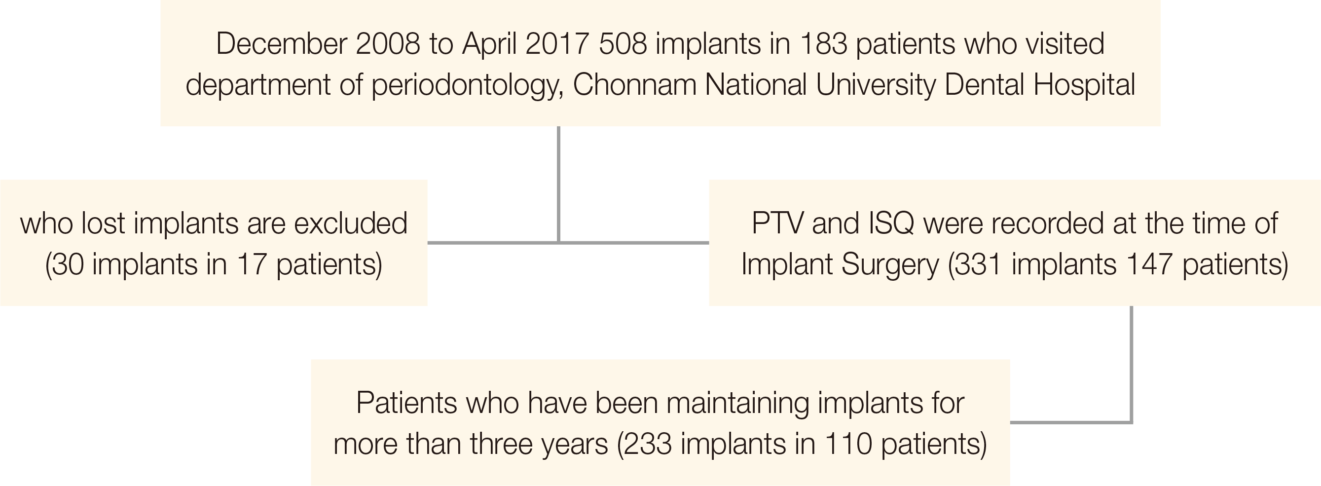
2008년 12월부터 2017년 4월까지 전남대학교 치과병원 치주과에서 10년 이상의 임플란트 수술에 숙련된 치주과 의사 한 명이 임플란트를 식립한 성인남녀 183명의 508개 임플란트를 대상으로 하였고 다음과 같은 포함 및 제외 기준으로 표본을 설정하였다.
치아 발거 원인은 고려하지 않았고, 전자의무기록 중 임플란트 1차수술과 임플란트 2차수술시 PTV, ISQ값이 모두 기록되지 않은 환자는 제외되었다. 임플란트 1차수술 후 3년 이상 유지되고 있는 환자를 표본으로 선택하고 임플란트가 상실된 환자는 Rosenberg 등15의 실패 시기 분류에 따라 총 30명이 제외되었다; Stage 1 (임플란트 식립 이후 2차수술을 하기까지의 기간) 11개, stage 2 (2차수술과 최종 보철물이 구강 내 완성되기까지의 기간) 8개, stage 3 (최종 보철물 완성 후 1년 이내) 1개, stage 4 (1년에서 5년 사이 탈락) 7개, stage 5 (5년 이후 기간 동안 탈락) 3개였다. 항응고제투여(고혈압, 심장질환, 혈관질환, 뇌질환), 간질환, 면역억제제(신장이식), 폐질환 (만성폐쇄성폐질환, 결핵), 골다공증 환자가 포함되었다.
이번 연구는 전남대학교 치과병원 생명연구윤리심의위원회의 윤리적, 과학적 기준에 따른 심의 후 진행되었다(CNUDH-2017-017).
2008년 12월부터 2017년 4월까지 전남대학교 치과병원 치주과에서 식립된 성인 183명의 508개 임플란트에 대한 진료 내역을 EMR을 통해 확인하였다. 보철물은 다수의 보철과 의사에 의해 수복되었다. 임플란트가 상실된 환자 17명의 임플란트 30개는 제외시키고, EMR 상에 임플란트 1차 수술과 임플란트 2차 수술 시 모두 periotest value (PTV)와 implant stability quotient (ISQ)가 기록되어 있으며 3년이상 임플란트를 유지중인 환자110명의 233개의 임플란트를 대상으로 하였다(Fig. 1).
군의 특성을 파악하기 위해 진료 기록부의 정보를 토대로 환자의 성별, 연령, 식립 부위에 따른 연구 집단을 기록하고, 임플란트 요소(임플란트 매식체의 직경, 길이, 임플란트-지대주 체결 방식, 임플란트 회사), 환자의 국소적, 전신적 요인(치주질환, 흡연, 당뇨, 항응고제의 섭취, 간질환, 면역억제제의 섭취, 골다공증), 추가적인 술식 및 유지관리 형태(골유도재생술(GBR) 유무, 상악동거상술 유무, supportive periodontal therapy (SPT) 유무)를 기록하고, PTV와 ISQ값을 기록하였다(Fig. 2).
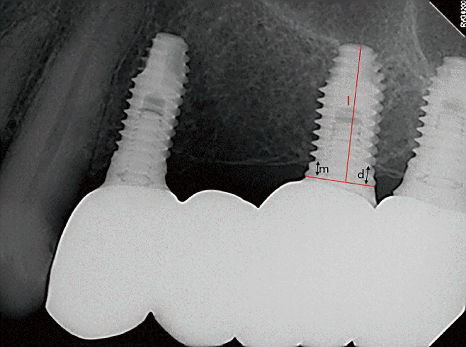
임플란트 식립 후 추적 검사 기간 동안 MBL을 평가하기 위하여 표준구내방사선영상이 사용되었으며, 최근 내원일을 기준으로 표준구내방사선 영상이 없는 경우는 파노라마 방사선 영상을 이용하여 분석을 시행하였다. INFINITT PACS M6 (INFINITT Healthcare Co., Ltd., Seoul, Korea) 프로그램을 이용하였다.
방사선 사진상의 확대/축소를 보정하기 위하여 환자의 표준구내방사선영상에서의 임플란트 매식체의 길이및 직경을 EMR에서 확인한 임플란트 매식체의 길이 및 직경과 대조하여 확대율을 계산하여 이를 임플란트 주위변연골 소실의 높이 보정에 사용하였다. MBL은 근심과 원심을 나눠서 측정하고 근·원심 평균값의 소수점 둘째 자리까지 기록하였다.
국제 구강 임플란트 전문의 학회(International Congress of Oral Implantologists)의 2007년 합의 회의에 제시된 기준인 success, satisfactory survival, compromised survival, failure로 분류하였다.16
임플란트 1차 수술 시 mount를 Periotest®로 PTV 측정하였고, Smartpeg® (Osstell AB, Gothenburg, Sweden)연결하여 RFA 측정해 ISQ로 기록하였다. 1 Stage 수술의 경우 mount를 제거한 fixture에 Healing abutment를 연결하여 PTV 측정하였다. 임플란트 2차 수술 시 또는 1 Stage 수술의 경우 수술 후 3개월차에 PTV, RFA 측정하였다.
기술 통계적 분석을 시행하여 연구 집단의 특성을 빈도와 백분율로 나타내기 위하여 빈도 분석을 시행하였다. 임플란트 매식체의 직경, 길이, 임플란트-지대주 체결 방식, 임플란트 회사, 전신질환의 유무, 치주질환의 유무, GBR 유무, 상악동거상술 유무, SPT 유무와 같은 인자들이 MBL과 3년이상 임플란트 성공률에 미치는 영향을 검증하기 위하여 다중회귀분석을 시행하였으며 후진제거법을 통해 불필요한 독립 변수들을 하나씩 제거하는과정을 반복하여 모형을 단순화하였다. 임플란트 1차, 2차 수술 시의 초기안정성지수(PTV, ISQ)와 3년 이상 성공적인 임플란트 성공률(success, satisfactory survival) 간의 비율 구성에 유의미한 차이가 있는지 알아보기 위하여 카이제곱 검정(교차분석)을 시행하였다. 3년이상 임플란트 장기 생존률(개월수)과 MBL에 대한 임플란트 초기 안정성 지표의 상관관계를 확인하기위해 피어슨 상관관계 분석을 시행하였다. IBM SPSS Statistics v23.0 (IBM SPSS Inc., New York, USA)을 이용하였으며 P < .05 수준에서 통계학적으로 유의한 것으로 간주하였다.

결과

이번 연구에서는 110명 환자에서 233개의 임플란트를 대상으로 하였고 임플란트 생존기간은 임플란트 1차 수술 이후를 기준으로 최소 36개월에서 최대 106개월이었다. 남성은 69명으로 총 151개의 임플란트가 식립되었고, 여성은 41명으로 총 82개의 임플란트를 식립되었다. 50대의 비율이 가장 높았으며 상악 구치부에 가장 많은 임플란트가 식립되었다(Table 1).
임플란트 매식체의 직경이 4 mm, 길이는 10 mm인 경우, 그리고 임플란트-지대주 연결방식이 external type인 경우가 가장 많이 식립되었다(Table 2). 치주염의 심도는 중도(severe)가 가장 많았고, GBR을 시행한 경우는 44.6%, 상악동거상술을 시행한 경우가 17.6%, SPT를 진행한 경우는 75.5%였다. 흡연자는 13.3%, 당뇨 환자는 14.2%, 항응고제 투약자는 19.7%, 간질환자는 4.3%, 면역억제제 투약자는 1.3%, 골다공증 환자는 4.7%였다(Table 3).
ICOI의 임플란트 성공률 분류 기준에 따라 방사선사진상 MBL을 기준으로 하여 success가 196개, satisfactory survival이 37개였다(Table 4).
임플란트 주위 변연골 흡수에 영향을 미치는 요소를검증하기 위해 다중회귀분석을 시행한 결과 회귀계수의 유의성 검증 결과, 임플란트-지대주 체결 방식(β = -.189, P < .05), SPT 유무(β = -.163, P < .05), 당뇨(β = -.164, P < .05), 골다공증(β = -.211, P < .005)은 변연골소실에 통계학적으로 유의한 음의 상관관계를 보였다. 즉 임플란트-지대주 체결 방식이 external type인 경우보다 in-ternal type이 변연골 흡수가 적었고, SPT를 진행한 경우와 당뇨나 골다공증 환자의 경우 변연골 소실이 많았다. 표준화 계수의 크기를 비교하면, 골다공증(β = -.211), 임플란트-지대주 체결방식(β = -.189), 당뇨(β = -.164), SPT 유무(β = -.163) 순으로 MBL에 큰 영향을 미치는 것으로 검증되었다(Table 5). 후진제거법을 이용한 다중회귀분석을 실시한 결과, 변연골 소실에 영향을 미치는 요소로 임플란트 매식체의 길이, 임플란트-지대주 체결 방식, GBR, SPT, 당뇨, 항응고제 복용, 골다공증이었다(P < 0.000)(Table 6).
3년 이상 임플란트 성공률에 미치는 영향에 대해 후진제거법으로 다중회귀분석을 시행한 결과 임플란트 장기성공률 중 success에 영향을 미치는 요소는 항응고제 복용이었다(Table 7). Satisfactory survival of implant에 영향을 미치는 요소는 없었다.
임플란트 성공률에 대하여 임플란트 1차 수술 시 PTV의 영향을 검증하기 위해 교차표를 산출한 결과 success는 good osseointegration이 182개(78.1%), borderline to use in clinic이 12개(5.2%), insufficient osseointegration이 2개(0.9%)으로 나타났고, satisfactory survival은 good osseointegration이 34개(14.6%), borderline to use in clinic이 3개(1.3%)였으며 카이제곱 검정을 실시한 결과, 유의한 차이를 보이지 않았다(Table 8). 임플란트 성공률에 대한 임플란트 1차 수술 시 ISQ 정도의 영향을 검증하기 위해 교차표를 산출한 결과 success는 high stability가 103개(44.2%), medium stability가 66개(28.3%), low stability가 25개(10.7%)로 나타났고, satisfactory survival은 high stability가 22개(9.5%), medium stabil-ity가 7개(3.0%), low stability가 8개(3.4%)로 나타났으며 카이제곱 검정의 결과 유의한 차이를 보이지 않았다(Table 9).
임플란트 성공률에 대한 임플란트 2차 수술 시 PTV와의 관련성 검증하기 위해 교차표를 산출하였을 때success에서는 good osseointegration이 191개(82.0%), borderline to use in clinic이 4개(1.7%)로 나타났고, satisfactory survival에서는 good osseointegration이 37개(15.9%)로 나타났으며 카이제곱 검정을 실시한 결과, 유의한 차이는 없었다(Table 10). 임플란트 성공률에 대한 임플란트 2차 수술 시 ISQ와의 관련성을 검증하기 위해 교차표를 산출한 결과 success에서는 high stability가 148개(63.4%), medium stability가 37개(15.9%), low stability가 9개(3.9%), un-measureable이 2개(0.9%)로 나타났고, satisfactory survival에서는 high stability가 26개(11.2%), medium stability가 7개(3.0%), low stability가 3개(1.3%)로 나타났으며 카이제곱 검정한 결과, 유의한 차이를 보이지 않았다(Table 11).
임플란트 장기 생존(개월 수)과 임플란트 1차 수술시 PTV, ISQ, 임플란트 2차 수술시 PTV, ISQ 간 상관관계를 확인하기 위해 피어슨 상관관계 분석한 결과 임플란트 장기 생존(개월 수)은 임플란트 2차 수술 시 PTV와 음의 상관관계를 보였다(r = .016, P < .05). 임플란트 1차 수술시 PTV, ISQ, 임플란트 2차 수술 시 ISQ는 임플란트 장기 생존(개월 수)과 유의한 상관관계를 보이지 않았다(Table 12). 변연골 흡수 정도와 임플란트 생존 개월수의 상관관계를 확인하기 위해 피어슨 상관관계 분석을실시한 결과 유의한 차이가 없었다(r = .073).

고찰

이번 연구는 2008년 12월부터 2017년 4월까지 전남대학교 치과병원 치주과에서 10년 이상 숙련된 한 명의 치주과 의사가 임플란트를 식립하고, 포함 및 제외 기준을 충족한 환자 110명의 233개의 임플란트를 대상으로 하여 임플란트의 장기간 유지에 미치는 요인들을 알아보기위해 시행하였다. 그 결과 임플란트-지대주 체결 방식이 external인 경우보다 internal인 경우 변연골 흡수가 적었고, SPT를 진행한 경우 변연골 흡수가 증가하였으며, 당뇨나 골다공증 환자의 경우 변연골 흡수가 많았다. 임플란트 2차 수술 시 PTV 값은 감소하고, ISQ 값은 증가하는 양상을 보였으며, 임플란트 2차 수술 시 높은 PTV는 임플란트의 3년 이상 장기 생존에 유의한 영향을 주었다. Peñarrocha 등17은 임플란트 주위골 흡수와 환자의 성별 및 연령, 임플란트 식립 부위 간의 연관성은 적다고 보고하였으며 이번 연구에서도 유사한 결과를 얻었다. ICOI의 임플란트 성공률 분류 기준에 따라 방사선영상에서 변연골 흡수를 기준으로 하여 success가 196개, satisfactory survival이 37개였고, compromised survival과 failure는 없었다. 10년 이상 숙련된 한 명의 치과의사가 임플란트 식립 후 임플란트 안정성이 충분히 확보된후에 보철물 제작 단계를 진행하였고, 3년 이상 임플란트가 생존하고 있는 환자를 표본으로 선택했기 때문이라고할 수 있을 것이다.
임플란트-지대주 체결 방식은 external type이 internal type보다 변연골 흡수가 많았다. 이는 Mederios 등,18 Lemos 등,19 Caricasulo 등20의 연구의 결과와 유사하다. Mederios 등18의 경우 메타분석에서 10개의 논문으로부터 총 2708개의 임플란트(2347개의 internal type과 361개의 external type)를 864명의 환자를 대상으로 하여 internal connections 임플란트가 external connections 임플란트보다 변연골 흡수가 더 적음을 보여주었다. Lemos 등19의 연구에서도 internal-connection의 경우 변연골 흡수가 더 적음을 보여주었다(Mean Difference: 0.44 mm; 95% Confidence interval (CI): 0.26 - 0.63 mm). 그렇지만 임플란트 성공률에는 차이가 없었다(P = 0.65; Risk Ratio (RR): 0.83; 95% CI: 0.38 - 1.84). Caricasulo 등20은 internal type이 단기, 중기 기간 동안 더 나은 변연골 수준을 나타낸다고 하였다.
SPT에 의한 적절한 구강위생관리는 치료 결과의 장기적 안정을 위해 가장 중요한 요소이다.21-24 동일한 표본 집단을 가진 이전의 Choi 등25의 연구에 따르면 유지치주치료를 받은 환자의 임플란트 생존율은 100%, 정기적인 유지치주치료를 받지 않은 환자의 생존율은 89.2%였다는 결과와 상반된다. Choi 등25의 연구는 임플란트 탈락 단계별로 분류하여 환자의 임플란트 생존율을 구하였고이번 연구에서는 임플란트가 탈락한 경우는 제외시켜 표본을 선택한 후 임플란트 주위골 흡수 정도에 따라 임플란트 생존율을 구하였기 때문에 결과가 다른 것으로 보인다. 또한 SPT는 치주질환이 심하거나 추가적으로 골이식 수술을 하는 등의 요인이 있었기 때문에 변연골 소실량이 많은 것으로 보인다.
당뇨와 골다공증의 기왕력은 변연골 흡수를 증가시킨다는 보고들이 있었다. 이는 임플란트 금기증은 아니지만 변연골 흡수를 증가시키므로 당뇨 및 골다공증을 재고하여야 한다고 하였다. 당뇨병 환자들의 임플란트 성공률과 예측 가능성에 대한 연구들 중 임플란트 성공률이 85.6%에서 94.3%라고 볼 때, 당뇨환자들의 실패율이 보통이라고 보고하였다.26-28 골다공증은 폐경기 변화(I형), 연령-연관 변화(II형), 혹은 의원성 이유(III형)에 기인한 1차성 골다공증과 다양한 질병들과 당뇨병, 알코올중독, 영양결핍, 그리고 흡연과 같은 다양한 상태들에 의해 발생하는 2차성 골다공증이 있다. 다양한 형태의 골다공증은 모두 무기질 밀도 감소라는 근본적인 문제들을 공유하며, 이러한 조건들은 임플란트 골유착과 이의유지를 저하시킬 수 있기 때문에 골다공증 환자에서 임플란트는 기능을 하지 못할 것이라는 전제는 타당하다.그러나 현재까지 명확한 증거는 없으며 논쟁은 계속되고있다.29,30 상악동거상술을 받은 49명의 환자들에 대한 후향적 분석에서, 골밀도가 낮은 11명의 환자들은 연령과성별에서 동등한 대조군과 비교했을 때 유의하게 낮은임플란트 성공률을 나타냈지만, 다른 비교기준들은 유의한 차이를 보이지 않는 보고가 있다.31
이번 연구에서는 임플란트 매식체의 직경, 길이, 임플란트 회사, 임플란트-지대주 체결 방식, 치주질환의 유무, GBR 유무, 상악동거상술 유무, SPT 유무, 전신질환의 유무(흡연, 당뇨, 항응고제 복용, 간질환, 면역억제제 복용, 골다공증)가 3년이상 임플란트 성공률 success와 satisfactory survival of implant에 영향을 주는 가에 관한 검증에서 통계학적인 유의성은 없었다. 임플란트가 상실된 환자는 Rosenberg 등15의 실패 시기에 따라 총 30명이 제외되었다. 이 논문은 10년이상 임플란트 수술에 숙련된 치과의사 1명에 의해 진행되었고, 임플란트 1차 수술 이후 3년이상 임플란트가 생존하고 있는 환자를 기준으로 진행되었다. 즉, 임플란트 성공률이 success, satisfactory survival에만 환자군이 존재하며 compromised survival, failure 환자군은 존재하지 않아 표본 분포 불균일에 의해 유의하지 않는 결과가 나온 것으로 사료된다.
임플란트 성공률에 대한 임플란트 1차, 2차 수술 시 PTV의 골유착 정도 차이와 ISQ의 임플란트 안정성 정도의 영향은 관련성을 보이지 않았다. 즉 임플란트 장기간 유지에 있어서 초기 PTV, ISQ값은 장기간 유지에는 영향을 미치지 않음을 알 수 있었다. 이는 Adell 등13의 연구에서 임플란트 초기 안정성이 골유착의 필수 조건이며, 골유착의 효과적인 예견지표라고 한 것과 관련이 있을 것이라고 판단하였다. 이 연구에서는 10년이상 수술을 시행한 숙련된 치과의사 1명이 전남대학교 치과병원 치주과에서 임플란트 1차 수술과 2차 수술시 PTV수치나 ISQ 수치를 매번 확인하고, 수치가 골유착에 불충분하다 싶으면 매식체의 두께나 길이를 다르게 하거나 보철물을 하기 전의 기간을 늘리는 방식으로 충분한 임플란트 초기 안정성을 얻기 위해 노력하기 때문에 임플란트안정성이 높은 구간에 대부분의 표본이 존재하는 것과도연관이 있을 것으로 판단된다.
임플란트 1차 수술 후 2차 수술시 PTV는 good osseointegration이 92.7%에서 97.9%로 늘었고, insufficient osseointegration 0.9%에서 0%으로 줄었고, ISQ는 high stability가 53.7%에서 74.6%가 되었고 low stability는14.1%에서 5.2%로 감소함을 보였다. 1차 수술 후 2차 수술시 임플란트 수술부위가 치유됨에 따라 PTV값은 전체적으로 감소하고, ISQ값은 전체적으로 증가하여 이는 1차 수술 후 치유기간동안 골유착이 증가한다는 의미를 갖는다는 것이다. Truhlar 등32은 2차수술시 PTV값은 -3.82 ± 3.04 (quality 1 bone), -3.70 ± 3.06 (quality 2 bone), -3.31 ± 3.18 (quality 3 bone), -1.29 ± 3.57 (quality 4 bone) 수치를 가지며, Periotest가 2차수술 시의 골유착 상태를 평가하는 데 적절한 기구라고 하였다. 이 연구에서는 골질은 고려하지 않았지만 2차 수술시 PTV 값이 골유착 정도를 평가하는데 유용하다는 결과와 일치한다. RFA는 전통적으로 임상 기준으로 임플란트 실패를검사할 수 있다.33 Vidyasagar34의 연구에서는 1차 수술시ISQ 66 ± 6.2 (range 52 to 79)가 2차 수술시 ISQ 65 ± 6.2 (range 52 to 79)값으로 변화하며, 하악에 식립한 임플란트가 상악 보다 더 높은 안정성을 보인다고 하였다. Vidyasagar의 연구와 유사하게 이번 연구에서도 2차수술 후 약간 증가하는 양상을 보였다.
임플란트 초기 안정성 지표(임플란트 1차 수술, 2차 수술 시 PTV, ISQ)와 임플란트 장기 생존(개월수)과의 상관관계는 임플란트 2차 수술시 PTV값과 장기 생존(개월 수)에 유의한 음의 상관관계를 보였는데 부하 전에 안정성이 장기간의 생존률에 영향을 줌을 알 수 있었다. 임플란트 2차 수술시 PTV값이 작을수록(-8 ≤ PTV < 0) 임플란트 장기 생존에 유의하다는 결과만 얻을 수 있었다. 이는 임플란트 식립 후 추적 검사 중에 높은 임플란트 안정성 지수 값을 갖는 임플란트가 성공적으로 골유착된다는 Sennerby와 Meredith14 연구 결과와 공통적으로 임플란트 2차 수술시의 PTV가 낮을수록 골유착이 커지며, 임플란트 장기 생존에 유의하다는 결과를 얻었다.
이번 연구의 장점은 10년 이상 임플란트 수술이 숙달된 한 명의 치과의사에서만 시행된 환자 중 3년 이상 임플란트가 유지되고 있는 임플란트를 대상으로 연구 집단의 특성을 파악하고, 최대 106개월의 장기 추적을 통해 임플란트 초기안정성의 영향을 파악하려고 시도해 본점이다. 술자 내 편차나 술자간 편차를 배제한 연구 집단을 이용할 수 있는 장점이 있다. 반면 이 연구의 한계점은 후향적연구였기 때문에 방사선 사진 추적에 어려움을 겪었고, 구내표준방사선사진이 없는 경우 파노라마 사진을 이용해 변연골 소실을 측정했기 때문에 변연골 소실량에있어 오차가 발생할 수 있었을 것이다. 또한 임플란트 초기안정성이 확보되지 않은 경우 다음 보철 단계를 늦추거나 다른 매식체로 교체하는 등 초기안정성을 확보하기 때문에, 초기안정성을 낮은 표본이 없어 임플란트 초기 안정성 지표와 임플란트 성공률 간의 유의성이 없다는 점이며 좀 더 많은 수의 표본이 필요하고 장기간 추적관찰이 필요하다.

결론

이 연구는 임플란트 수술이 시행되어 최소 3년이상 임플란트를 유지하고 있는 환자에서 장기간 임플란트 유지에 영향을 미칠 수 있는 요인이 무엇인지 파악하고, 임플란트 초기안정성 지표가 임플란트 성공에 미치는 영향을파악하며, 장기간 유지되는지 알아보기 위해 시행되었다.
ICOI 임플란트 성공률 분류 기준에 따라 방사선사진상 변연골 흡수에 대하여 success가 196개(84.1%), satisfactory survival이 37개(15.9%)였다. 변연골 흡수에 임플란트 체결방식, SPT 유무, 당뇨, 골다공증이 음의 상관관계를 보였다. 표준화 계수 크기를 비교했을 때, 골다공증, 임플란트 체결방식, 당뇨, 넷 순으로 변연골 흡수에 큰 영향을 미쳤다. 후진제거법을 이용한 다중회귀분석시 임플란트 매식체의 길이, 체결방식, GBR, SPT, 당뇨, 항응고제, 골다공증이 변연골 흡수에 영향을 미쳤다. 임플란트 성공률에 있어 1차, 2차 수술 시의 임플란트 안정성 지수(PTV, ISQ)의 상관관계에 유의성이 없었다. 2차수술시 전체적으로 PTV 수치가 감소하였고, ISQ는 증가하였다. 변연골 흡수 정도와 임플란트 생존 개월수 간의 상관관계는 없었다. 임플란트 2차 수술시 PTV 감소는 osseointegration의 증가와 임플란트의 3년 이상 장기 생존에 유의한 영향을 주었다.

References

1. Brånemark PI, Svensson B, van Steenberghe D. 1995; Ten-year survival rates of fixed prostheses on four or six implants ad modum Brånemark in full edentulism. Clin Oral Implants Res. 6:227–31. DOI: 10.1034/j.1600-0501.1995.060405.x. PMID: 8603114.
crossref
2. Mengel R, Schröder T, Flores-de-Jacoby L. 2001; Osseointegrated implants in patients treated for generalized chronic periodontitis and generalized aggressive periodontitis: 3- and 5-year results of a prospective long-term study. J Periodontol. 72:977–89. DOI: 10.1902/jop.2001.72.8.977. PMID: 11525449.
crossref
3. Buser D, Ingimarsson S, Dula K, Lussi A, Hirt HP, Belser UC. 2002; Long-term stability of osseointegrated implants in augmented bone: a 5-year prospective study in partially edentulous patients. Int J Periodontics Restorative Dent. 22:109–17. PMID: 12019706.
4. Brägger U, Hirt-Steiner S, Schnell N, Schmidlin K, Salvi GE, Pjetursson B, Matuliene G, Zwahlen M, Lang NP. 2011; Complication and failure rates of fixed dental prostheses in patients treated for periodontal disease. Clin Oral Implants Res. 22:70–7. DOI: 10.1111/j.1600-0501.2010.02095.x. PMID: 21158932.
crossref
5. Jung RE, Zembic A, Pjetursson BE, Zwahlen M, Thoma DS. 2012; Systematic review of the survival rate and the incidence of biological, technical, and aesthetic complications of single crowns on implants reported in longitudinal studies with a mean follow-up of 5 years. Clin Oral Implants Res. 23:2–21. DOI: 10.1111/j.1600-0501.2012.02547.x. PMID: 23062124.
6. Lang NP, Zitzmann NU. 2012; Clinical research in implant dentistry: evaluation of implant-supported restorations, aesthetic and patient-reported outcomes. J Clin Periodontol. 39:133–8. DOI: 10.1111/j.1600-051X.2011.01842.x. PMID: 22533953.
crossref
7. Pjetursson BE, Thoma D, Jung R, Zwahlen M, Zembic A. 2012; A systematic review of the survival and complication rates of implant-supported fixed dental prostheses (FDPs) after a mean observation period of at least 5 years. Clin Oral Implants Res. 23 Suppl 6:22–38. DOI: 10.1111/j.1600-0501.2012.02546.x. PMID: 23062125.
8. Chiapasco M. 2004; Early and immediate restoration and loading of implants in completely edentulous patients. Int J Oral Maillofac Implants. 19:76–91. PMID: 15635948.
9. Cochran DL, Nevins M. 2012; Biologic width: a physiologically and politically resilient structure. Int J Periodontics Restorative Dent. 32:371–3. PMID: 22577641.
10. Moy PK, Medina D, Shetty V, Aghaloo TL. 2005; Dental implant failure rates and associated risk factors. Int J Oral Maxillofac Implants. 20:569–77. PMID: 16161741.
11. Ahmed A, Chambers MS, Goldschmidt MC, Habib A, Lei X, Jacob RF. 2012; Association between microbial flora and tissue abnormality around dental implants penetrating the skin in reconstructed oral cancer patients. Int J Oral Maillofac Implants. 27:684–94. PMID: 22616064.
12. Kasat V, Ladda R. 2012; Smoking and dental implants. J Int Soc Prev Community Dent. 2:38–41. DOI: 10.4103/2231-0762.109358. PMID: 24478965. PMCID: PMC3894084.
crossref
13. Adell R, Lekholm U, Rockler B, Brånemark PI. 1981; A 15-year study of osseointegrated implants in the treatment of the edentulous jaw. Int J Oral Surg. 10:387–416. DOI: 10.1016/S0300-9785(81)80077-4. PMID: 6809663.
crossref
14. Sennerby L, Merdith N. 2008; Implant stability measurements using resonance frequency analysis: biological and biomechanical aspects and clinical implications. Periodontol 2000. 47:51–66. DOI: 10.1111/j.1600-0757.2008.00267.x. PMID: 18412573.
crossref
15. Rosenberg ES, Cho SC, Elian N, Jalbout ZN, Froum S, Evian CI. 2004; A comparison of characteristics of Implant failure and survival in periodontally compromised and periodontally healthy patients: a clinical report. Int J Oral Maillofac Implants. 19:873–9. PMID: 15623064.
16. Misch CE, Perel ML, Wang HL, Sammartino G, Galindo-Moreno P, Trisi P, Steigmann M, Rebaudi A, Palti A, Pikos MA, Schwartz-Arad D, Choukroun J, Gutierrez-Perez JL, Marenzi G, Valavanis DK. 2008; Implant Success, Survival, and Failure: the International Congress of Oral Implantologists (ICOI) Pisa Consensus Conference. Implant Dent. 17:5–15. DOI: 10.1097/ID.0b013e3181676059. PMID: 18332753.
crossref
17. Peñarrocha M, Palomar M, Sanchis JM, Guarinos J, Balaguer J. 2004; Radiologic study of marginal bone loss around 108 dental implants and its relationship to smoking, implant location, and morphology. Int J Oral Maxillofac Implants. 19:861–7. PMID: 15623062.
18. de Medeiros RA, Pellizzer EP, Vechiato Filho AJ, Dos Santos DM, da Silva EV, Goiato MC. 2016; Evaluation of marginal bone loss of dental implants with internal or external connections and its association with other variables: A systematic review. J Prosthet Dent. 116:501–6. DOI: 10.1016/j.prosdent.2016.03.027. PMID: 27422232.
crossref
19. Lemos CAA, Verri FR, Bonfante EA, Santiago Júnior JF, Pellizzer EP. 2018; Comparison of external and internal implant-abutment connections for implant supported prosthesis. A systematic review and meta-analysis. J Dent. 70:14–22. DOI: 10.1016/j.jdent.2017.12.001. PMID: 29221955.
crossref
20. Caricasulo R, Malchiodi L, Ghensi P, Fantozzi G, Cucchi A. 2018; The influence of implant-abutment connection to peri-implant bone loss: A systematic review and meta-analysis. Clin Implant Dent Relat Res. 20:653–64. DOI: 10.1111/cid.12620. PMID: 29761902.
crossref
21. Knowles JW, Burgett FG, Nissle RR, Shick RA, Morrison EC, Ramfjord SP. 1979; Results of periodontal treatment related to pocket depth and attachment level. Eight years. J Periodontol. 50:225–33. DOI: 10.1902/jop.1979.50.5.225. PMID: 287778.
crossref
22. Ramfjord SP, Caffesse RG, Morrison EC, Hill RW, Kerry GJ, Appleberry EA, Nissle RR, Stults DL. 1987; Four modalities of periodontal treatment compared over 5 years. J Periodontal Res. 22:222–3. DOI: 10.1111/j.1600-0765.1987.tb01573.x. PMID: 2955103.
crossref
23. Ramfjord SP, Morrison EC, Burgett FG, Nissle RR, Shick RA, Zann GJ, Knowles JW. 1982; Oral hygiene and maintenance of periodontal support. J Periodontol. 53:26–30. DOI: 10.1902/jop.1982.53.1.26. PMID: 6948947.
crossref
24. Lindhe J, Nyman S. 1984; Long-term maintenance of patients treated for advanced periodontal disease. J Clin Periodontol. 11:504–14. DOI: 10.1111/j.1600-051X.1984.tb00902.x. PMID: 6384275.
crossref
25. Choi SJ, Jeong HN, Kim OS. 2018; Effect of supportive periodontal therapy on long-term implant survival rate. Oral Biol Res. 42:228–34. DOI: 10.21851/obr.42.04.201812.228.
crossref
26. Balshi SF, Wolfinger GJ, Balshi TJ. 2007; An examination of immediately loaded dental implant stability in the diabetic patient using resonance frequency analysis (RFA). Quintessence Int. 38:271–9. PMID: 17432781.
27. Fiorellini JP, Chen PK, Nevins M, Nevins ML. 2000; A retrospective study of dental implants in diabetic patients. Int J Periodontics Restorative Dent. 20:366–73. PMID: 11203576.
28. Kapur KK, Garrett NR, Hamada MO, Roumanas ED, Freymiller E, Han T, Diener RM, Levin S, Ida R. 1998; A randomized clinical trial comparing the efficacy of mandibular implant-supported overdentures and conventional dentures in diabetic patients. Part I: Methodology and clinical outcomes. J Prosthet Dent. 79:555–69. DOI: 10.1016/S0022-3913(98)70177-5. PMID: 9597609.
crossref
29. Baxter JC, Fattore L. 1993; Osteoporosis and osseointegration of implants. J Prosthodont. 2:120–5. DOI: 10.1111/j.1532-849X.1993.tb00393.x. PMID: 8242165.
crossref
30. Dao TT, Anderson JD, Zarb GA. 1993; Is osteoporosis a risk factor for osseointegration of dental implants? Int J Oral Maxillofac Implants. 8:137–44. DOI: 10.1097/00008505-199404000-00014. PMID: 8359868.
31. Blomqvist JE, Alberius P, Isaksson S, Linde A, Hansson BG. 1996; Factors in implant integration failure after bone grafting: an osteometric and endocrinologic matched analysis. Int J Oral Maxillofac Surg. 25:63–8. DOI: 10.1016/S0901-5027(96)80015-4. PMID: 8833304.
32. Truhlar RS, Morris HF, Ochi S, Winkler S. 1994; Assessment of implant mobility at second-stage surgery with the Periotest: DICRG Interim Report No. 3. Dental Implant Clinical Research Group. Implant Dent. 3:153–6. DOI: 10.1097/00008505-199409000-00002. PMID: 7749399.
33. Friberg B, Sennerby L, Meredith N, Lekholm U. 1999; A comparison between cutting torque and resonance frequency measurements of maxillary impalnts. A 20-month clinical study. Int J Oral Maxillofac Surg. 28:297–303. DOI: 10.1016/S0901-5027(99)80163-5.
crossref
34. Vidyasagar L, Salms G, Apse P, Teibe U. 2004; Dental implant stability at stage I and II surgery as measured using resonance frequency analysis. Stomatologija. 6:67–72.

Fig. 1
Flow chart of the final study sample.
PTV, periotest value; ISQ, implant stability quotient.
jdras-38-2-97-f1.tif
Fig. 2
Measurement of marginal bone loss.
jdras-38-2-97-f2.tif
Table 1
Description of the study population and implant placed site
Variable Subjects (N) Percentage (%) Numbers of implants (N = 233)
Gender Male 69 64.8 151
Female 41 35.2 82
Age 20 - 29 2 0.9 2
30 - 39 5 3.9 9
40 - 49 14 13.7 32
50 - 59 37 33.0 77
60 - 69 32 29.2 68
70 - 79 18 17.2 40
90 - 99 2 2.1 5
Placed sites Maxillary anteriors 10.3 24
Maxillary posteriors 40.3 94
Mandibular anteriors 6.9 16
Mandibular posteriors 42.5 99

anteriors: central incisors, lateral incisors, canines.

posteriors: premolars, molars.

Table 2
The characteristics of placed implants
Percentage (%) Number (N = 233)
Fixture diameter (mm) 3/3.25/3.5 0.4/0.9/3.0 1/2/7
3.75/4.0/4.5 3.9/50.1/2.1 9/125/5
5.0/6.0 35.6/0.4 83/1
Fixture length (mm) 6.0/7.0 0.4/0.4 1/1
8.5/10.0 3.9/56.7 9/132
11.5/12.0 33.0/0.4 77/1
13.0 5.2 12
Implant brand Osstem 51.5 120
Straumann 1.3 3
3i 33.9 79
MegaGen 13.3 31
Abutment connection type External 96.6 225
Internal 3.4 8
Table 3
Implants distribution by periodontal status, systemic condition, additional procedures and SPT
Percentage (%) Number (N = 233)
Periodontitis status Healthy 2.1 5
Slight 3.0 7
Moderate 39.1 91
Severe 55.8 130
Smoking Smoker 13.3 31
Non-smoker 86.7 202
Diabetes patient 14.2 33
Non-patient 85.8 200
Anticoagulant taking Yes 19.7 46
No 80.3 187
Liver disease patient 4.3 10
Non-patient 95.7 223
Immunosuppressant taking Yes 1.3 3
No 98.7 230
Osteoporosis Patient 4.7 11
Non-patient 95.3 222
GBR Operate 44.6 104
Non-operate 55.4 129
SE Operate 17.6 41
Non-operate 82.4 192
SPT Operate 75.5 176
Non-operate 24.5 57

GBR, guided bone regeneration; SE, sinus elevation; SPT, supportive periodontal therapy.

Table 4
Marginal bone loss according to success and satisfactory survival
Percentage (%) Numbers of implants Mean (mm)(Min - Max)
Success 84.1 196 0.94 (-1.07 - 1.98)
Satisfactory survival 15.9 37 2.58 (2.02 - 3.71)
Total 100.0 233 1.20 (-1.07 - 3.71)
Table 5
Factors affecting peri-implant marginal bone loss
Dependent variables Independent variables B S.E. β t P VIF
Marginal bone loss (Constant) 3.778 1.693 2.232 0.027
Implant diameter 0.076 0.106 0.049 0.721 0.472 1.205
Implant length 0.110 0.057 0.133 1.938 0.054 1.229
Implant system -0.017 0.027 -0.044 -0.621 0.535 1.314
Implant type -0.861 0.309 -0.189 -2.782 0.006* 1.203
Periodontal disease -0.067 0.085 -0.053 -0.782 0.435 1.211
GBR -0.193 0.133 -0.116 -1.457 0.147 1.649
SE -0.079 0.178 -0.036 -0.443 0.658 1.741
SPT -0.315 0.133 -0.163 -2.373 0.018* 1.231
Smoking 0.170 0.173 0.070 0.984 0.326 1.306
Diabetes -0.391 0.178 -0.164 -2.197 0.029* 1.462
Anticoagulant 0.260 0.157 0.124 1.650 0.100 1.487
Liver disease -0.336 0.275 -0.082 -1.222 0.223 1.175
Immunosuppressant 0.077 0.492 0.010 0.157 0.875 1.165
Osteoporosis -0.828 0.259 -0.211 -3.196 0.002* 1.143
F = 3.128 (P < 0.000), R2 = 0.167, adjR2 = 0.114, D - W = 1.788

*: P < .05.

B, beta estimator; S.E., standard error; β, standardized coefficient; VIF, variance inflation factor; GBR, guided bone regeneration; SE, sinus elevation; SPT, supportive periodontal therapy.

Table 6
Factors affecting peri-implant marginal bone loss (Backward Elimination)
Dependent variables Independent variables B S.E. β t P VIF
Marginal bone loss (Constant) 3.466 .850 4.078
Implant length .099 .052 .120 1.890 0.942 1.061
Implant type -.846 .295 -.185 -2.863 0.902 1.109
GBR -.205 .106 -.123 -1.939 0.944 1.060
SPT -.292 .125 -.151 -2.337 0.901 1.110
Diabetes -.351 .163 -.147 -2.160 0.813 1.231
Anticoagulant .260 .147 .124 1.761 0.757 1.320
Osteoporosis -.804 .255 -.205 -3.154 0.891 1.122
F = 5.669 (P < 0.000), R2 = 0.150, adjR2 = 0.123, D - W = 1.749

Backward Elimination : All independent variables are entered into the equation first and each one is deleted one at a time if they do not contribute to the regression equation.

B, beta estimator; S.E., standard error; β, standardized coefficient; VIF, variance inflation factor; GBR, guided bone regeneration; SPT, supportive periodontal therapy.

Table 7
Factors affecting success of implants (Backward Elimination)
Dependent variables Independent variables B S.E. β t P VIF
Success of implant (Constant) 1.310 .183 7.165 0.000
Anticoagulant -.207 .100 -.147 -2.075 0.039 1.000
F = 4.306 (P < 0.039), R2 = 0.022, adjR2 = 0.017, D - W = 1.715

B, beta estimator; S.E., standard error; β, standardized coefficient; VIF, variance inflation factor.

Table 8
Description of the relationship between PTVs at implant first surgery and implant successPTV, periotest value.
Implant stability scale Implant quality scale Total χ2
Success Satisfactory survival
PTV (1st surgery) Good osseointegration (-8 < PTV ≤ 0) Number of implant 182 34 216 0.57
Percentage (%) 78.1 14.6 92.7
Borderline to use in clinic (0 < PTV ≤ 10) Number of implant 12 3 15
Percentage (%) 5.2 1.3 6.4
Insufficient osseointegration (10 < PTV) Number of implant 2 0 2
Percentage (%) 0.9 0.0 0.9
Total Number of implant 196 37 233
Percentage (%) 84.1 15.9 100

PTV, periotest value.

Table 9
Description of the relationship between ISQ values at implant first surgery and implant success
Implant stability scale Implant quality scale Total χ2
Success Satisfactory survival
ISQ (1st surgery) High stability (ISQ ≥ 70) Number of implant 103 22 125 4.27
Percentage (%) 44.2 9.5 53.7
Medium stability (60 ≤ ISQ < 70) Number of implant 66 7 73
Percentage (%) 28.3 3.0 31.3
Low stability (ISQ < 60) Number of implant 25 8 33
Percentage (%) 10.7 3.4 14.1
Un-measureable Number of implant 2 0 2
Percentage (%) 0.9 0.0 0.9
Total Number of implant 196 37 233
Percentage (%) 84.1 15.9 100.0

ISQ, implant stability quotient.

Table 10
Description of the relationship between PTVs at implant second surgery and implant success
Implant stability scale Implant quality scale Total χ2
Success Satisfactory survival
PTV (2nd surgery) Good osseointegration (-8 < PTV ≤ 0) Number of implant 191 37 228 0.80
Percentage (%) 82.0 15.9 97.9
Borderline to use in clinic (0 < PTV ≤ 10) Number of implant 4 0 4
Percentage (%) 1.7 0.0 1.7
Un-measureable Number of implant 1 0 1
Percentage (%) 0.4 0.0 0.4
Total Number of implant 196 37 233
Percentage (%) 100.0 100.0 100.0

PTV, periotest value.

Table 11
Description of the relationship between ISQ values at implant second surgery and impalnt success
Implant stability scale Implant quality scale Total χ2
Success Satisfactory survival
ISQ (2nd surgery) High stability (ISQ ≥ 70) Number of implant 148 26 174 0.86
Percentage (%) 63.4 11.2 74.6
Medium stability (60 ≤ ISQ < 70) Number of implant 37 7 44
Percentage (%) 15.9 3.0 18.9
Low stability (ISQ < 60) Number of implant 9 3 12
Percentage (%) 3.9 1.3 5.2
Un-measureable Number of implant 2 1 3
Percentage (%) 0.9 0.4 1.3
Total Number of implant 196 37 233
Percentage (%) 84.1 15.9 100.0

ISQ, implant stability quotient.

Table 12
Correlation between implant stability at first and second surgery and long-term survival rate
Month 1st, PTV 2nd, PTV 1st, ISQ 2nd, ISQ
Long-term survival rate (month) 1
Implant 1st surgery, PTV -.135 1
Implant 2nd surgery, PTV -.031 .016 1
Implant 1st surgery, ISQ -.089 .101 -.006 1
Implant 2nd surgery, ISQ -.072 .017 -.008 -.012 1

PTV, periotest value; ISQ, implant stability quotient.

TOOLS
Similar articles