

 PDF
PDF Citation
 Print
Print


Introduction
The paracondylar process (PC) (also termed the estiloid, paramastoid, parajugular, and/or paraoccipital process) is a bony extension from the inferior side of the occipital bone [1]. The vertebral artery, facial nerve, and contents of the jugular foramen are close to it. A PC is found in approximately 0.5% to 2% of the population [2], including some variants in which it articulates with the epitransverse process of the atlas, causing neck ankylosis and limiting the range of motion of the head [2]. Although it might be palpated during physical examination, imaging is required to confirm its presence [3]. Appreciating the anatomical location and variations of the PC is clinically important for those performing surgeries in this region, and for understanding its role in symptomatic cases. Here, we report an extremely rare case of three PCs associated with other anatomical variations of the skull.
Go to : 
Case Report
We report on the skull of an adult male found to have multiple skull base variations. The skull reported here is from the skeleton which is part of the osteological collection curated at Tulane University School of Medicine. The specimen was Caucasian and approximately 70-year-old at death. Three bony excrescences from the occipital bone were identified in the paracondylar region (Fig. 1). The smallest of these processes was 7 mm long (P1) and was just medial to the left mastoid process of the temporal bone and posterior to the styloid process (Fig. 1). One bony process was attached to the occipital condyle and was 12 mm (P3) in length (Figs. 1–3). The longest of these processes was what is also referred to as a paramastoid process and this was 17 mm long (P2) and arose from the jugular process of the occipital bone (Figs. 1, 2). This process (P2) articulated with an epitransverse process of the ipsilateral C1 vertebra forming a joint (Figs. 4–6). In addition, extending from this process and forming a secondary joint with the superior articular process of C1 was an additional bony excrescence (Figs. 2, 6). The skull specimen also exhibited a torus occipitalis transversus (Figs. 4, 7) and an ipsilateral septated hypoglossal canal. None of the variant processes in the paracondylar region was present on the contralateral side. The remainder of the skeleton from which this specimen was derived did not have other obvious anatomical variations or other variant bony excrescences.
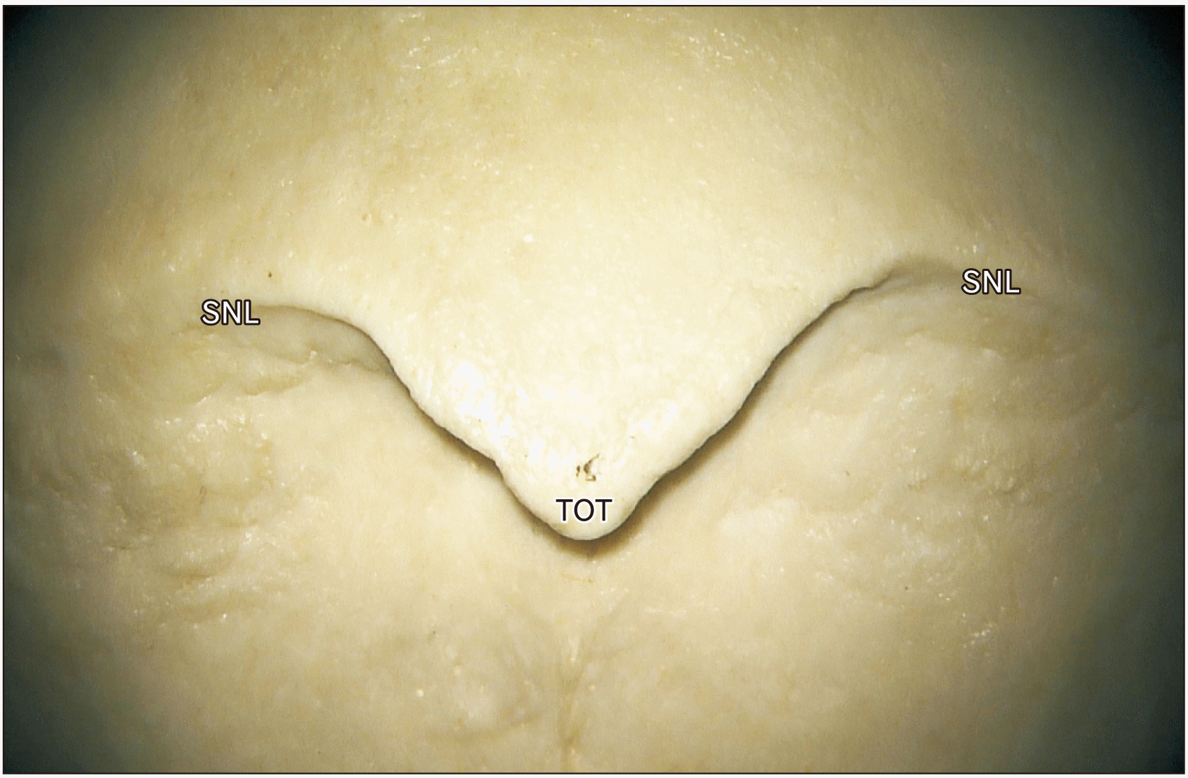
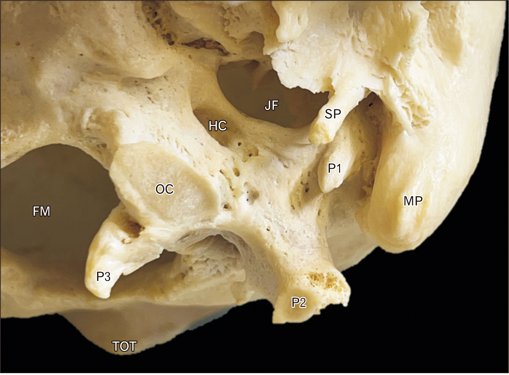 | Fig. 1Inferior view of the left sided skull base in the specimen described herein. Note the three odd bony protuberances: P1, medial to the mastoid process (MP); P2, the more typical ‘paracondylar process’; and P3, extending inferomedial to the occipital condyle. Also note the torus occipitalis transversus (TOT), foramen magnum (FM), occipital condyle (OC), hypoglossal canal (HC), jugular foramen (JF), and styloid process (SP).
|
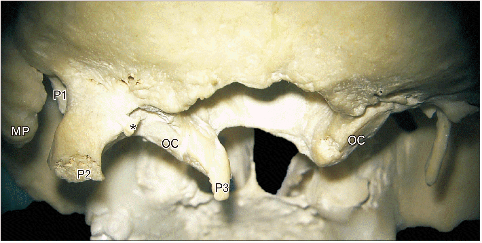 | Fig. 2Posterior view of the skull base. Note the three odd bony protuberances: P1, medial to the mastoid process (MP); P2, the more typical ‘paracondylar process’; and P3, extending inferomedial to the occipital condyle (OC). *Additional bony excrescence.
|
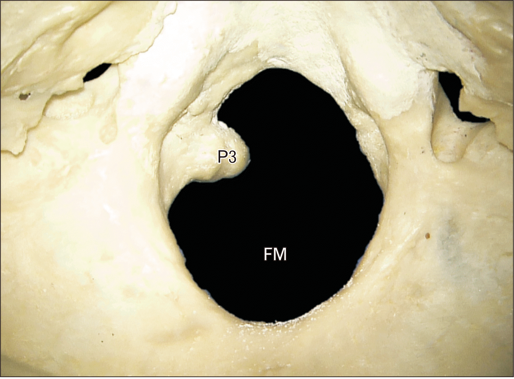 | Fig. 3Internal view through the foramen magnum (FM) noting the medial extension (P3) from the left occipital condyle.
|
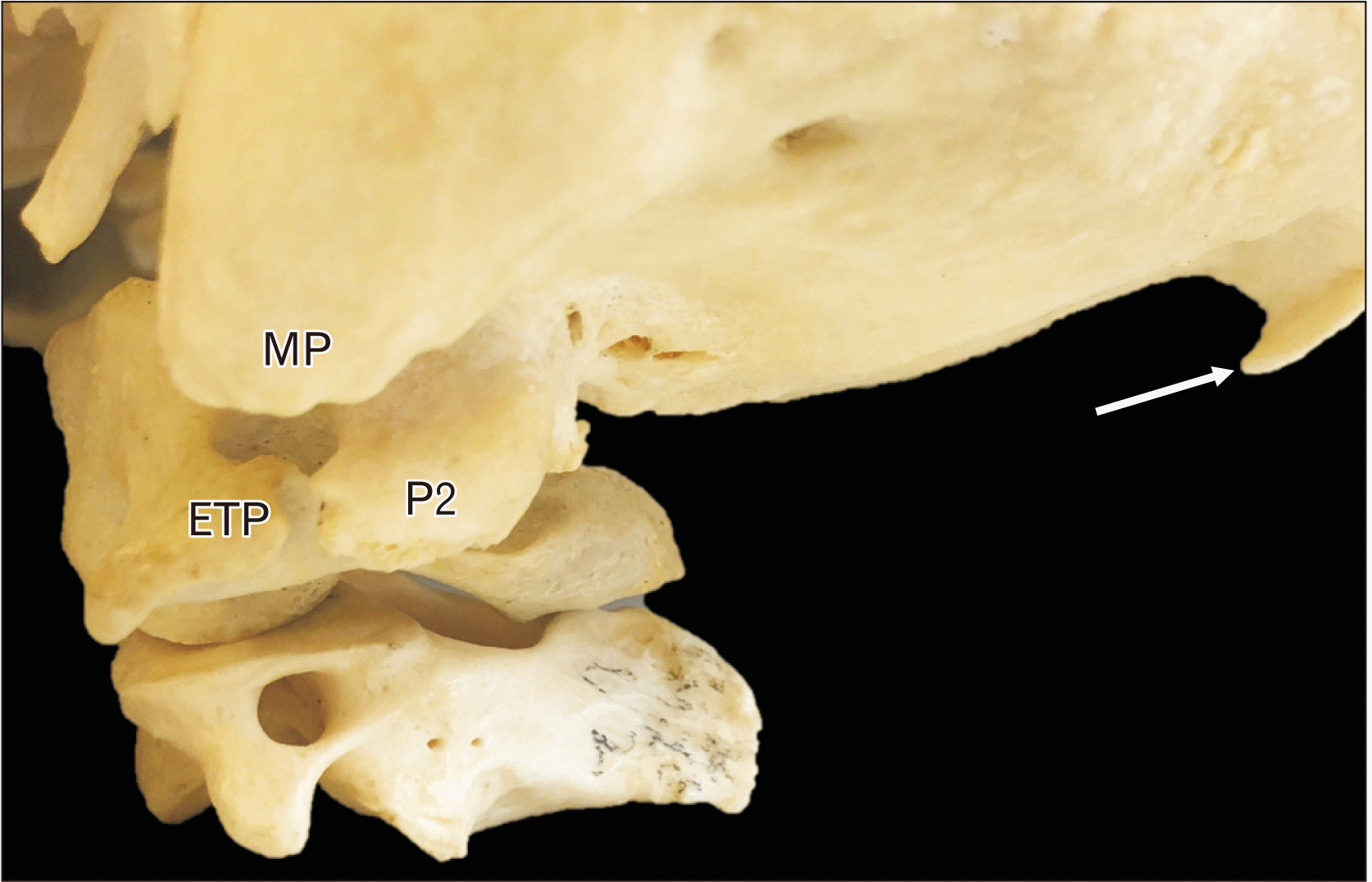
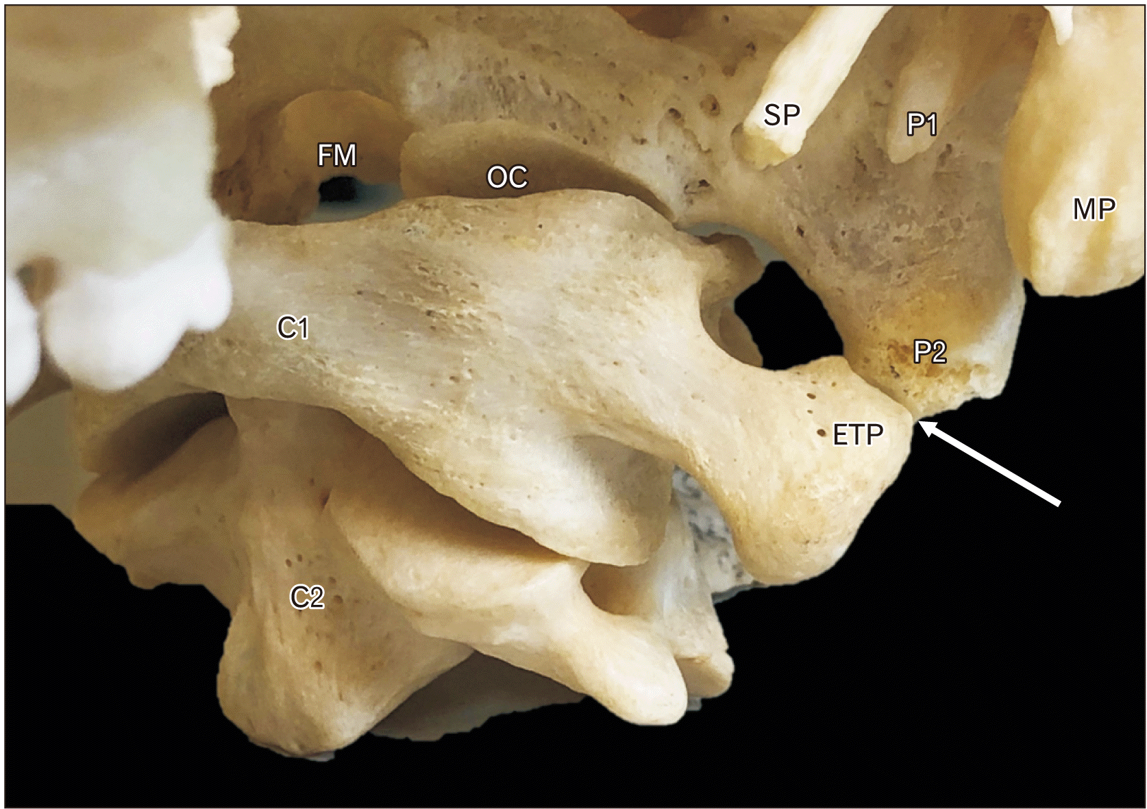 | Fig. 4Articulation between the upper two cervical vertebrae (C1 and C2) and the skull reported here. Note the epitympanic process (ETP) of the left transverse process of the atlas (C1) articulating with the paracondylar process (P2) of the skull (arrow). The abnormal bony process (P1) medial to the mastoid process (MP) is also seen. For reference, note the styloid process (SP), occipital condyle (OC), and foramen magnum (FM).
|
 | Fig. 5Lateral view. Note the joint formed between the epitympanic process of C1 (ETP) and the paracondylar process (P2) of the occipital bone. Note the mastoid process (MP) and torus occipitalis transversus (arrow).
|
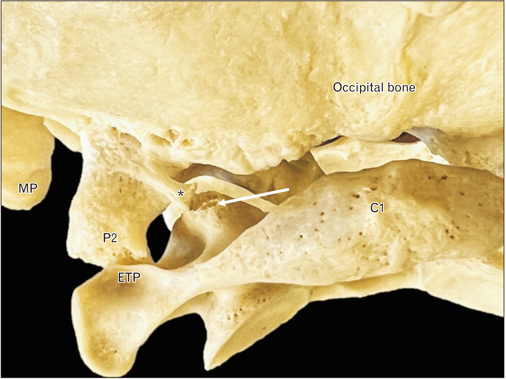 | Fig. 6Posterior view of the skull noting the two points of articulation between the paracondylar process (P2). The more lateral articulation is between the paramastoid process and epitympanic process (ETP) of C1. The more medial articulation (arrow) is between a process (*) from the P2 and the superior articular process of C1 (arrow). MP, mastoid process.
|
Go to :
Discussion
Whether the PC is formed congenitally or is acquired is a matter for debate [2]. It is hypothesized that the PC forms around the 4th week of embryo development because of malformation of the first cervical and the fourth occipital sclerotomes [1]. During development, the first cervical sclerotome and the caudal part of the fourth occipital sclerotome fuse to form the proatlas [1]. In a process called caudal shifting, in which a vertebra mimics its caudal neighbor, the cervical sclerotome fails to separate, wholly or partly, from the occipital sclerotome [1]. This variable degree of separation between the sclerotomes can cause numerous variations of PCs, explaining symptomatic cases. The alternative hypothesis, that the PC is acquired and not congenital, implicates either excessive pull by the rectus capitis lateralis muscle during infancy, or trauma [2].
Clinical significance
The identification of a PC, as mentioned earlier, can result in symptoms such as decrease movement of the neck at the craniocervical junction. However, most patients found to have a PC are asymptomatic. However, a variant PC is clinically more significant if it is connected to the epitransverse process of the atlas (Figs. 4–6), causing symptoms of neck ankylosis and a limited range of head motion [3]. The repeated contact of the PC and the atlas can cause synovial joint degeneration, leading to various degrees of headaches and neck pain [1]. The large PC in our case that articulated with the epitransverse process of the atlas would also prove to be an obstacle for the needle for clinicians who attempt to inject the atlantooccipital joint in patients with arthritic pain or occipital neuralgia of an arthrological origin. As seen in our case, the addition of two other PC would certainly have limited, in life, the patient’s ability to rotate the head, especially with the more medial of the PC that extended from the occipital condyle. This bony excrescence would inhibit normal movement at the atlantooccipital joint thus limited the amount of flexion/extension at this joint.
The PC can be seen on computed topography (CT) or magnetic resonance imaging (MRI). In one case, both methods were used. The CT images showed a bony mass articulating with the transverse process of the atlas, while the MRI showed a bony extension from the skull base [3]. From the locations of these bony masses, McCall et al. [3] were able to identify a right sided PC. Lastly, as it has, to our knowledge, not been reported before, the most medial of the PC in our specimen, originating from the occipital condyle itself, would probably result in an imaging dilemma and potentially, misdiagnosis.
Surgical significance
The PC can also be seen during surgery at the base of the occipital bone. Once identified, it must be carefully maneuvered around owing to its close proximity to the jugular process, vertebral artery, and facial nerve [4]. Additionally, suboccipital approaches to the skull base e.g., transcondylar approaches for resection of, for example, tumors near the foramen magnum e.g., meningioma, can be inhibited by the presence of a PC. The large PC in our case shown that articulated with the atlas at two points would definitely obscure the view of the occipital condyle for the surgeon. Therefore, intraoperative removal of this PC with drilling would be necessary in order to safely approach the ipsilateral occipital condyle.
In conclusions, knowledge of the bony variations at the skull base are important to those who operate in this region or review and interpret radiological imaging. Although the single PC has been reported before including some scant illustrations of an additional PC near the mastoid process as we describe [5], to our knowledge, three simultaneous PCs, in general, and a PC which is an extension of the occipital condyle, have not been reported in the extant medical literature. Therefore, we believe the present case is very unusual and of archival value.
Go to :
 XML Download