Journal List > Anat Cell Biol > v.55(2) > 1516078771

Romeh, El-Safty, El-Mehi, and Faried: Antioxidant, anti-inflammatory, and anti-fibrotic properties of olive leaf extract protect against L-arginine induced chronic pancreatitis in the adult male albino rat

Abstract

Chronic pancreatitis (CP) is an inflammatory disease affects the pancreas with upcoming fibrosis and notable parenchymal destruction. CP poses a high risk for pancreatic carcinoma. The present study aimed to investigate, for the first time up to our knowledge, the effect of olive leaf extract on L-arginine induced CP with referral to some of its underlying mechanisms. Forty adult male albino rats were divided equally into four groups; control, olive leaf extract treated (200 mg/kg orally once daily), CP group (300 mg L-arginine/100 g body weight intraperitoneally, once daily for 3 weeks then every 3 days for the subsequent 3 weeks), and CP treated with olive leaf extract group. At the end of the experiment, body weight, serum glucose, serum insulin, homeostatic model assessment of insulin resistance (HOMA-IR), serum amylase and lipase as well as tissue superoxide dismutase (SOD), and malondialdehyde (MDA) levels were assessed. Pancreatic tissues were subjected to histological and immuno-histochemical studies. The CP group revealed significant decrease in body weight and increase in serum glucose, serum insulin, HOMA-IR score, serum amylase, and serum lipase levels. Significant increase in MDA level and decrease in SOD level were detected. Marked degenerative changes and fibrosis were detected. Upregulation of alpha smooth muscle actin (α-SMA), transforming growth factor beta (TGF-β), caspase-3, and interleukin-6 (IL-6) immunoreactions were implicated in CP pathogenesis. Olive leaf extract alleviated all the examined parameters via its-antioxidant, anti-inflammatory, and anti-fibrotic properties. Olive leaf extract can protect against CP and restore pancreatic functions.

Introduction

Chronic pancreatitis (CP) is an inflammatory disease affects the pancreas with upcoming fibrosis and notable parenchymal destruction [1]. Unfortunately, the incidence of this disease is still in-increase [2]. The commonest cause of CP is prolonged alcohol consumption, the second one is idiopathic and the third is gallstones [3]. The patients with CP possessed frequent attacks of acute pancreatitis with subsequent exocrine and endocrine dysfunction [4]. Patients with CP complains of increased blood glucose and insulin levels. This indicates insulin resistance reflected by increased homeostatic model assessment of insulin resistance (HOMA-IR) index [1]. The CP has a serious negative impact on the social and psychological health [5]. Further, the risk of pancreatic cancer in CP patients increased almost eightfold [6].
L-arginine is an α-amino acid used in the biosynthesis of proteins [7]. L-arginine plays an important role in cell division, wound healing, removing ammonia from the body, immune function, and the release of hormones. It is important in the regulation of blood pressure [8]. Despite medical treatment, the lethality of severe acute pancreatitis and CP with L-arginine is still high (20%–30%) [9]. When given as intraperitoneal injections, L-arginine is easily administered and damages pancreatic cells. So, it can be used as a method to induce CP by repeated injections over a longer period [10].
In spite of decades of research, knowledge about the specific pathogenesis of CP is still limited. So, the treatment of this disease remains empirical, and the current therapeutic plans of this disease are symptomatic and supportive including nutrition and pain control [11]. However, inflammation and free radicals are forming together the scaffold of disease etiology [12].
Pancreatic stellate cells (PSCs) can regulate the normal architecture of the pancreas by balancing the matrix degradation process and fibrogenesis [13]. An inflamed pancreas induced the PCSs’ proliferation and activation with secretion of cytokines and extracellular matrix (ECM) proteins [14]. Activated PSCs have been accepted as a key precursor of pancreatic fibrosis, which is a hallmark of CP. Hence, it is evident that targeting PSCs may be a promising treatment for CP [15].
For a long time, phytotherapy as olive tree has been used to treat many diseases [16]. The main ingredient of olive leaf is oleuropein, which has a high antioxidant activity. Thus, olive leaf is considered one of the most active natural antioxidants [17]. In addition, the olive leaf contains the highest concentration of olive plant polyphenols more than the fruit or oil [18]. Olive leaf extract (OLE) is used as a supplementation or as a drug. It has many benefits including anti-atherosclerotic, anti-arrhythmic [19], anti-tumor, anti-hypertensive, antioxidant [20], anti-inflammatory [21], and anti-fibrotic [22] properties. Thus, the research community's interest in olive leaves and their extracts has intensified in recent years because of the olive's great potential for use in medicine and the pharmaceutical industry [23].
Taken together, this study was designed to examine, for the first time up to our knowledge, the usefulness of OLE administration in preventing L-arginine induced CP in a rat model.

Materials and Methods

Animals

The current study was conducted on 40 healthy adult male Sprague–Dawley rats (3 months old, weighing 180–200 g). They were kept in the animal house of the Faculty of Medicine, Menoufia University, Egypt. The animals were maintained under controlled temperature and humidity conditions and provided with water and a balanced diet ad libitum. Strict care and hygiene measures were taken to maintain a healthy environment for all rats all the time. All experimental procedures were approved by the ethical research committee of the Menoufia University Faculty of Medicine, Egypt (MU29122020).

Experimental substances

L- Arginine

It was purchased from Sigma Chemical Co. (St. Louis, MO, USA) in the form of powder and dissolved in normal saline (40% wt/vol, pH 7.4) [24].

Olive leaf extract

The leaves of olive (Olea europaea L.) were collected from Al Arish, Egypt. The plant material was authenticated in the Faculty of Agriculture, Menoufia University, Egypt. The extract was prepared at the Faculty of Science, Menoufia University, Egypt using the method of Al-Attar and Abu Zeid [25] with some modifications. The powder of dried olive leaves (200 g) was prepared and added to 7 L of hot water. After 3 hours, the mixture was slowly boiled for 30 minutes. The mixture was then cooled to room temperature and subjected to an electric mixer for 20 minutes. Thereafter, the solution was filtered. The filtrate of olive leaves was evaporated in an oven at 40°C to produce dried residues (active principles). The yield mean of the leaves extract was 20.3% with references to the powdered samples.

Experimental design

After an adaptation period of one week, rats were randomly divided into four equal groups (ten rats each):
  • Group I (control group): kept without any treatment.

  • Group II (OLE treated group): received a daily oral dose of aqueous OLE by gastric intubation at a dose 200 mg/kg, dissolved in 2 ml distilled water, for 10 weeks [26].

  • Group III (CP group): received distilled water, in an amount equal to that used for dissolving OLE, for 4 weeks to be followed by CP induction by intraperitoneal injection of 300 mg L-arginine/100 g body weight, dissolved in normal saline (40% wt/vol, pH 7.4), once daily for 3 weeks. Subsequently, L-arginine injections were given at the same dose every 3 days for the subsequent 3 weeks [24].

  • Group IV (CP treated with OLE group): received OLE, at the same dose and route of administration, 4 weeks before L-arginine injection. Supplementation with OLE continued during the period of CP induction for further 6 weeks.

Specimen collection

At the end of the experimental period (10 weeks), the rats body weight was estimated, and the blood samples were collected from the rats’ tail dorsal veins for the biochemical evaluation. Rats were anaesthetized using an intraperitoneal injection of ketamine (90 mg/kg)/xylazine (15 mg/kg) and then sacrificed by cervical dislocation. The anterior abdominal wall was incised longitudinally, and the pancreas was dissected out and flushed with physiological saline. Each specimen was divided into two portions; one was fixed in 10% neutral buffered formalin for the histological and immunohistochemical assessment, and the other was immediately frozen at −70°C to be used for tissue biochemical evaluation.

Evaluation methods

Biochemical study

Estimation of blood glucose & serum parameters: Fasting blood glucose level was evaluated using blood glucose test strips (Accu-Chek; Diagnostic Solutions Inc., Irvine, CA, USA) and One Touch Basic Blood Glucose Meter (Life Scan Inc., Milpitas, CA, USA). The other blood samples were allowed to clot then centrifuged at 4,000 rpm for 10 minutes. The collected serum was used for assessment of serum levels of insulin [27], amylase [28], and lipase [29].
HOMA-IR was calculated using this formula: HOMA-IR=fasting glucose (mg/dl)×fasting insulin (µU/ml)/405 [27].
Assessment of lipid peroxidation & tissue antioxidant status: The frozen pancreatic tissue was homogenized in ice-cold phosphate buffer (140 mmol/L KCl, 20 mmol/L phosphate, PH 7.4) and centrifuged at 4,500 rpm for 15 minutes at 4°C. According to Kono [30], the supernatant was used to measure the levels of superoxide dismutase (SOD) and malondialdehyde (MDA) in the tissues.

Histological study

After fixation in 10% neutral formalin for 24 hours, the pancreatic specimens were dehydrated using ascending grades of alcohol then cleared and embedded in paraffin. Microtome was used to cut sections of 5 μm thick. Sections were subjected to hematoxylin and eosin (H&E) staining for routine histological examination and Mallory trichrome stain for detection of collagen fiber deposition.
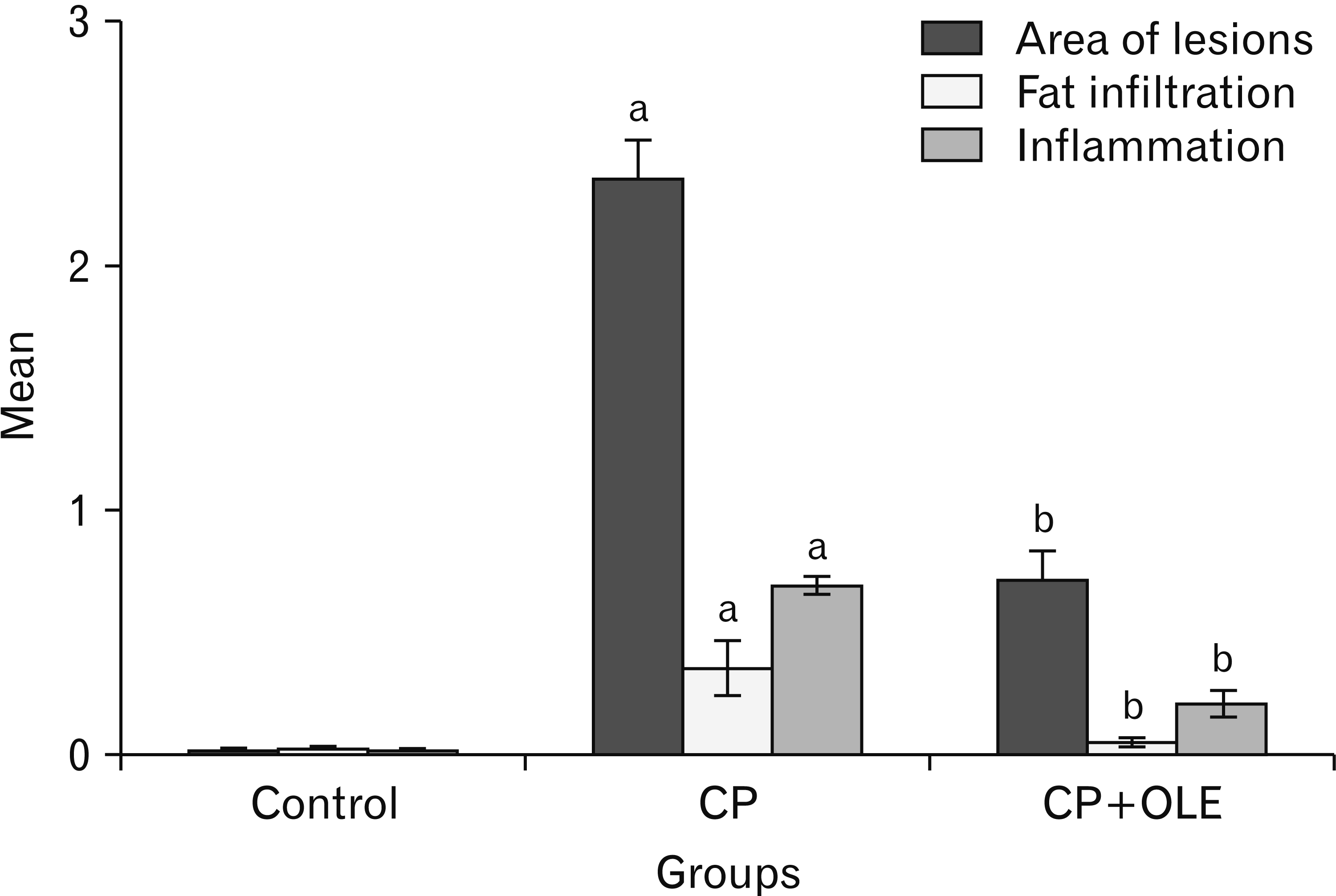
Histologic scoring of area of lesions, inflammation, and fatty infiltration were performed by Image J software (Bethesda, MD, USA) in at least three sections/animal according to Liu et al. [31] (Table 1).
For immunohistochemical staining, paraffin sections were placed in xylene for 1–2 minutes, then rehydrated with degraded ethanol (100%, 95%, and 70% ethanol) twice for 5 minutes each time, and then inserted in distilled water for another 5 minutes. The slices were inserted in phosphate buffered saline (PBS) and blocked in 0.1% H2O2 for 30 minutes to impede the endogenous peroxidase activity. The sections were then maintained in a blocking solution (10% normal goat serum) at room temperature (22°C) for 60 minutes. Then incubation with the primary antibodies: caspase-3 (rabbit polyclonal antibody, 1:400; Abcam, Cambridge, MA, USA), interleukin‐6 (IL-6) (a mouse monoclonal antibody, 1:400; Abcam), alpha smooth muscle actin (α-SMA) (a rabbit polyclonal antibody, 1:100; Abcam), and transforming growth factor beta (TGF-β) (a mouse monoclonal antibody, 1:500; Midco Trade) was performed. After PBS, the sections were incubated with biotinylated secondary antibody for 20 minutes at room temperature. After rinsing with PBS, streptavidin-Horseradish peroxidase solution was placed on the slices for 10 minutes; 3,3-diaminobenzoic acid dissolved in PBS was used for secondary antibody binding; and 0.03% H2O2 was added immediately before use. Finally, the sections were dipped with PBS, and the slides were counterstained with two drops of hematoxylin. The slides were then dehydrated with ascending ethanol (70%, 95%, and 100%) for 5 minutes, then cleared with xylene and then covered with coverslips using a mounting solution [32].

Quantitative assessment

Using Image J software version K 1.45, the following parameters were measured:
  1. The area % of collagen deposition.

  2. The area % of caspase-3, IL-6, α-SMA, and TGF-β immunoreactions.

For each parameter, five non-overlapping fields (×40) were randomly taken from every specimen using Leica DML B2/11888111 microscope equipped with Leica DFC450 camera.

Statistical analysis

The collected data were expressed as the mean±SD. The data was analyzed using SPSS version 23 (IBM Corp., Armonk, NY, USA). One-way analysis of variance (ANOVA) followed by Bonferroni post-hoc test was used to assess the significance of differences between groups. When P-value <0.05, the result was considered statistically significant [33].

Results

There was no significant difference in all the examined parameters between the control and OLE treated groups.

Body weight results

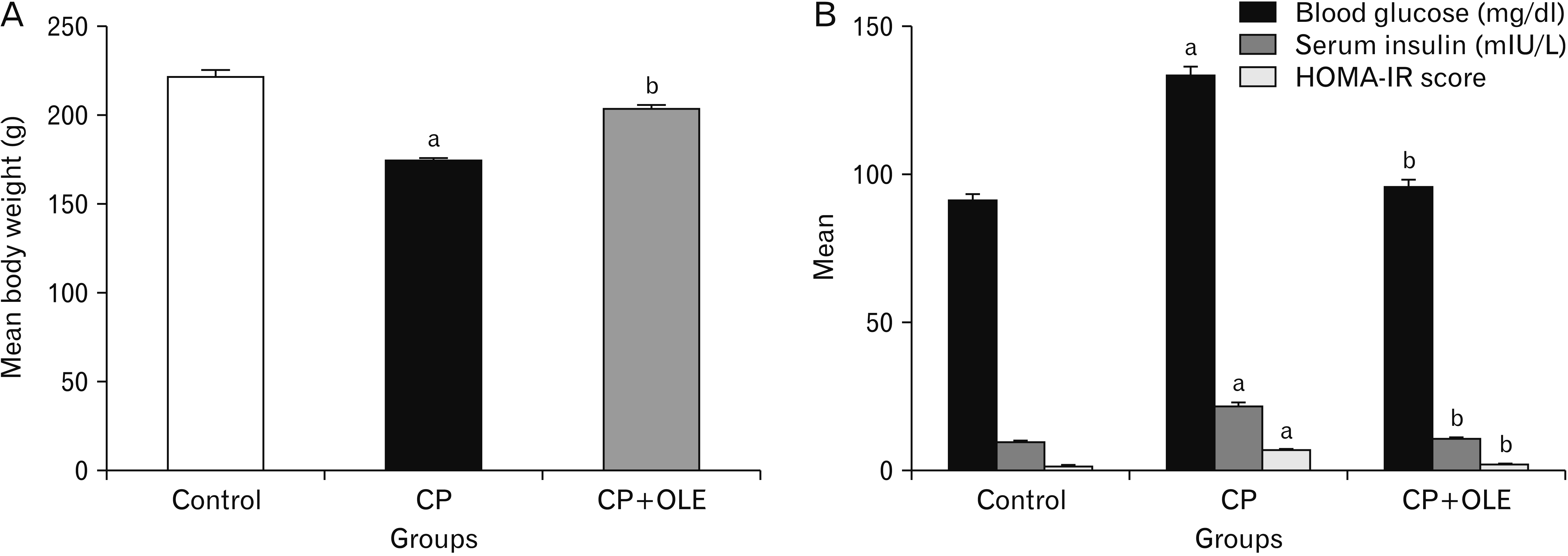
At the end of the experiment, there was a significant decrease (P<0.001) in the body weight in the CP group (174.25±2.39 vs. 221.3±5.3) compared with the control group. On the other hand, the CP group treated with OLE showed a significant increase (P<0.001) in its weight (203.16±3.26 vs. 174.25±2.39) compared with the CP group (Fig. 1A).

Biochemical results

Compared with the control group, the blood glucose level of the CP group increased significantly (P<0.001) (133.33±4.16 vs. 91.05±2.73). Compared with the untreated CP group, this level was significantly reduced (P<0.001) in the CP group treated with OLE (95.67±3.48 vs. 133.33±4.16) (Fig. 1B).
Regarding the serum insulin level, a significant increase (P<0.001) in its level was detected in the CP group (21.24±2.24 vs. 9.38±0.301) compared with the control group, while the CP group treated with OLE exhibited a significant decrease (P<0.001) in the serum insulin level (10.31±0.461 vs. 21.24±2.24) compared with the untreated CP group (Fig. 1B). So, there was a significant increase (P<0.001) in HOMA-IR score (insulin resistance) in the CP group (6.77±0.302 vs. 1.24±0.168) compared with the control group. But HOMA-IR score in the CP group treated with OLE exhibited a significant decrease (P<0.001) compared with the untreated CP group (2.01±0.213 vs. 6.77±0.302) (Fig. 1B).
The serum amylase and lipase levels showed a significant increase (P<0.001) in the CP group compared with the control group (204.25±3.66 vs. 87.53±0.996 and 53.22±1.47 vs. 29.2±0.638 respectively), while the CP group treated with OLE exhibited a significant decrease (P<0.001) in the serum amylase and lipase levels (103.05±2.71 vs. 204.25±3.66 and 33.91±2.37 vs. 53.22±1.47 respectively) compared with the untreated CP group (Fig. 2A, B).
The assessment of MDA and SOD levels in the pancreatic homogenates revealed a significant increase (P<0.001) in MDA level (13.08±0.292 vs. 1.169±0.067) and a significant decrease (P<0.001) in SOD level (0.694±0.055 vs. 2.409±0.298) in the CP group compared with the control group. In addition, the CP group treated with OLE showed a significant decrease (P<0.001) in the MDA level (6.84±0.58 vs. 13.08±0.292) and a significant increase (P<0.001) in SOD level (1.52±0.14 vs. 0.694±0.055) when compared with the untreated CP group (Fig. 2C, D).

Histological results

Light microscopic study

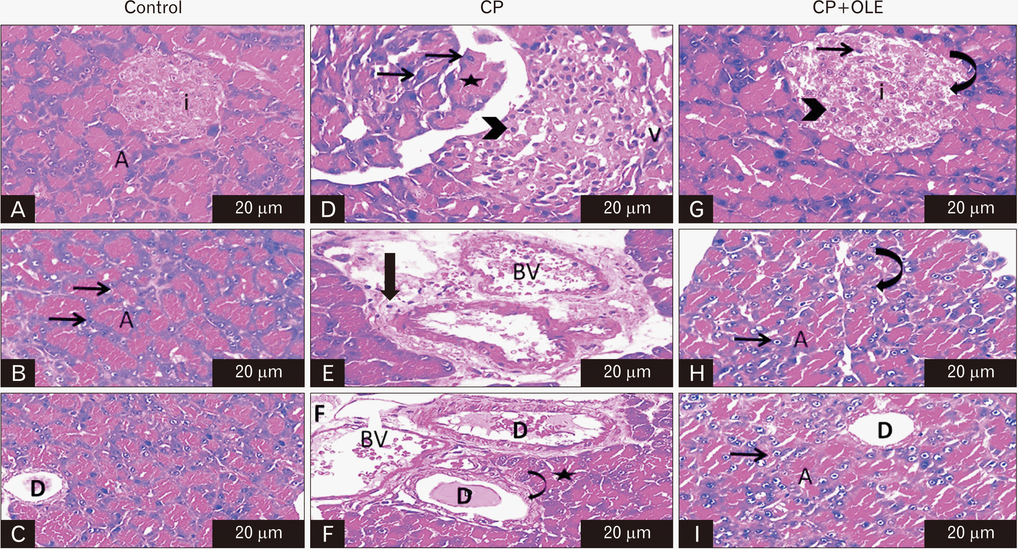
H&E stained sections from the control group showed that the pancreas was divided into many small lobules of different sizes and shapes, separated by interlobular connective tissue. Each lobule consisted of the main exocrine part; a compound duct system and pancreatic acini with basal basophilic part and an eosinophilic apical part. The acinar cells had vesicular nuclei with prominent nucleoli. Masses in the endocrine part (islets of Langerhans) were unevely distributed in-between the pancreatic acini (Fig. 3A–C).
The CP group showed marked degenerative alterations. Most of the acinar cells showed vacuolation of the cytoplasm and decreased zymogen granules. Dilatation of the interlobular pancreatic duct was observed. Dilated congested blood vessels were also seen. Inflammatory cellular infiltration among the degenerated pancreatic acini was seen. Islets of Langerhans showed many vacuoles and most of the cells appeared with deeply stained pyknotic nuclei (Fig. 3D–F).
The CP group treated with OLE showed an improvement of most of the previously mentioned histopathological findings in the CP group. The pancreatic lobules appeared almost normal. The majority of the acinar cells appeared nearly normal, while some cells appeared with deeply stained pyknotic nuclei. Islets of Langerhans were almost normal, but some vacuoles and deeply stained pyknotic nuclei were also seen (Fig. 3G–I).
Statistically, the scores of area % of lesions, fat infiltration and inflammation were significantly increased (P<0.001) in the CP group compared with the control group (2.35±0.32 vs. 0.015±0.005, 0.35±0.158 vs. 0.017±0.009 and 0.686±0.06 vs. 0.03±0.02 respectively). These results were significantly decreased (P<0.001) in the CP group treated with OLE compared with the untreated CP group (0.78±0.217 vs. 2.35±0.32, 0.143±0.31 vs. 0.35±0.158 and 0.206±0.07 vs. 0.686±0.06 respectively) (Fig. 4).
The Mallory trichrome stained sections of the control group showed slight collagen deposition between the pancreatic lobules and in the islets of Langerhans (Fig. 5A, B). Sections of the CP group showed excessive collagen deposition around the ductal system and between the degenerated pancreatic acini. In addition, islets of Langerhans showed a large amount of collagen fiber deposition (Fig. 5C, D). The sections of the CP group treated with OLE showed fine collagen fibers between the pancreatic lobules, around the duct system, and in the islets of Langerhans (Fig. 5E, F).
Statistically, a significant increase (P<0.001) in the area % of collagen deposition in the CP group compared with the control group (98.08±1.32 vs. 0.686±0.06). In addition, there was also a significant decrease (P<0.001) in the area % in the CP group treated with OLE compared to the untreated CP group (36.55±3.06 vs. 98.08±1.32) (Fig. 5G).

Immunohistochemical results

Immunohistochemically, the area % of caspase-3 and IL-6 immunoreactions showed a significant increase (P<0.001) in the CP group compared with the control group (98.02±1.88 vs. 0.28±0.03 and 96.68±1.54 vs. 1.3±0.21 respectively). In addition, there was also a significant decrease (P<0.001) in the surface area % of caspase-3 and IL-6 immunoreactions in the CP group treated with OLE compared with the untreated CP group (3.7±0.49 vs. 98.02±1.88 and 17.47±0.86 vs. 96.68±1.54 respectively) (Fig. 6E–H).
Further, compared with the control group, the area % of α-SMA and TGF-β immunoreactions in the CP group increased significantly (P<0.001) (96.95±1.62 vs. 0.05±0.02 and 97.24±1.51 vs. 0.23±0.03 respectively) (Fig. 6I–L). In addition, compared with the untreated CP group, the area % of α-SMA and TGF-β immunoreactions in the CP group treated with OLE was significantly reduced (P<0.001) in (36.55±3.06 vs. 96.95±1.62 and 11.16±0.52 vs. 97.24±1.51 respectively) (Fig. 6M–P).

Discussion

Considering the growing incidence of pancreatitis and inflammatory pancreatic disorder as well as life-changing processes that affect patients with CP, it becomes highly necessary to search for effective methods to prevent and mitigate the disease [34]. The use of natural agents, particularly plant-based drugs, is favored for preventing and/or improving CP compared to other complementary and alternative approaches [35]. This study revealed, for the first time to our knowledge, that OLE is an effective natural agent in preventing CP induced by L-arginine in an experimental rat model. Interestingly, rats treated with OLE showed no toxicity to the pancreas from the serum enzyme assay and morphological studies, showing that OLE is a relatively safe agent for the long-term administration.
In this study, the CP model was induced by daily injection of L-arginine followed by intermittent injections. At the end of the experiment, although the pancreatic tissues of the CP and the CP+OLE groups were different in extension, they showed histopathological features of CP such as pancreatic fibrosis and atrophy of the pancreatic exocrine cell mass. So, CP induction was successful in this experimental model. This was supported by Al-attar and Alsalmi [26] who stated that this protocol was a reliable method for inducing CP. They mentioned that daily injection of L-arginine produced reproducible lesions similar to human CP, and those intermittent injections were required to maintain them.
Moreover, significant elevation of serum levels of the digestive pancreatic enzymes, amylase and lipase, was detected in this study. This further confirmed that CP was developed in this experimental rat model as previously reported by Khan and Matar [36] who stated that these two enzymes’ levels, in experimental studies, are important markers in establishing the efficacy of investigational agents for development of pancreatitis. This could explain weight loss observed in the CP group as a result of the decreased absorption of essential substances in the ingested food caused by the deteriorated function of the exocrine pancreas [37]. OLE supplementation could significantly reduce such elevated activities of these enzymes, suggesting that OLE possibly has a certain degree of protective influence against L-arginine induced pancreatitis and altered pancreatic acinar cell functions [38].
Pancreatic fibrosis is a very important histological finding of pancreatic diseases, such as CP, leading to progressive loss of the lobular morphology and architecture of the pancreas, damage of the large ducts, and severe changes in the arrangement and structure of the islets of Langerhans as previously reported by Bombardo et al. [39]. In this work, similar findings were detected in pancreatic specimens of CP which further supported the development of the disease. Sections of the pancreas from the OLE treated group, on the other hand, showed restored tissue architecture with a significant reduction in fibrosis, suggesting that OLE might have a direct effect on the pancreas in ameliorating CP injuries. Moreover, the antifibrotic effect of OLE was previously reported in the liver of cadmium intoxicated rats [20]. Geyikoglu et al. [40] explained that OLE bioactive constituents such as oleuropein and hydroxy-tyrosol reversed fibrotic and collagen deposition via anti-inflammatory and antioxidant mechanisms. The appearance of fat droplets in the acinar cells was an indicative criterion for the development of pancreatitis in rats [41].
Fibrosis is a marked sign that interstitial PSCs have been activated in CP due to the upregulation in the release of mast cell degranulation and lipid peroxidation products [39]. TGF-β is a major pro-fibrogenic cytokine that was significantly increased in the pancreatic specimens of CP in this study. Increased expression of TGF-β has been observed in the damaged acinar cells adjacent to areas of fibrosis [42]. TGF-β upregulated in CP specimens, promotes PSC activation, and stimulates ECM synthesis [43, 44]. Moreover, the expression of α-SMA, which indicates the activation of PSCs, was significantly upregulated in CP in this work as well as in previous studies [45, 46]. OLE administration led to marked reduction of TGF-β and α-SMA expression, implying that targeting the PSCs could be one of the important mechanisms for the anti-fibrogenic effect of OLE. In accordance with this, rhein and ellagic acid, phytochemicals extracted from plant-based foods, treatment deactivated PSCs by reducing the expression of their fibrogenic markers α-SMA and TGF-β which are associated with pancreatic fibrosis [34].
Decreased antioxidant levels and increased oxidative stress play key roles in the pathogenesis of CP [47]. Significant oxidative stress and lipid peroxidation were detected in our animal model of CP as manifested by the decrease of SOD and increase of MDA level as well as caspase-3 expression. Increased reactive oxygen species (ROS) production can lead to damage of the cellular macromolecules such as proteins, DNA and lipids producing lipid peroxidation [48]. Treatment of the rats with OLE significantly attenuated CP associated oxidative stress and lipid peroxidation. This antioxidant activity has been reported in multiple studies [49-51]. According to recent reports, apart from their antioxidant properties, polyphenols have been found to have far-reaching modulatory effects on key mitochondrial pathways thus protecting the cell from the toxic effects of ROS [52]. The anti-apoptotic effect of OLE was confirmed by the declined expression of caspase-3 in the pancreas of CP rats. This reproduced the results of previous research [17].
The underlying inflammation of the pancreatic parenchyma and the associated subsequent fibrotic destruction are common pathogenic factors in CP and represent targets for protection by modulating the pancreatic inflammation [53, 54]. Laboratory research emphasized that the proliferation of ROS is a trigger for inflammation in CP because they activate the signaling cascades that convert the highly damaged acinar cells into chemokines and cytokines production sites [55]. This was consistent with the results of this study, which revealed inflammatory cell infiltration and significant upregulation of IL-6 expression in CP specimens. After oral administration of OLE, this inflammation was significantly reduced. Based on the results of this study, the anti-inflammatory effects of OLE and its bioactive ingredients have been reported in many studies [56, 57].
In the current work, significant increase in plasma glucose levels associated with hyperinsulinemia in rats with CP indicated insulin resistance (IR) which was confirmed by the significant upregulation of HOMA-IR. Eriksson [58] postulated that excessive ROS production led to IR by reducing the insulin signaling pathway. Hennig et al. [59] stated that phosphorylation of insulin receptor substrate following the increase of proinflammatory cytokine production, causing antagonistic properties in insulin, could lead to insulin resistance. The antioxidant and anti-inflammatory properties of OLE could thus explain the improved IR, hyperglycemia and hyperinsulinemia associated with extract administration in this work. de Bock et al. [18] reported similar effects of OLE and confirmed that OLE could attenuate IR by reducing the expression of tumor necrosis factor-α, IL-6, and IL-1β while improving insulin signal transduction to lower blood glucose levels.
Summing up our findings, we can conclude that olive leaf extract could effectively prevent CP and restore pancreatic functions by attenuating oxidative stress, fibrosis, inflammation and apoptosis and enhancing the antioxidant defenses. From a future perspective, further studies can be applied on animals with fully developed pancreatitis to prove its curative effect. Moreover, further detailed experiments and clinical trials will be required before establishing the potential clinical application of olive leaf extract.

Notes

Author Contributions

Conceptualization: GHR. Data acquisition: GHR. Data analysis: GHR. Drafting of the manuscript: GHR. Critical revision of the manuscript: FENAHES, AESEM, MAF. Approval of the final version of the manuscript: all authors.

Conflicts of Interest

No potential conflict of interest relevant to this article was reported.

References

1. Whitcomb DC, Frulloni L, Garg P, Greer JB, Schneider A, Yadav D, Shimosegawa T. 2016; Chronic pancreatitis: an international draft consensus proposal for a new mechanistic definition. Pancreatology. 16:218–24. DOI: 10.1016/j.pan.2016.02.001. PMID: 26924663. PMCID: PMC6042966.
crossref
2. Hu FB. 2011; Globalization of diabetes: the role of diet, lifestyle, and genes. Diabetes Care. 34:1249–57. DOI: 10.2337/dc11-0442. PMID: 21617109. PMCID: PMC3114340.
3. Otsuki M, Yamamoto M, Yamaguchi T. 2010; Animal models of chronic pancreatitis. Gastroenterol Res Pract. 2010:403295. DOI: 10.1155/2010/403295. PMID: 21197438. PMCID: PMC3010641.
crossref
4. Brock C, Nielsen LM, Lelic D, Drewes AM. 2013; Pathophysiology of chronic pancreatitis. World J Gastroenterol. 19:7231–40. DOI: 10.3748/wjg.v19.i42.7231. PMID: 24259953. PMCID: PMC3831204.
crossref
5. Melki G, Laham L, Karim G, Komal F, Kumar V, Barham S, Grossman M, Kuru S, Mohamed H, Garris R, Baddoura W. 2019; Chronic pancreatitis leading to pancreatogenic diabetes presenting in diabetic ketoacidosis: a rare entity. Gastroenterology Res. 12:208–10. DOI: 10.14740/gr1203. PMID: 31523331. PMCID: PMC6731042.
crossref
6. Zheng Z, Chen Y, Tan C, Ke N, Du B, Liu X. 2019; Risk of pancreatic cancer in patients undergoing surgery for chronic pancreatitis. BMC Surg. 19:83. DOI: 10.1186/s12893-019-0537-1. PMID: 31286902. PMCID: PMC6615265. PMID: 7706b5000bb245f9b3d232deaffec6b4.
crossref
7. Dong JY, Qin LQ, Zhang Z, Zhao Y, Wang J, Arigoni F, Zhang W. 2011; Effect of oral L-arginine supplementation on blood pressure: a meta-analysis of randomized, double-blind, placebo-controlled trials. Am Heart J. 162:959–65. DOI: 10.1016/j.ahj.2011.09.012. PMID: 22137067.
crossref
8. Meshram A, ivastava N Sr. 2015; Diverse potential and pharmacological studies of arginine. J Proteins Proteomics. 6:237–43.
9. Malins LR, Cergol KM, Payne RJ. 2013; Peptide ligation-desulfurization chemistry at arginine. Chembiochem. 14:559–63. DOI: 10.1002/cbic.201300049. PMID: 23426906.
crossref
10. Sharma S, Rana SV, Nada R, Malhotra S, Rana S, Bhasin DK. 2017; Wheatgrass (Triticum Aestivum): an effective anti-oxidant in L-arginine induced chronic pancreatitis model of rat: a dose dependent study. Imp J Interdiscip Res. 3:245–51.
11. Braganza JM, Lee SH, McCloy RF, McMahon MJ. 2011; Chronic pancreatitis. Lancet. 377:1184–97. DOI: 10.1016/S0140-6736(10)61852-1. PMID: 32798493.
crossref
12. Mayerle J, Hoffmeister A, Werner J, Witt H, Lerch MM, Mössner J. 2013; Chronic pancreatitis--definition, etiology, investigation and treatment. Dtsch Arztebl Int. 110:387–93. DOI: 10.3238/arztebl.2013.0387. PMID: 23826027. PMCID: PMC3698906.
13. Apte MV, Pirola RC, Wilson JS. 2012; Pancreatic stellate cells: a starring role in normal and diseased pancreas. Front Physiol. 3:344. DOI: 10.3389/fphys.2012.00344. PMID: 22973234. PMCID: PMC3428781.
crossref
14. Tang D, Wang D, Yuan Z, Xue X, Zhang Y, An Y, Chen J, Tu M, Lu Z, Wei J, Jiang K, Miao Y. 2013; Persistent activation of pancreatic stellate cells creates a microenvironment favorable for the malignant behavior of pancreatic ductal adenocarcinoma. Int J Cancer. 132:993–1003. DOI: 10.1002/ijc.27715. PMID: 22777597.
crossref
15. Pothula SP, Xu Z, Goldstein D, Pirola RC, Wilson JS, Apte MV. 2016; Key role of pancreatic stellate cells in pancreatic cancer. Cancer Lett. 381:194–200. DOI: 10.1016/j.canlet.2015.10.035. PMID: 26571462.
crossref
16. Eidi A, Eidi M, Darzi R. 2009; Antidiabetic effect of Olea europaea L. in normal and diabetic rats. Phytother Res. 23:347–50. DOI: 10.1002/ptr.2629. PMID: 18844257.
17. El SN, Karakaya S. 2009; Olive tree (Olea europaea) leaves: potential beneficial effects on human health. Nutr Rev. 67:632–8. DOI: 10.1111/j.1753-4887.2009.00248.x. PMID: 19906250.
18. de Bock M, Derraik JG, Brennan CM, Biggs JB, Morgan PE, Hodgkinson SC, Hofman PL, Cutfield WS. 2013; Olive (Olea europaea L.) leaf polyphenols improve insulin sensitivity in middle-aged overweight men: a randomized, placebo-controlled, crossover trial. PLoS One. 8:e57622. DOI: 10.1371/journal.pone.0057622. PMID: 23516412. PMCID: PMC3596374. PMID: a0492c205ccf45dca7c6c494aa26b436.
crossref
19. Burja B, Kuret T, Janko T, Topalović D, Živković L, Mrak-Poljšak K, Spremo-Potparević B, Žigon P, Distler O, Čučnik S, Sodin-Semrl S, Lakota K, Frank-Bertoncelj M. 2019; Olive leaf extract attenuates inflammatory activation and DNA damage in human arterial endothelial cells. Front Cardiovasc Med. 6:56. DOI: 10.3389/fcvm.2019.00056. PMID: 31157238. PMCID: PMC6531989.
crossref
20. Boss A, Bishop KS, Marlow G, Barnett MP, Ferguson LR. 2016; Evidence to support the anti-cancer effect of olive leaf extract and future directions. Nutrients. 8:513. DOI: 10.3390/nu8080513. PMID: 27548217. PMCID: PMC4997426.
crossref
21. Al-Basher GI. 2018; Anti-fibrogentic and hepatoprotective potential of methanolic olive extract on cadmium induced toxicity in rats. Life Sci J. 15:1–11.
22. Lin Z, Zheng LC, Zhang HJ, Tsang SW, Bian ZX. 2015; Anti-fibrotic effects of phenolic compounds on pancreatic stellate cells. BMC Complement Altern Med. 15:259. DOI: 10.1186/s12906-015-0789-y. PMID: 26223780. PMCID: PMC4520255.
crossref
23. Lins PG, Marina Piccoli Pugine S, Scatolini AM, de Melo MP. 2018; In vitro antioxidant activity of olive leaf extract (Olea europaea L.) and its protective effect on oxidative damage in human erythrocytes. Heliyon. 4:e00805. DOI: 10.1016/j.heliyon.2018.e00805. PMID: 30255162. PMCID: PMC6148714.
24. Fredstrom SB, Jessurun J, Gallaher DD. 2009; Pancreatitis induced in rats by repetitive administration of L-arginine. Pancreas. 38:344–5. DOI: 10.1097/MPA.0b013e318184ff83. PMID: 19307932.
crossref
25. Al-Attar AM, Abu Zeid IM. 2013; Effect of tea (Camellia sinensis) and olive (Olea europaea L.) leaves extracts on male mice exposed to diazinon. Biomed Res Int. 2013:461415. DOI: 10.1155/2013/461415. PMID: 23691503. PMCID: PMC3652132.
26. Al-Attar AM, Alsalmi FA. 2019; Effect of Olea europaea leaves extract on streptozotocin induced diabetes in male albino rats. Saudi J Biol Sci. 26:118–28. DOI: 10.1016/j.sjbs.2017.03.002. PMID: 30622415. PMCID: PMC6318816.
crossref
27. Judzewitsch RG, Pfeifer MA, Best JD, Beard JC, Halter JB, Porte D Jr. 1982; Chronic chlorpropamide therapy of noninsulin-dependent diabetes augments basal and stimulated insulin secretion by increasing islet sensitivity to glucose. J Clin Endocrinol Metab. 55:321–8. DOI: 10.1210/jcem-55-2-321. PMID: 7045153.
crossref
28. Foo AY, Bais R. 1998; Amylase measurement with 2-chloro-4-nitrophenyl maltotrioside as substrate. Clin Chim Acta. 272:137–47. DOI: 10.1016/S0009-8981(98)00009-6. PMID: 9641355.
crossref
29. Haffter D, Meyer N, Scholer A, Gyr K. 1983; Diagnostic value of the determination of serum amylase and serum lipase in suspected acute onset of acute or chronic pancreatitis. Schweiz Med Wochenschr. 113:184–8. German. PMID: 6188210.
30. Kono Y. 1978; Generation of superoxide radical during autoxidation of hydroxylamine and an assay for superoxide dismutase. Arch Biochem Biophys. 186:189–95. DOI: 10.1016/0003-9861(78)90479-4. PMID: 24422.
crossref
31. Liu J, Xia Q, Zhang Q, Li H, Zhang J, Li A, Xiu R. 2009; Peroxisome proliferator-activated receptor-gamma ligands 15-deoxy-delta(12,14)-prostaglandin J2 and pioglitazone inhibit hydroxyl peroxide-induced TNF-alpha and lipopolysaccharide-induced CXC chemokine expression in neonatal rat cardiac myocytes. Shock. 32:317–24. DOI: 10.1097/SHK.0b013e31819c374c. PMID: 19174742.
32. Koivukoski S. 2015. Assessing goblet cell metaplasia and expression of peptidase inhibitor 15 in mouse prostate tissue [Thesis]. Tampere University;Tampere:
33. Christley RM. 2010; Power and error: increased risk of false positive results in underpowered studies. Open Epidemiol J. 3:16–9. DOI: 10.2174/1874297101003010016.
crossref
34. Valente R, Waldthaler A, Scandavini CM, Vujasinovic M, Del Chiaro M, Arnelo U, Löhr JM. 2020; Conservative treatment of chronic pancreatitis: a practical approach. Scand J Surg. 109:59–68. DOI: 10.1177/1457496920905559. PMID: 32192418.
crossref
35. Ramakrishnan P, Loh WM, Gopinath SCB, Bonam SR, Fareez IM, Mac Guad R, Sim MS, Wu YS. 2020; Selective phytochemicals targeting pancreatic stellate cells as new anti-fibrotic agents for chronic pancreatitis and pancreatic cancer. Acta Pharm Sin B. 10:399–413. DOI: 10.1016/j.apsb.2019.11.008. PMID: 32140388. PMCID: PMC7049637.
crossref
36. Khan FY, Matar I. 2007; Chylous ascites secondary to hyperlipidemic pancreatitis with normal serum amylase and lipase. World J Gastroenterol. 13:480–2. DOI: 10.3748/wjg.v13.i3.480. PMID: 17230625. PMCID: PMC4065911.
crossref
37. Dasgupta T, Hebbel RP, Kaul DK. 2006; Protective effect of arginine on oxidative stress in transgenic sickle mouse models. Free Radic Biol Med. 41:1771–80. DOI: 10.1016/j.freeradbiomed.2006.08.025. PMID: 17157180. PMCID: PMC1948977.
crossref
38. Zari TA, Al-Attar AM. 2011; Therapeutic effects of olive leaves extract on rats treated with a sublethal concentration of carbendazim. Eur Rev Med Pharmacol Sci. 15:413–26. PMID: 21608437.
39. Bombardo M, Chen R, Malagola E, Saponara E, Hills AP, Graf R, Sonda S. 2018; Inhibition of class I histone deacetylases abrogates tumor growth factor β expression and development of fibrosis during chronic pancreatitis. Mol Pharmacol. 94:793–801. DOI: 10.1124/mol.117.110924. PMID: 29880639.
crossref
40. Geyikoglu F, Emir M, Colak S, Koc K, Turkez H, Bakir M, Hosseinigouzdagani M, Cerig S, Keles ON, Ozek NS. 2017; Effect of oleuropein against chemotherapy drug-induced histological changes, oxidative stress, and DNA damages in rat kidney injury. J Food Drug Anal. 25:447–59. DOI: 10.1016/j.jfda.2016.07.002. PMID: 28911689.
crossref
41. Bello M, Basilio-Antonio L, Fragoso-Vázquez J, Avalos-Soriano A, Correa-Basurto J. 2017; Molecular recognition between pancreatic lipase and natural and synthetic inhibitors. Int J Biol Macromol. 98:855–68. DOI: 10.1016/j.ijbiomac.2017.01.150. PMID: 28212930.
crossref
42. Schneider E, Schmid-Kotsas A, Zhao J, Weidenbach H, Schmid RM, Menke A, Adler G, Waltenberger J, Grünert A, Bachem MG. 2001; Identification of mediators stimulating proliferation and matrix synthesis of rat pancreatic stellate cells. Am J Physiol Cell Physiol. 281:C532–43. DOI: 10.1152/ajpcell.2001.281.2.C532. PMID: 11443052.
crossref
43. McCarroll JA, Naim S, Sharbeen G, Russia N, Lee J, Kavallaris M, Goldstein D, Phillips PA. 2014; Role of pancreatic stellate cells in chemoresistance in pancreatic cancer. Front Physiol. 5:141. DOI: 10.3389/fphys.2014.00141. PMID: 24782785. PMCID: PMC3988387.
crossref
44. Gong Z, Yuan Y, Lou K, Tu S, Zhai Z, Xu J. 2002; Mechanisms of Chinese herb emodin and somatostatin analogs on pancreatic regeneration in acute pancreatitis in rats. Pancreas. 25:154–60. DOI: 10.1097/00006676-200208000-00007. PMID: 12142738.
crossref
45. Mews P, Phillips P, Fahmy R, Korsten M, Pirola R, Wilson J, Apte M. 2002; Pancreatic stellate cells respond to inflammatory cytokines: potential role in chronic pancreatitis. Gut. 50:535–41. DOI: 10.1136/gut.50.4.535. PMID: 11889076. PMCID: PMC1773172.
crossref
46. Omary MB, Lugea A, Lowe AW, Pandol SJ. 2007; The pancreatic stellate cell: a star on the rise in pancreatic diseases. J Clin Invest. 117:50–9. DOI: 10.1172/JCI30082. PMID: 17200706. PMCID: PMC1716214.
crossref
47. Buchwalow I, Schnekenburger J, Tiemann K, Samoilova V, Bankfalvi A, Poremba C, Schleicher C, Neumann J, Boecker W. 2013; L-arginine-NO-cGMP signalling pathway in pancreatitis. Sci Rep. 3:1899. DOI: 10.1038/srep01899. PMID: 23712581. PMCID: PMC3664897.
crossref
48. Abdin AA, El-Hamid MA, El-Seoud SH, Balaha MF. 2010; Effect of pentoxifylline and/or alpha lipoic acid on experimentally induced acute pancreatitis. Eur J Pharmacol. 643:289–96. DOI: 10.1016/j.ejphar.2010.06.020. PMID: 20599924.
crossref
49. Al-Azzawie HF, Alhamdani MS. 2006; Hypoglycemic and antioxidant effect of oleuropein in alloxan-diabetic rabbits. Life Sci. 78:1371–7. DOI: 10.1016/j.lfs.2005.07.029. PMID: 16236331.
crossref
50. Abd El-Azim AO. 2014; Antioxidant effect of olive leaf extract on methotrexate-induced hepatic injury in rats. Canadian J Clin Nutr. 2:4–14. DOI: 10.14206/canad.j.clin.nutr.2014.01.02.
crossref
51. Al-Attar AM, Shawush NA. 2015; Influence of olive and rosemary leaves extracts on chemically induced liver cirrhosis in male rats. Saudi J Biol Sci. 22:157–63. DOI: 10.1016/j.sjbs.2014.08.005. PMID: 25737646. PMCID: PMC4336450.
crossref
52. Lobo V, Patil A, Phatak A, Chandra N. 2010; Free radicals, antioxidants and functional foods: impact on human health. Pharmacogn Rev. 4:118–26. DOI: 10.4103/0973-7847.70902. PMID: 22228951. PMCID: PMC3249911.
crossref
53. Cohen Z, Wilson J, Ritter L, McDonagh P. 2004; Caspase inhibition decreases both platelet phosphatidylserine exposure and aggregation: caspase inhibition of platelets. Thromb Res. 113:387–93. DOI: 10.1016/j.thromres.2004.03.020. PMID: 15226093.
54. Mohammed HA, Okail HA, Ibrahim MA, Emam NM. 2018; Influences of olive leaf extract in the kidney of diabetic pregnant mice and their offspring. J Basic Appl Zool. 79:1–13. DOI: 10.1186/s41936-018-0024-8.
crossref
55. Pretnar-Oblak J, Sabovic M, Vidmar G, Zaletel M. 2007; Evaluation of L-arginine reactivity in comparison with flow-mediated dilatation and intima-media thickness. Ultrasound Med Biol. 33:1546–51. DOI: 10.1016/j.ultrasmedbio.2007.04.011. PMID: 17618037.
crossref
56. Badr A, Fouad D. 2016; Anti-apoptotic and anti-inflammatory effects of olive leaf extract against cisplatin-induced nephrotoxicity in male rats. Int J Pharmacol. 12:675–88. DOI: 10.3923/ijp.2016.675.688.
crossref
57. Rahman SH, Ammori BJ, Larvin M, McMahon MJ. 2003; Increased nitric oxide excretion in patients with severe acute pancreatitis: evidence of an endotoxin mediated inflammatory response? Gut. 52:270–4. DOI: 10.1136/gut.52.2.270. PMID: 12524412. PMCID: PMC1774972.
crossref
58. Eriksson JW. 2007; Metabolic stress in insulin's target cells leads to ROS accumulation - a hypothetical common pathway causing insulin resistance. FEBS Lett. 581:3734–42. DOI: 10.1016/j.febslet.2007.06.044. PMID: 17628546.
crossref
59. Hennig R, Kekis PB, Friess H, Adrian TE, Büchler MW. 2002; Pancreatic polypeptide in pancreatitis. Peptides. 23:331–8. DOI: 10.1016/S0196-9781(01)00605-2. PMID: 11825647.

Fig. 1
(A) A histogram showing a significant decrease in the body weight in the CP group compared to the control group (a)P<0.001). The body weight was significantly improved in the CP group treated with OLE compared to the untreated CP group (b)P<0.001). (B) A histogram revealing a significant upregulation in blood glucose level, serum insulin level, and HOMA-IR score in the CP group compared to the control group (a)P<0.001). These values were significantly decreased in the CP group treated with OLE compared to the untreated CP group (b)P<0.001). CP, chronic pancreatitis; OLE, olive leaf extract; HOMA-IR, homeostatic model assessment of insulin resistance.
acb-55-2-205-f1.tif
Fig. 2
(A, B) Histograms showing a significant decrease in serum amylase and lipase levels in the CP group compared to the control group (a)P<0.001). These values were significantly improved in the CP group treated with OLE compared to the untreated CP group (b)P<0.001). (C, D) Histograms revealing a significant increase in MDA level and decrease in SOD level in the CP group compared to the control group (a)P<0.001). These values were significantly attenuated in the CP group treated with OLE (b)P<0.001) compared to the untreated CP group. CP, chronic pancreatitis; OLE, olive leaf extract; SOD, superoxide dismutase; MDA, malondialdehyde.
acb-55-2-205-f2.tif
Fig. 3
(A–I) Representative photomicrographs of H&E-stained sections of rat pancreas in the different experimental groups (H&E ×40, scale bars=20 μm). (A–C) Control group: showing normal pancreatic acini “A” with vesicular nuclei (arrows) and normal islets of Langerhans “i”. Normal intralobular pancreatic duct “D” is seen. (D–F) CP group exhibiting marked vacuolations “V” and pyknotic nuclei (arrowhead) of the islets of Langerhans “i” and disorganized pancreatic acini (asterisk). Congested blood vessel “BV”, inflammatory infiltrate (thick arrow), thick walled (curved arrow) intralobular pancreatic ducts “D”, decreased zymogen granules (asterisk) and fatty degeneration “F” are seen. (G–I) CP group treated with OLE revealing regaining of nearly normal β-cells (arrowhead), but pyknotic nuclei (arrow), islets of Langerhans “i”, and some vacuoles (curved arrow) can be also seen. Restored pancreatic acini “A” with many vesicular nuclei (arrow) except for some pyknotic nuclei (curved arrow) are seen; Restored intralobular pancreatic duct “D” is also noted. CP, chronic pancreatitis; OLE, olive leaf extract.
acb-55-2-205-f3.tif
Fig. 4
A histogram showing a significant increase in the scores of areas of lesions, fat infiltration, and inflammation in the CP group compared to the control group (a)P<0.001). These scores were significantly improved in the CP group treated with OLE compared to the untreated CP group (b)P<0.001). CP, chronic pancreatitis; OLE, olive leaf extract.
acb-55-2-205-f4.tif
Fig. 5
(A–F) Representative photomicrographs of Mallory trichrome stained sections of rat pancreas in the different experimental groups (×40, scale bars=20 μm). (A, B) Control group showing fine collagen fiber deposition (arrow) in between the pancreatic acini and around the normal pancreatic duct (thick arrow). (C, D) CP group showing massive collagen fiber deposition (arrow) within islets of Langerhans “i” and around the pancreatic duct (curved arrow). (E, F) CP group treated with OLE revealing moderate amount of collagen deposition (arrow) within islets of Langerhans and around the pancreatic duct (thick arrow). (G) A histogram revealing a significant increase in the area % of collagen fiber deposition in the CP group compared to the control group (a)P<0.001). This area % was significantly decreased in the CP group treated with OLE (b)P<0.001) compared to the untreated CP group. CP, chronic pancreatitis; OLE, olive leaf extract.
acb-55-2-205-f5.tif
Fig. 6
Representative photomicrographs of immunostained pancreatic sections from the different studied groups showing increase in caspase-3 (A–C), IL-6 (E–G), α-SMA (I–K), and TGF-β (M–O) positive immunoreactions in the CP group compared to the control group (arrow). CP group treated with OLE reveals decrease in caspase-3, IL-6, α-SMA, and TGF-β immunoreaction compared to the untreated CP group (×40, scale bars=20 µm). (D, H, L, P) Histograms showing a significant increase in caspase-3, IL-6, α-SMA, and TGF-β positive immunoreactions in the CP group compared to the control group (a)P<0.001). CP group treated with OLE exhibits decrease in caspase-3, IL-6, α-SMA, and TGF-β immunoreactions compared to the untreated CP group (b)P<0.001). CP, chronic pancreatitis; OLE, olive leaf extract; IL, interleukin; TGF, transforming growth factor; SMA, smooth muscle actin.
acb-55-2-205-f6.tif
Table 1
Quantitative histologic grading score for chronic pancreatitis
Parameter Score
0 1 2 3
Area of lesions None <20% 20%–50% >50%
Inflammation None <20% 20%–50% >50%
Fatty infiltration None <5% 5%–20% >20%
TOOLS
Similar articles