Introduction
The root canal morphology of the mandibular second molars in many cases is double roots consisting of a mesial and distal root, though some have one or greater than two roots. In cases with C-shaped root, a gutter-shaped root (GSR), a fused root on the buccal side, is often encountered. For studies of root morphology and internal anatomical structures of teeth, extracted teeth have been used [
1-
7], as well as transparent root canal models [
1-
5] and molding samples [
6] when examining the number of root canals. Recently, dental cone-beam computed tomography (CBCT) has come to be frequently used in clinical settings to determine the number of root canals as well as morphology, with several reports of morphologic observation findings of hard tissues using CBCT presented [
8-
17].
There are also various reports related to number of roots. Teeth with a single root were found in 5%–6% of examined cases [
1,
4,
5], while those with double roots were found in 75.8% to 94%, and with three roots were found in 1.2% to 9% [
1-
3,
5]. Additionally, development of a GSR was reported to be seen in a wide range from 3% to 39.2% [
1,
2,
7,
11,
18], with ethnic variations also shown. One report found that teeth with a GSR were more frequent in females [
18], indicating a possible sex difference. Also, studies of development of a supernumerary root on the lingual side of mandibular molars have been presented [
19,
20], while root morphology has been speculated to be related to plaque accumulation [
21]. As for the root canal morphology of GSR cases, examinations based on the classification of Fan et al. [
22] have been conducted.
In the present study, CBCT images were examined to determine the root morphology of mandibular second molars in Japanese and development of a GSR, as well as to classify root canal morphology in GSR cases.
Go to :

Materials and Methods
Reconstructed CBCT images obtained from 173 cases (males 89, females 84, age range 14–81 years, mean 40.4 years) including root canal treatment-naive mandibular second molars were used. All imaging examinations were performed with a CBCT (3DX Multi Image Micro CT FDP; MORITA, Kyoto, Japan) at Ohu University Hospital. CBCT images were obtained with scanning performed for 18 seconds at 80 kV and 5 mA. Inclusion criteria for the analyzed samples were as follows [
17]:
Entire mandibular second molar included in imaging range
Dental root growth completed
No root resorption or radiolucent area in root apex
No canal filling or prosthesis in post or crown
Clear and complete images available
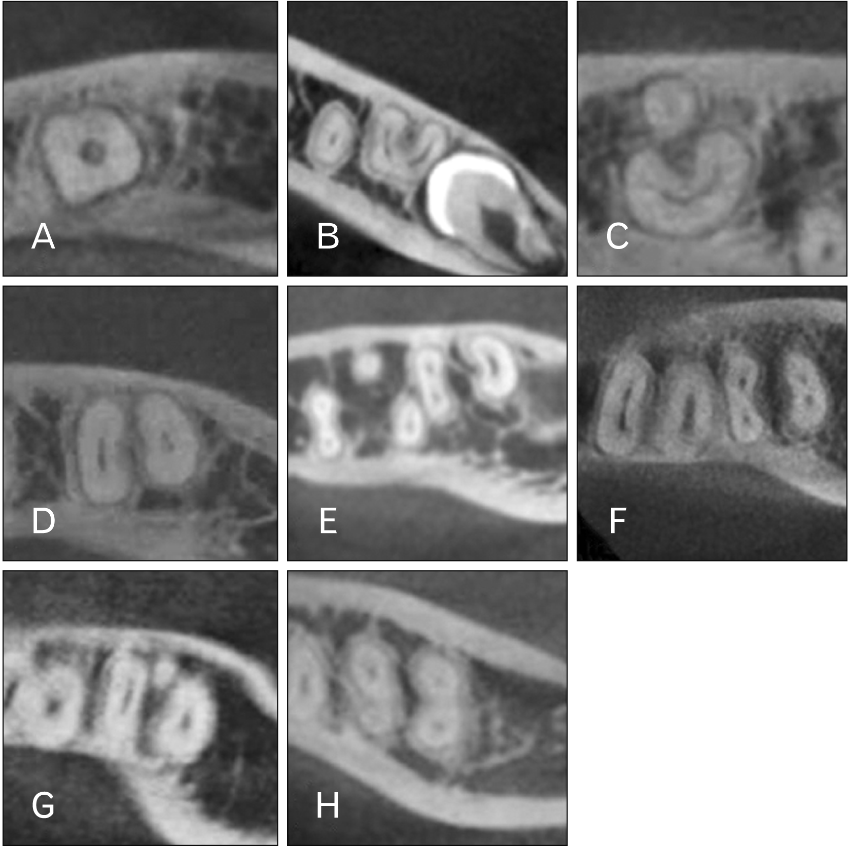
Sliced images at one-third of the root apex were made by reconstructing images vertical to the tooth axis using the imaging software package (One Volume Viewer; MORITA) (
Fig. 1). Root morphology assessments were done with the sliced images.
 | Fig. 1Cone-beam computed tomography images demonstrating various root and root canal morphological features. (A, B) Single root. (A) Conical single root. (B) Gutter-shaped root. (C–F) Double-root. (C) Gutter-shaped root and root on lingual side. (D) Double root with two root canals. (E) Double root with three root canals. (F) Double root with four root canals. (G) Three roots with three root canals. (H) Four roots with four root canals. 
|
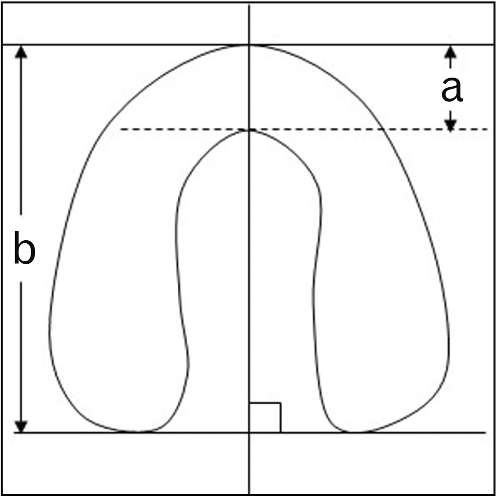
Observations of root morphology included determination of number of roots and calculation of the ratio of fused roots related to root thickness of GSR cases. Using the public domain software package ImageJ [
23], measurements of the length of the fused site and root thickness on the line through the most protruded point or most recessed point on the buccal side of the fused site, which connects at a right angle to the line between the most protruded points on the lingual sides of the mesial and distal roots, were made, with the results presented as percentage (
Fig. 2).
 | Fig. 2Method for fused site of root. a, thickness at fused site; b, thickness of root canal. a/b = Ratio of fused site of root canal divided by root canal thickness. 
|
For root canal morphology observations of GSR cases, binarized images were created with ImageJ. The range of the mandibular second molar root canal was selected in each sliced image and binarization was performed based on the image data within the root (
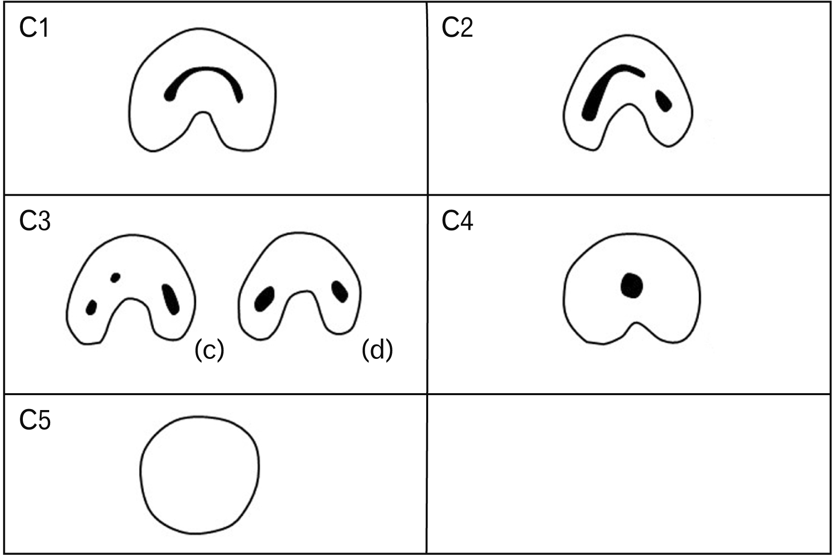
Fig. 3). The number of root canals was determined using the binarized images and classified into six types, using the Melton classification modified by Fan et al. (
Fig. 4), as follows [
22]:
 | Fig. 3Binarization process. (A) Sliced image showing mandibular second molar root canal. (B) Image showing periradicular region of interest. (C) Binarized image showing region of interest. 
|
 | Fig. 4Classification for root canal morphology of gutter-shaped root. C1, C-shaped; C2, Semicolon-like shape; C3(c), Separated three-root canals; C3(d), Separated two-root canals with smaller root canals, different from that shown in C3(c); C4, Single root; C5, Unobservable root canal. Cited from Fan et al. J Endod 2004;30:899-903, with permission [ 22]. 
|
C1: Uninterrupted C-shaped root canal
C2: Interrupted C-shaped and semicolon-like root canal
C3(c): Separated three-root canals
C3(d): Separated two-root canals
C4: Round or oval-shaped single canal
C5: Unobservable root canal
The ratio of the fused site to root thickness in GSR cases was compared between sexs using a t-test.
The present study protocol was approved by the Ethics Committee of Ohu University (No. 234).
Go to :

Results
As for root morphology results, a single root was found in 61 cases (35.3%), one of which was a simple conical root, while 107 cases (61.8%) had double roots, one of which had a root on the lingual side of the GSR, 4 cases (2.3%) had three roots, and one (0.6%) had four roots (
Table 1).
Table 1
Number of mandibular second molar root canals and ratio of development of gutter-shaped root
|
Number of the roots |
Number (%) |
|
One |
61 (35.3) |
|
Two |
107 (61.8) |
|
Three |
4 (2.3) |
|
Four |
1 (0.6) |

A GSR was found in 61 cases (35.3%), of which 60 (34.7%) showed a single root and one (0.6%) double roots on the lingual side of the GSR. The rate of development in males was 24.7% (22/89) and in females was 46.4% (39/84). The average ratio of the fused site of the root to root thickness at the one-third root apex was 47.8% in all cases, 47.0% in males and 49.6% in females, with no significant difference between sexs (
Table 2).
Table 2
Cases and development of gutter-shaped root
|
Sex |
Samples |
C-shaped root (n) |
Prevalence (%) |
With fused roots (%) |
|
Male |
89 |
22 |
24.7 |
47.0 |
|
Female |
84 |
39 |
46.4 |
49.6 |
|
Total |
173 |
61 |
35.3 |
48.7 |

C1 (uninterrupted C-shaped root canal) was the most common, accounting for 47.5% of these cases, followed by C2 (interrupted C-shaped and semicolon like-root canal) in 23.0%, C3(d) (separated two-root canals) in 19.7%, and C3(c) (separated three-root canals) and C4 (round or oval-shaped single canal), which were each found in 4.9%, while no cases of C5 (unobservable root canal) were found. The most common morphology of GSR in both sexs was C-shaped type, while the second common in males was semicolon type and in females were separated two-root canals (
Table 3).
Table 3
Classification of root canal morphology in gutter-shaped root cases
|
Shape |
Male |
Female |
Total |
|
C1(a) |
9 (40.9) |
20 (51.3) |
29 (47.5) |
|
C2(b) |
8 (36.4) |
6 (15.4) |
14 (23.0) |
|
C3(c) |
1 (4.5) |
2 (5.1) |
3 (4.9) |
|
C3(d) |
3 (13.6) |
9 (23.1) |
12 (19.7) |
|
C4(e) |
1 (4.5) |
2 (5.1) |
3 (4.9) |
|
Total |
22 |
39 |
61 |

Go to :

Discussion
Various studies have reported regarding the number of roots seen in mandibular second molars, in which those with a single root were noted in 5% to 6% of examined cases [
1,
4,
5], while two roots have been reported in a range of 75.8% to 94% [
1-
5,
7], and three roots in 1.2% to 9% [
1-
3,
5]. As for a GSR, development has been reported to occur in a wide range of 3% to 39.2% of examined cases [
1,
2,
7,
11,
18], with greater frequency noted in Asian patients. It was reported a GSR in 3% of Iranian patients [
1], while other reports noted 7.53% of cases examined in Indian [
2], 39.2% of cases in Japanese [
18], and 39.2% of cases in Chinese [
11]. The results showing GSR development in 35.3% of the present patients are similar to those for other Asian populations.
Another study found a difference between sexs for development of a GSR, with cases of a fused mesial root and distal root more prevalent in females [
18]. In the present observation study, prevalence was also higher in females (39 of 84; 46.4%) as compared to males (22 of 89; 24.7%). Another report noted that the crown diameters and crown areas of the mandibular molar was significantly greater in males [
24]. In females, the distance between the roots is generally shorter, thus more easily leading to fusion and development of a GSR. Kato et al. [
21] reported that teeth extracted due to periodontal disease showed a higher ratio of fused roots. In the present findings, there was no significant difference for ratio of the fused site to root thickness at the one-third of the root apex between sexs.
Zare Jahromi et al. [
1] reported that roots with a conical outer shape tended to show a simple root morphology, while those with a wider shape often had a more complex morphology. However, precise root canal morphology details cannot be obtained with the methods employed in that study. According to the guidelines regarding use of imaging for endodontics issued by the American Association of Endodontics (AAE) and the American Academy of Oral and Maxillofacial Radiology (AAOMR), CBCT is a helpful tool for diagnosis of complex or irregular root canal morphology at the time of pulp extirpation [
25]. Therefore, appropriate use of CBCT imaging including a lower exposure dose along with proper the field of view (FOV) settings is considered to be helpful for determining root canal morphology.
CBCT provides grayscale images without a CT value, thus various methods have been reported for measurement, including single rater observation [
12], confirmation of reliability of obtained data with a κ-test after repeated measurements [
13,
14], and obtaining consensus from several different raters [
15,
16]. Another report showed that deviation between raters can be avoided by using an image processing method for evaluation of grayscale images [
26]. In the present study, ImageJ [
23] was used for determining root canal morphology details by binarization after selecting the root outline of CBCT sliced images.
Development of a C-shaped root canal was reported to be more often seen in females than males [
18], while a contrasting report found no significant difference [
10]. In the present observation study, the most common root canal morphology at the one-third of the root apex side was a C-shaped root canal in both sexs. In other studies, a single root canal was found in 3% of examined cases, while double root canals were found in 6% to 11.1%, three root canals in 44.4% to 54%, and four root canals in 33.3% to 34% [
1,
27].
Development of a C-shaped canal has been reported to occur in a wide range of examined cases, from 19.1% to 38.6% [
11,
28]. In our study, the most common root canal morphology in males was a C-shaped root canal in 40.9%, followed by a semicolon-shaped root canal in 36.4%, while in females, the most common was a C-shaped root canal in 51.3%, followed by a root canal with two roots in 23.1%. Suzuki et al. [
18] conducted a study of Japanese and reported that on the one-third of the root apex side, the most common finding in males was a tooth with two root canals, followed by that with three root canals, while in females a tooth with two root canals was the most common, followed by a C-shaped root canal with a single root. That greater occurrence of a C-shaped root canal in females was confirmed by the present results. In the study by Suzuki et al. [
18], it was noted that the number of root canals tended to increase in the root apex direction. However, in our study, the most common type in both sexs was a C-shaped root canal with a single root canal.
A supernumerary root of the mandibular molar on the lingual side was termed “radix entomolaris” [
19,
20], while that on the buccal side is known as “radix paramolaris.” Radix entomolaris is considered to be a cause for plaque accumulation like that seen with a GSR [
21] and also reported to be a characteristic of Asian patients [
20]. Song et al. [
8] found that 0.4% of GSR cases had a root on the distal lingual side and a study conducted in Indian noted that 13.3% had radix entomolaris in the mandibular first molars [
21]. In the present study of mandibular second molar morphology, there was only one case (0.6%) with a supernumerary root on the lingual side of the GSR.
This investigation was conducted to observe morphological features of roots and root canals of mandibular second molars in patients undergoing treatment in Japanese using CBCT findings. The numbers of roots and canal features varied, most of the mandibular second molars were found to have two roots, with a GSR noted in 35.5%. Morphological assessment with CBCT is helpful for precise determination of root canal morphology and presence of a GSR in mandibular second molars.
Go to :


 PDF
PDF Citation
 Print
Print


 XML Download