Abstract
Cubital fossa is the site where the venous accesses are frequently made. Superficial veins at this site display variations in their pattern among different populations. Knowledge of different venous pattern in the cubital fossa is important for diagnostic, surgical and therapeutic procedures. The purpose of this study was to report variations of the cubital superficial vein patterns in the southern Ethiopian subjects. An institution based cross-sectional study design was employed among 401 randomly selected patients presented at the triage room of Arba Minch General Hospital from January 15 to February 15, 2021. A questionnaire was used to collect socio-demographic data and images of the common and variant superficial venous patterns were recorded. Descriptive statistical analysis was performed. P<0.05 was considered as statistical significance. In the present study, a total of 802 cubital fossae from 401 study participants were examined. Five patterns of superficial veins were identified. Type 2 was the most common pattern and observed in 55.0% of cubital fossae (42.1% right and 67.8% left cubital fossae). The least common, type 5 variant was detected in 2.6% cubital fossae (2.7% right and 2.5% left). Statistically significant association based on sex and laterality was noted. The current study concluded that type 2 and type 3 patterns were more frequent superficial venous patterns in the cubital fossa and more common in males than female. Awareness of these uncommon cubital venous patterns and their incidence is very useful for those performing venipuncture or venisection especially under emergency conditions.
The superficial and the deep venous system of the upper limb drains deoxygenated blood from the arm, forearm, and hand. Superficial veins are close to the surface of the body, and located within the subcutaneous tissue of the upper limb [1]. The main superficial veins of the cubital fossa include cephalic, basilic, median cubital, and median antebrachial. The cephalic vein is the longest vein of the upper limb, which normally originates from the lateral end of the dorsal venous network of the hand [2]. It runs upwards through the roof of the anatomical snuffbox and winds around the lateral border of the distal part of the forearm. The vein continues upwards in front of the elbow and along the lateral border of the biceps brachii. The cephalic vein pierces the deep fascia at the lower border of the pectoralis major, runs in the deltopectoral triangle and pierces the clavipectoral fascia, and joins the axillary vein [3]. The basilic vein is always present and begins from the medial end of the dorsal venous arch. It runs upwards along the back of the medial border of the forearm, winds round this border near the elbow, and continues upwards in front of the elbow. The basilic vein of the arm is anatomically compatible for use in arteriovenous fistulas for hemodialysis. The superficial segment of this vein may also be used in general, vascular and endovascular surgery to introduce a catheter above the cubitus [4]. The median cubital vein is a large communicating vein between the cephalic and basilic veins. It begins from the cephalic vein about 3 cm below the bend of the elbow, runs obliquely upwards and medially, and ends in the basilic vein about 3 cm above the medial epicondyle [5]. The median antebrachial vein receives the superficial venous network of the palm and ascends on the anterior aspect of the forearm and enters the basilic or median cubital veins [6].
The cubital fossa is the site where the venous accesses are frequently made [7]. Superficial veins in this site are the most common veins usually used to get blood samples, blood transfusions, and intravenous injections. Venipuncture has a great failure rate up to 30% and frequently requires several tries. Reasons such as lack of knowledge, difficulty in observing and assessing the superficial venous patterns in cubital fossa compromise the achievement of the procedures. The variability in the patterns of these veins is challenging to precisely perform the procedure for health professionals as only the common patterns are expected. Under normal circumstances the superficial veins are visible through the skin, however due to conditions that affect hemodynamics like in shocked patients, these veins will collapse and the visibility would diminish [8, 9]. Health professionals might injure the nearby neurovascular structures while trying to access into the veins. This injury causes local inflammation, hematoma, thrombus, infection, bruising and sensory changes [10, 11].
The superficial veins of the cubital fossa have various patterns of anastomosis than commonly known; these variations were found up to 20% in previous studies [12]. The most common pattern of cubital venous arrangement is formed, when the median vein of the forearm (median antebrachial vein) continues with the two terminal branches of the median cephalic and median basilic vein which connects the cephalic and the basilic vein, respectively [13]. It is postulated that genetic and hydrodynamic factors have an important role in the variations of venous patterns in the cubital fossa [8]. Awareness of sex and ethnic differences, typical and unusual venous patterns may help to achieve more direct access to these vessels, especially in emergency conditions. Moreover, knowledge of different venous pattern in the cubital fossa is important for diagnostic, surgical and therapeutic procedures. Taking into account the wide range of superficial cubital venous patterns found in different populations [14], the present study aimed to contribute for clinical trial such as venipuncture on patterns of superficial veins in the cubital fossa and its variations with sex and laterality.
The present study was conducted at Arba Minch General Hospital, located in Arba Minch town, southern Ethiopia. An institution based cross-sectional study design was employed among 401 randomly selected patients presented at the triage room from January 15 to February 15, 2021. Study participant with known vascular disease, scar, and wound over the cubital fossa were excluded from the study. A questionnaire was used to collect socio-demographic data (age, sex, and ethnicity) and images of the common and variant superficial venous patterns were recorded. After obtaining informed verbal consent, study participants were requested to expose their both upper limbs distal to the mid arm and extend elbow joint in supine position. To enhance the visibility of the veins, participants were clenching their fists on and off, massaging of the forearm and lightly tapping of the tissue to encourage vasodilation before applying a tourniquet. Then, a tourniquet was applied 10 cm proximal to elbow crease for about 3 to 5 minutes with active flexion and extension of the fingers until the veins were exposed for observation. The tourniquet was firm enough to occlude the veins but allowed for the pulsation of radial artery. Once the veins were exposed, the researcher and data collectors have recorded the observed patterns of the superficial veins of cubital fossa on the questionnaire.
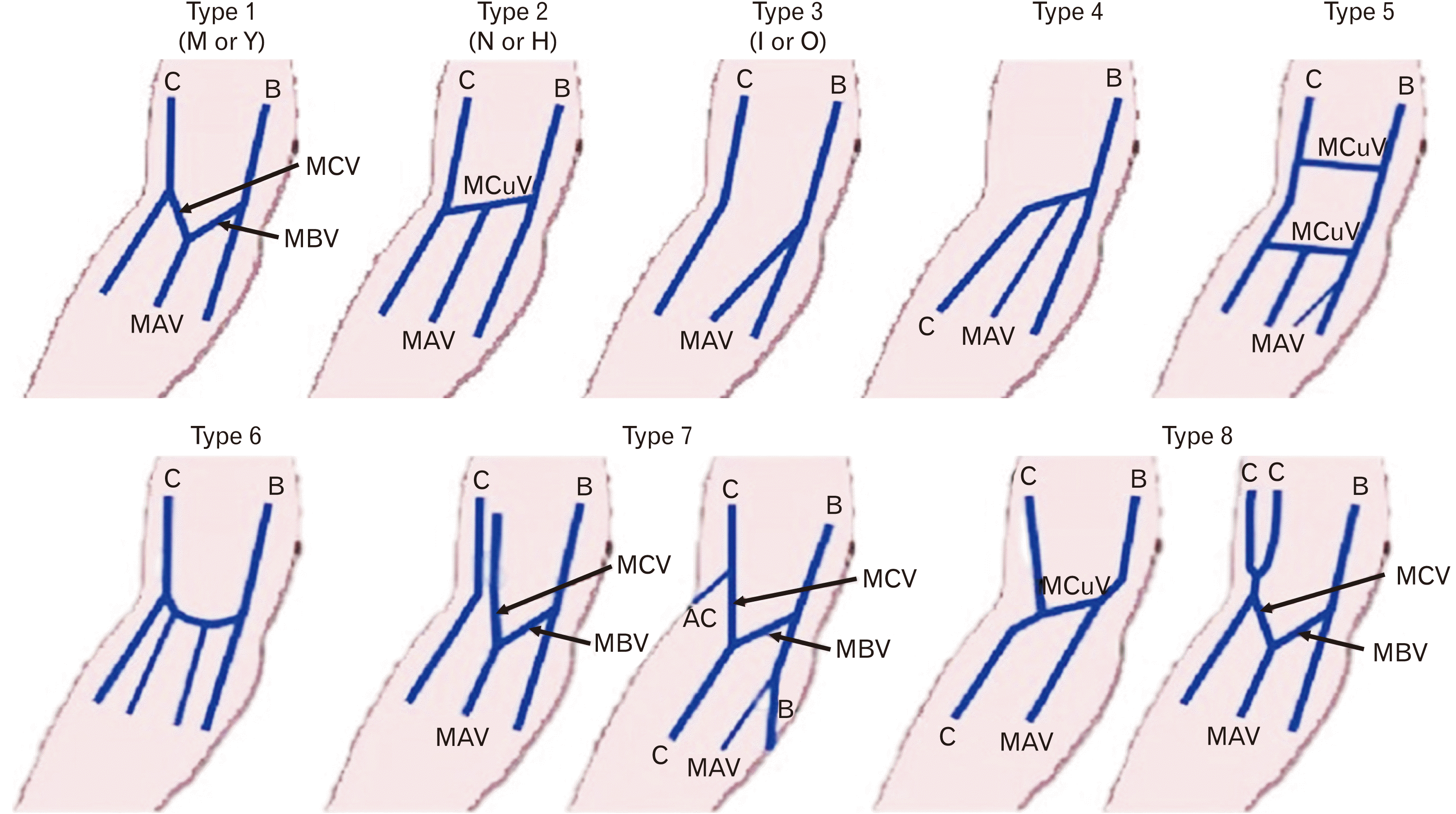
The present study adopts the classification of cubital superficial venous patterns by [15], as it is the most complete and versatile (Fig. 1).
These classifications are
Type 1: “M’’ shape pattern, in which the median vein of the forearm (median antebrachial vein) continues with the two terminal branches of the median cephalic and median basilic vein which connects the cephalic and the basilic vein, respectively.
Type 2: the “N’’ shape arrangement is formed when the median cubital vein diverges from the cephalic vein a few centimeters below the elbow joint and passes obliquely upward to ulnar side to join basilic vein a few centimeters above the elbow joint and obtain tributaries from the median vein of the forearm.
Type 3: the ‘I’ or’’ O’’ type pattern, in which only the basilic and the cephalic vein present. There is no communication between cephalic and basilic vein at the median antebrachial vein joins the basilic vein.
Type 4: In which the cephalic vein run from ulnar to radial side to join the basilic vein. No proximal cephalic vein.
Type 5: In which two median cubital veins over and under the crease of the elbow, where median antebrachial vein of the forearm drains into median cubital or basilic vein.
Type 6: This type of pattern happens, where the cephalic and the basilic vein joined by the arched vein with a proximal oriented concavity in to which two or more veins are drained from the forearm.
Type 7: Type 1 “M” like subtype where the median cephalic vein does not connect to the cephalic vein.
Type 8: Above the crease of the elbow, arrangement with double brachial cephalic vein, very minor type of venous pattern.
All the collected data was coded, cleaned, and entered into Epi-data version 4.4 (The EpiData Association, Odense, Denmark) and exported to SPSS version 25 (IRB Corp., Armonk, NY, USA) for analysis. For comparisons, chi-square Fisher exact test was used to determine significance at P<0.05.
This study was approved by Arba Minch University College of Medicine and Health Sciences’ institutional research ethics review board (Ref.no: IRB/551/12; Issue date: 26/11/2020). Verbal consent was requested from study participants.
The respondents age ranged from 18 to 59 years with a mean age of 32.91±10.4 years.
Of the total participants, 77.6% were males and majority of participants (75.3%) were ethnic Gamo (Table 1).
In the current study, a total of 802 cubital fossae were analyzed. From the total of 802 cubital fossae five patterns of superficial veins were identified. Type 2 was the most common pattern and observed in 441 (55.0%) of cubital fossae. It was seen in 42.1% right and 67.8% left cubital fossae. Type 1 pattern was identified in 25.7% cubital fossae (38.7% right and 12.7% left). Type 3 pattern was identified in 10.1% cubital fossae (8.9% right and 11.2% left side). Fifty three cubital fossae (6.6%) exhibited type 4 variant (7.5% right and 5.7% left cubital fossae). Type 5 variant was detected in 2.6% cubital fossae (2.7% right and 2.5% left cubital fossae) (Table 2).
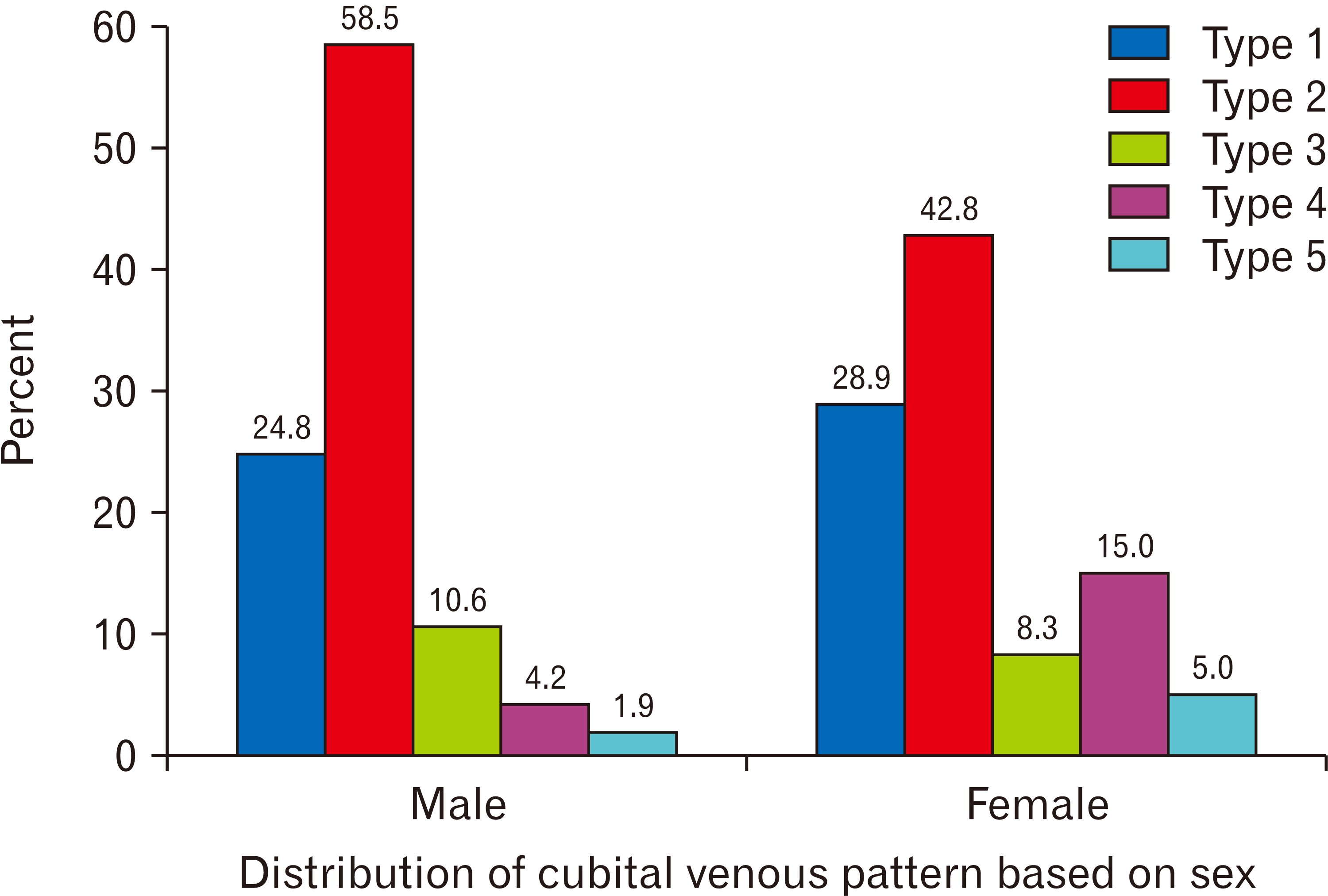
A total of 622 cubital fossae from 311 male participants and 180 cubital fossae from 90 female participants were analyzed for possible differences of venous pattern. In both sexes of study participants, five patterns of superficial veins were identified. The common patterns of superficial veins in males and females were type 2; it was seen in 364 (58.5%) cubital fossae and 77 (42.8%) cubital fossae of males and females respectively (Fig. 2).
Type 1 come in second with 154 (24.8%) and 52 (28.9%) males and females cubital fossae respectively. Type 3 pattern was higher in males study participants and it was observed in 66 cubital fossae (10.6%). On the other hand type 4 pattern was reported with high frequency in 27 (15.0%) females cubital fossae compared to males. Type 5 patterns were recorded below 10% be sides, it was observed in 12 males cubital fossae and 9 females cubital fossae (Fig. 3).
The present study clearly showed that the occurrence of type 2 and type 3 venous patterns are more common in males than females. On the other hand type 1 and type 4 patterns were more common in females than males (Fig. 2). Out of 401 study subjects, 72.1% had identical type of venous pattern in both arms, while the remaining 27.9% had different patterns in each arm. The occurrences of type 1 patterns are higher in right hands (38.7%) than left hands (12.7%). Sex and laterality fulfills the assumptions of chi-squared test. Noteworthy, the result of Pearson chi-square test revealed that sex and laterality were found to have statistically significant association (P<0.05) with venous pattern (Table 2).
Variations of the superficial veins of the upper limb are well-known. In embryonic life the veins arise from capillary plexus, which increase by sprouting anastomosing and then fuse, enlarge forming fewer and larger channels. Genetic and hydrodynamic factors plays an important role in the final pattern of arrangement of veins, which may results variations in venous pattern of the cubital fossa [16]. The superficial veins of the upper limb are commonly used in the clinical practice for venipuncture and arteriovenous fistulas for hemodialysis patients, as they are easy to reach and drain a significant portion of the upper limb blood [17].
In the present study, five patterns of superficial veins of the cubital fossa were identified. The common patterns were type 2 (55.0%) and type 1 (25.7%). Moreover, sex and laterality were significantly associated (P<0.05) with cubital venous patterns. In this study, a total of 441 (55.0%) cubital fossae exhibited type 2 in which the cephalic vein gives off median cubital vein which passes upward to medial side to join basilic vein. This finding is consistent with the venous pattern mentioned in a standard anatomical text books and several previous studies [2, 12, 18-20]. However, type 2 venous pattern was not found to be common in studies conducted by [8] and [13].
According to the present study, the second common venous arrangement was type 1 in which a single median antebrachial vein splits into median cephalic vein and median basilic vein and joins the cephalic and basilic vein, respectively. The type 1 pattern was found in 25.7% cubital fossae. Similar findings were reported by [2, 13, 21]. However, the prevalence of this venous pattern was found lower in studies conducted by [12] and [19]. The possible reasons for the difference in the occurrences of common venous patterns between studies might be explained by racial differences across the populations (Table 3).
Several studies were reported that most health professionals are aware of only type 1 and type 2 superficial venous patterns in the cubital fossa [12]. However, in the current study additional three cubital venous patterns were found. 10.1% cubital fossae show type 3 in which the median antebrachial vein join basilic vein without communication between cephalic and basilic vein. In line with present findings, the study conducted by Bekel et al. [12], found type 3 pattern in 18.6%. In type 4, cephalic vein run from radial to ulnar side to join the basilic vein and it receive tributaries from the front of the forearm; there was no proximal cephalic vein and it was depicted in 6.6% of the cubital fossae. In line with our finding studies conducted among Indians and Malaysians by [16, 19] showed the prevalence of type 4 patterns in less than 10%. The least common superficial venous pattern of the cubital fossa was type 5 (2.6%), in which the cephalic and basilic vein connected by arched vein with a proximal oriented concavity. Similar low incidence of this pattern was reported in previous studies [9, 20, 21]. The observed difference of venous patterns among studies may be due to numerous classifications of venous patterns and subjective opinions of the researchers, for example findings from dissected cadavers showed extraordinarily low incidences of type 1 and type 2 superficial venous patterns in the cubital fossa [7, 16].
The present study showed that superficial venous patterns in cubital fossa were related with sex. The occurrences of type 2 and type 3 venous patterns were more common in males than females (Fig. 2). Similarly to our findings, several previous studies reported that superficial cubital venous patterns were also statistical significant association with sex [8, 12, 13, 22]. In contrast, some studies [2, 16, 21] reported that sex had no effects on the patterns of cubital veins. This discrepancy may be due to sample size difference between the studies. For example study conducted in Korea stated that due to small subjects, including sex influence and statistically interpret the difference was not possible [2]. A recent study by Żytkowski et al. [23] and Del Sol et al. [24] reported a rare case where a duplicated median cubital vein crossed by medial cutaneous nerve of the forearm, such proximity to the nerves might increase the risk for iatrogenic nerve injury during venipuncture.
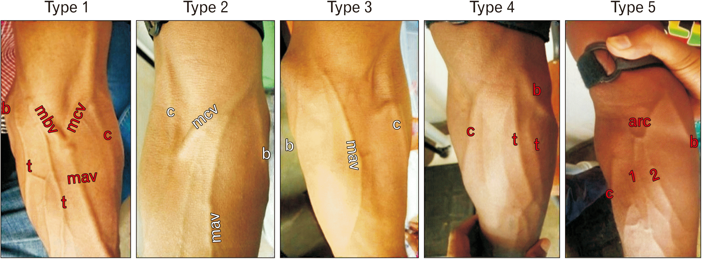
Findings from a meta-analysis study indicated that whether the investigation was cadaveric or clinical, the commonest pattern of superficial veins in the cubital fossa was type 2 followed by type 1. Similarly this most common pattern (type 2) also follows to standard text book of anatomy, with the cephalic vein gives of median cubital vein which is passes up ward and medially to join the basillic vein (Fig. 3) [1]. This view was not only supported by study done in the North West Ethiopians as a commonest pattern (58.5%) [12], but also studies conducted in Koreans, Indian, Poland and Malaysians populations with the percentage of 50.1%, 51%, 43.7%, and 66.5% respectively [2, 6, 16, 21].
The present study had some limitation of missing small vessels and not able to examine the positional relationship with the cutaneous nerve in the cubital region.
In conclusion, the current study observed that type 2 and type 3 patterns were more frequent superficial venous patterns in the cubital fossa and more common in males than female. The venous patterns in the present study are in line with the findings of former studies. However, some rare venous patterns were also identified. Repeated observations of anatomical variations deepen existing knowledge, can help to overcome the subjectivity of descriptions by individual researchers, and can also be useful for clinicians in their daily practice, especially under emergency conditions.
Notes
References
1. Standring S. 2016. Gray's anatomy: the anatomical basis of clinical practice. 41st ed. Elsevier;Edinburg:
2. Lee H, Lee SH, Kim SJ, Choi WI, Lee JH, Choi IJ. 2015; Variations of the cubital superficial vein investigated by using the intravenous illuminator. Anat Cell Biol. 48:62–5. DOI: 10.5115/acb.2015.48.1.62. PMID: 25806123. PMCID: PMC4371182.

3. Drake RL, Vogl W, Mitchell AWM. 2010. Gray's anatomy for students. 2nd ed. Churchill Livingstone/Elsevier;Philadelphia:
4. Darabi MR, Shams A, Bayat P, Bayat M, Babaee S, Ghahremani B. 2015; A case report: variation of the cephalic and external jugular veins. Anat Sci J. 12:203–5. PMID: aa4a05e4ef64407ea2e18abbca0d4008.
5. Jain T, Yadav SK. 2015; Case study: variation of superficial veins pattern of upper limb found in dissection. Int Ayurvedic Med J. 3:2223–5.
6. Jasiński R, Poradnik E. 2003; Superficial venous anastomosis in the human upper extremity--a post-mortem study. Folia Morphol (Warsz). 62:191–9. PMID: 14507046.
7. Mikuni Y, Chiba S, Tonosaki Y. 2013; Topographical anatomy of superficial veins, cutaneous nerves, and arteries at venipuncture sites in the cubital fossa. Anat Sci Int. 88:46–57. DOI: 10.1007/s12565-012-0160-z. PMID: 23131916.

8. Vučinić N, Erić M, Macanović M. 2016; Patterns of superficial veins of the middle upper extremity in Caucasian population. J Vasc Access. 17:87–92. DOI: 10.5301/jva.5000429. PMID: 26109546.

9. Horowitz SH. 1994; Peripheral nerve injury and causalgia secondary to routine venipuncture. Neurology. 44:962–4. DOI: 10.1212/WNL.44.5.962. PMID: 8190306.

10. Newman B. 2001; Venipuncture nerve injuries after whole-blood donation. Transfusion. 41:571–2. DOI: 10.1046/j.1537-2995.2001.41040571.x. PMID: 11316914.

11. Stevens RJ, Mahadevan V, Moss AL. 2012; Injury to the lateral cutaneous nerve of forearm after venous cannulation: a case report and literature review. Clin Anat. 25:659–62. DOI: 10.1002/ca.21285. PMID: 22025401.

12. Bekel AA, Bekalu AB, Moges AM, Gebretsadik MA. 2018; Anatomical variations of superficial veins pattern in Cubital fossa among North West Ethiopians. Anat J Afr. 7:1238–43. DOI: 10.4314/aja.v7i2.174144.

13. Ukoha UU, Oranusi CK, Okafor JI, Ogugua PC, Obiaduo AO. 2013; Patterns of superficial venous arrangement in the cubital fossa of adult Nigerians. Niger J Clin Pract. 16:104–9. DOI: 10.4103/1119-3077.106777. PMID: 23377482.

14. Newman BH, Waxman DA. 1996; Blood donation-related neurologic needle injury: evaluation of 2 years' worth of data from a large blood center. Transfusion. 36:213–5. DOI: 10.1046/j.1537-2995.1996.36396182137.x. PMID: 8604504.

15. Yammine K, Erić M. 2017; Patterns of the superficial veins of the cubital fossa: a meta-analysis. Phlebology. 32:403–14. DOI: 10.1177/0268355516655670. PMID: 27343223.

16. Vasudha TK. 2013; A study on superficial veins of upper limb. Nat J Clin Anat. 2:204–8. DOI: 10.4103/2277-4025.297895. PMID: b47b9fe6acfb46138e543bc1b9fe2b6a.

17. Elamurugan E, Hemachandar R. 2017; Brachiocephalic arteriovenous fistula for hemodialysis through the median antecubital vein. Indian J Nephrol. 27:177–80. DOI: 10.4103/0971-4065.179333. PMID: 28553035. PMCID: PMC5434681.

18. Yamada K, Yamada K, Katsuda I, Hida T. 2008; Cubital fossa venipuncture sites based on anatomical variations and relationships of cutaneous veins and nerves. Clin Anat. 21:307–13. DOI: 10.1002/ca.20622. PMID: 18428997.

19. Dharap AS, Shaharuddin MY. 1994; Patterns of superficial veins of the cubital fossa in Malays. Med J Malaysia. 49:239–41. DOI: 10.1097/00006534-199808000-00075. PMID: 7845272.
20. Wasfi FA, Dabbagh AW, AlAthari FM, Salman SS. 1986; Biostatistical study on the arrangement of the superficial veins of the cubital fossa in Iraqis. Acta Anat (Basel). 126:183–6. DOI: 10.1159/000146212. PMID: 3751488.

21. Hamzah AA, Ramasamy S, Adnan AS, Khan AH. 2014; Pattern of superficial venous of the cubital fossa among volunteers in a tertiary hospital. Trop Med Surg. 2:1000164. DOI: 10.4172/2329-9088.1000164.

22. AlBustami F, Altarawneh I, Rababah E. 2014; Patterns of superficial venous arrangement in the cubital fossa of adult Jordanians. Jordan Med J. 48:269–74. DOI: 10.12816/0025077.
23. Żytkowski A, Tubbs RS, Iwanaga J, Olszewska A, Kunikowska B, Wysiadecki G. 2021; Duplication of the median cubital vein- case report with commentaries on clinical significance. Transl Res Anat. 24:100114. DOI: 10.1016/j.tria.2021.100114.
24. Del Sol M, De Angelis MA, Bolini PAD. 1988; Formacoes venosas de fossa cubital crianca. Pediatria Mod. 23:225–31. Portuguese.
Fig. 1
Diagrammatic illustration of superficial veins patterns in cubital fossa. C, cephalic vein; B, basillic vein; MCuV, median cubital vein; MCV, median cephalic vein; MBV, median basillic vein; MAV, median ante brachial vein.

Fig. 3
Five types of superficial venous patterns of study participants. 1 and 2, tributaries draining to the arched median vein; c, cephalic vein; b, basilic vein; mcv, median cephalic vein; mbv, median basilic vein; mav, median antebrachial vein; t, other tributaries; arc, arched vein with proximal concavity.

Table 1
Socio-demographic characteristics of adult patients attended in Arba Minch general Hospital, Gamo zone, Southern Ethiopia, 2021 (n=401)
| Variables | Frequency (n) | Percent (%) |
|---|---|---|
| Sex | ||
| Male | 311 | 77.6 |
| Female | 90 | 22.4 |
| Age (yr) | ||
| ≤19 | 29 | 7.2 |
| 20–29 | 200 | 49.9 |
| ≥30 | 172 | 42.9 |
| Ethnicity | ||
| Gamo | 302 | 75.3 |
| Gofa | 50 | 12.5 |
| Wollayita | 35 | 8.7 |
| Others | 14 | 3.5 |
Table 2
Variations of superficial veins patterns with sex and body side among adult study participants in Arba Minch General Hospital, Southern Ethiopia, 2021 (n=802)
| Variable | Pattern of superficial veins at the cubital fossa | ||||||
|---|---|---|---|---|---|---|---|
| Type 1 | Type 2 | Type 3 | Type 4 | Type 5 | Total | P-value | |
| Male | 154 (24.8) | 364 (58.5) | 66 (10.6) | 26 (4.2) | 12 (1.9) | 622 (100) | 0.000* |
| Female | 52 (28.9) | 77 (42.8) | 15 (8.3) | 27 (15.0) | 9 (5.0) | 180 (100) | |
| Right | 155 (38.7) | 169 (42.1) | 36 (8.9) | 30 (7.5) | 11 (2.7) | 401 (100) | 0.001* |
| Left | 51 (12.7) | 272 (67.8) | 45 (11.2) | 23 (5.7) | 10 (2.5) | 401 (100) | |
| Total | 206 (25.7) | 441 (55.0) | 81 (10.1) | 53 (6.6) | 21 (2.6) | 802 (100) | |
Table 3
Different patterns of cubital superficial veins noted in our population in comparison with the same in other studies
| Author | Population | Sample size (arms) | Study type | Type 1 | Type 2 | Type 3 | Type 4 | Type 5 | Type 6 | Type 7 | Type 8 |
|---|---|---|---|---|---|---|---|---|---|---|---|
| Present study | Ethiopian | 802 | Clinical | 206 (25.7) | 441 (55.0) | 81 (10.1) | 53 (6.6) | 21 (2.6) | - | - | - |
| Ukoha et al. [13] | Nigerian | 270 | Clinical | 75 (27.8) | 76 (28.2) | 11 (4.1) | 14 (5.2) | - | - | 14 (5.2) | 80 (29.6) |
| Bekel et al. [12] | Ethiopian | 800 | Clinical | 468 (58.5) | 149 (18.6) | 112 (14.0) | 71 (8.9) | - | - | - | - |
| Dharap and Shaharuddin [19] | Malaysia | 532 | Clinical | 86 (16.2) | 362 (68.0) | 44 (8.3) | 12 (2.2) | 1 (0.6) | 26 (4.9) | - | - |
| Vučinić et al. [8] | Serbs | 338 | Clinical | 177 (52.4) | 125 (37.0) | 17 (5.0) | 6 (1.8) | 10 (2.95) | - | - | - |
| Mikuni et al. [7] | Japanese | 128 | Cadaver | 1 (0.78) | 104 (82.0) | 9 (7.0) | - | - | - | - | 14 (11.0) |
| Del Sol et al. [24] | Brazil | 40 | Cadaver | 12 (30.0) | 12 (30.0) | 10 (25.0) | 4 (10.0) | - | - | - | 2 (5.0) |
| Vasudha [16] | Indian | 50 | Cadaver | 44 (88.0) | 2 (4.0) | 2 (4.0) | 2 (4.0) | - | - | - | - |
| Jasiński and Poradnik [6] | Polish | 80 | Cadaver | 26 (32.5) | 35 (43.7) | - | 13 (16.2) | - | - | - | 6 (7.5) |


 PDF
PDF Citation
 Print
Print


 XML Download