

 PDF
PDF Citation
 Print
Print


Introduction
Frontal sinuses are paired irregular pneumatic cavities located between the outer and inner tables of the frontal bone, posterior to the superciliary arches [1]. Frontal sinus development begins around the fourth or fifth week of gestation, and continues during the intrauterine and postnatal periods through puberty or even early adulthood [2]. The length, height, and width of the frontal sinuses increase continuously until 20 years of age, after which their size and shape are maintained except when disease or aging occur [3].
The frontal sinuses have been frequently studied using plain-film radiography as a screening method [4]. Although this technique is widely used, computed tomography (CT) plays an important role in evaluating diseases and detecting anatomical variations of the frontal sinus, and may gradually replace conventional radiographs [5]. The main advantage of CT over plain radiography is the absence of overlapping structures, allowing clear, three-dimensional (3D) visualizations of anatomical structures [6]. CT has also become an essential tool for planning frontal sinus surgeries and for navigation during functional endoscopic sinus surgery [7].
Studies of the frontal sinus using 3D reconstructions of CT data have recently increased [8]. There have been several reports on the use of 3D analysis for evaluating the complex anatomy of the frontal sinus and planning frontal sinus surgeries [9, 10]. Various forensic science studies have focused on using the frontal sinus for identifying individuals, since the morphology of the frontal sinuses us inherently variable [11, 12]. Some studies have performed frontal sinus measurements using 3D reconstruction analysis. Hacl et al. [8] reported the height, width, depth and volume of the frontal sinus in 36 facial CT, and analyzed the side and sex differences. Yun et al. [3] reviewed 352 facial CT scans from patients younger than 23 years to evaluate the development of the frontal sinus over time using width, height, length, and volume data. However, few studies of the frontal sinus have used 3D analysis of CT data to compare the linear and volumetric measurements between adult age groups.
Therefore, the purpose of this study was to use 3D reconstruction analysis to investigate the frontal sinuses and determine differences in its linear and volumetric measurements between age groups, nose sides, and sexes in adults.
Materials and Methods
Participants
Facial CT scans were selected retrospectively from patients hospitalized in the Department of Plastic and Reconstructive Surgery at the Konkuk University Chungju Hospital. Facial CTs of 349 subjects were selected, with an age range of 21 to 80 years in order to exclude developing bones. Of these 349 subjects, 68 were excluded due to frontal sinus fracture, surgery, or agenesis. The final sample therefore included 281 subjects, comprising 173 males and 108 females. The sample population was divided into two age groups: young adults aged 21 to 40 years, and old adults aged 41 to 80 years.
CT scanning, 3D rendering and measurements
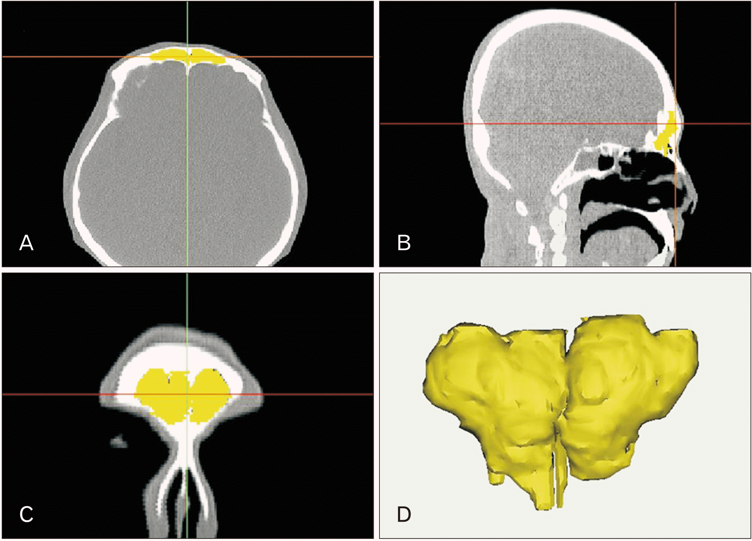
Transverse serial facial CT images were acquired with a slice thickness of 1 mm (Hispeed G; GE Healthcare, Niskayuna, NY, USA). The raw CT files were converted to clinical images in DICOM format, and then reconstructed into 3D-rendering files using Mimics software (version 20.0; Materialise, Leuven, Belgium) (Fig. 1), which was used to manually segment images of the frontal sinuses.
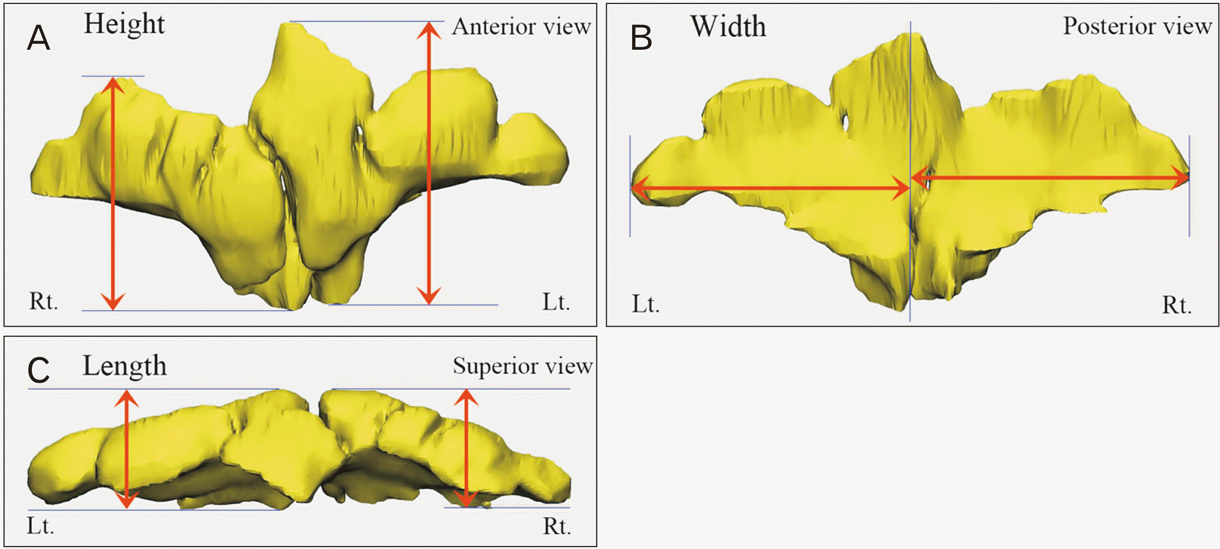
Three perpendicular reference planes (midsagittal, coronal, and Frankfort) were drawn on each rendered 3D file using 3-matic software (version 12.0; Materialise). The Frankfort plane is defined as the plane passing through three points: the left orbitale and both porion points. The orbitale is the lowest point of the lower margin of the orbit, and the right and left porions are the uppermost points of the roof of each external acoustic opening. The width (midsagittal plane), anteroposterior length (coronal plane), and height (Frankfort plane) of the frontal sinuses were measured using the planes as reference (Fig. 2). Widths of the left and right frontal sinuses were measured independently as the longest horizontal distance from the midsagittal plane (Fig. 1A). The total width of the frontal sinus was obtained by the sum of the widths of the two sides. The anteroposterior length of the frontal sinus was measured on the coronal plane as the distance between the most-anterior and most-posterior points from the center of the glabella (Fig. 1B). The anteroposterior lengths of the left and right sides were measured both independently and together to obtain the total length. Height was measured on the Frankfort plane as the vertical distance between the lowest and highest points (Fig. 1C). Height was measured both independently between the left and right sides and as a total height, measured from the lowest to the highest point on either side. Segmented frontal sinus volumes were calculated automatically using 3-matic software (Fig. 1D).
Statistical analysis
The side-, sex-, and age-related differences were determined using independent samples t-test. All data analysis was performed using IBM SPSS Statistics for Windows, version 24.0 (IBM Corp., Armonk, NY, USA). The cutoff for statistical significance was set at P<0.05.
Results
Facial CTs of 281 adults aged between 21 and 80 years (mean age of 38.3-year-old) were analyzed. Table 1 lists the width, height, length, and volume of the frontal sinuses by side, sex, and age group.
Almost all of these linear measurements were larger in the young adult group than in the old adult group for both sexes. Almost all measurements in males (including volume) differed significantly between the age groups (P<0.05). Almost all female measurements were larger in the young adult group, but the only height of the frontal sinus showed a significant difference between the age groups (P<0.05).
The mean frontal sinus volumes in males were 10.2 and 7.0 cm3 in the young and old adult groups, respectively (P<0.05). In females, there was no statistically significant difference in mean volume between age groups (P>0.05).
All of the linear and volumetric measurements are compared between the sexes are listed in Table 2. All dimensions except for the total height of the frontal sinus were significantly larger in males than in females regardless of age group (P<0.05). Sex differences were more predominant in young adults (Table 2).
Differences between frontal sinus sides in both sexes are presented in Table 3. The mean width of the right frontal sinus in males was 2 mm larger than the left frontal sinus (P<0.05). However, there were no significant differences in the width, height, and length between the two sides of the frontal sinus (P>0.05), except for the width of the frontal sinus in both sexes.
Discussion
Various previous studies have obtained anatomical measurements of the width, height, and anteroposterior length of the frontal sinus using plain radiographs and two-dimensional CT [13-15]. Many environmental and genetic factors have been demonstrated to influence the morphology of the frontal sinus between different populations [16]. Kirk et al. [15] measured the frontal sinus on 39 adult radiographs in Canada and reported a mean total width and height of 71.29 and 41.84 mm, respectively. In the present study, the mean total width and height were 54.7 and 31.5 mm, respectively, regardless of sex or age. The much larger measured dimensions in that Canadian sample compared with our study might have been due to differences in the size and shape of the skull between the samples.
Tatlisumak et al. [14] investigated the width, height, and anteroposterior length of each frontal sinus from 300 paranasal CT scans in Turkey, and compared the results between sexes and age groups. They reported that all linear parameters of the frontal sinus were significantly larger in males than in females. Another study analyzing 98 CT images found that the frontal sinus tended to be larger in males than in females, but this difference was not always statistically significant [17]. All linear parameters of the frontal sinus in our study were larger in males than in females, which is in line with previous studies (Table 1). We also compared these results between age groups: in the young adult group, the width, height, and length of each side were typically 4–9 mm larger in males than in females (P<0.05) (Table 2), whereas in the old adult group, almost all parameters of the frontal sinus were larger in males (P<0.05). Based on the results from both the present and previous studies, frontal sinus measurements can be considered to be very useful for differentiating between the sexes regardless of age. However, the differences for both sexes were smaller in the old than in the young adult group, which was due to contraction in the body size with aging being dominant in males. The correlation between contraction of the frontal sinus and aging should receive more attention in future studies.
Independent pneumatization of either the right or left side of the frontal sinus can cause an asymmetric appearance [18]. The septum separating the two sides of the sinus usually deviates from the midline [19]. Several studies have found the width of the left frontal sinus to be larger in both sexes and that the left sinus is more likely to be damaged when performing a supraorbital craniotomy [14, 17]. We measured the width of each side of the frontal sinus as the longest horizontal distance from the midsagittal plane, unlike previous studies that measured each width as the distance from the septum. We found that the frontal sinus was slightly wider on the right than on the left side in both sexes. The left side was higher and longer in males than in females, without a significant difference.
There has recently been an increasing number of studies on the frontal sinuses using volumetric measurements. These volumetric analyses using 3D reconstruction of CT data have mostly been used in forensic medicine and anthropology for determining the sex and identifying individuals [11, 12, 20]. 3D reconstruction analysis can also be used in clinical practice since it improves the clinician’s understanding of complex structures, diagnoses, and surgical planning [21]. Park et al. [22] reported that the air cavity volume was a significant parameter for evaluating the paranasal sinuses after investigating the CT data of 260 patients younger than 25 years. Those authors reported that the mean volume of the fully-grown frontal sinuses was 3.46 cm3. However, this volume is much smaller than our results (Table 1), which might be due to technical or resolution problems.
Hacl et al. [8] measured the volume of the frontal sinus from 36 facial CT scans, and found that it was 4.2 and 5.2 cm3 for the right and left frontal sinuses, respectively, in males, and 2.9 and 3.1 cm3 in females. However, these differences were not statistically significant. Another study performing volumetric measurements of the frontal sinus found mean volumes of 4.5 and 5.0 cm3 for the right and left sides, respectively, in males, and 2.4 and 2.6 cm3, respectively [23]. There was a significant difference between males and females but not between sides. We also compared the frontal sinus volume between age groups, sides, and sexes. There were significant differences between sexes and age groups, with higher values for males and young adults. The frontal sinus volume was much larger in males than in females. The differences were greater for sex than for age due to differences in the total skull size. Especially, the superciliary arch as a part of the anterior wall of the frontal sinus is more prominent in males than in females, therefore, it probably affected in size difference of the frontal sinus.
To the best of our knowledge, there have been few 3D volumetric analyses of the frontal sinuses determining differences between adult age groups, although one study did perform linear measurements in several age groups [14]. In that study the subjects were divided into five groups according to age (all >20 years old), and the linear measurements were generally largest among those aged 31–40 years, with a tendency to decrease with aging, which was similar to our findings. McLaughlin et al. [24] suggested that the frontal sinuses expand until 40 years of age because of the mechanical stress from mastication and growth hormones. The etiology of the frontal sinus becoming smaller with age should be a focus of future research.
Our results obtained through the 3D analysis of facial CT data indicated that the size of the frontal sinus is related to sex and age. The width, anteroposterior length, height, and volume of the frontal sinus were mostly larger in males and young adults than in females and old adults. The present analysis of the frontal sinus is the most accurate that we know of, and the comparisons according to sex and age were significant. These 3D measurements of the frontal sinus are useful for clinicians in understanding the complex anatomy of the frontal sinus and when they are performing surgical procedures.
 XML Download